


Serviços Personalizados
Artigo
 pdf em Inglês
 Artigo em XML
 Referências do artigo
 Enviar este artigo por email
Links relacionados
Compartilhar

 Permalink
RSBO (Online)
versão On-line ISSN 1984-5685
RSBO (Online) vol.9 no.2 Joinville Abr./Jun. 2012
Original Research Article
Effectiveness of an electronic apex locator used after preflaring of cervical and middle third
João Marcelo da Silva Teixeira I; Myrna Bastos Barcellos I; Marco André de Berrêdo Pinho I; Carlos Augusto de Melo Barbosa I; Rivail Antônio Sérgio Fidel I; Sandra Rivera Fidel I
I Department of Integrated Clinical Procedures (Proclin), School of Dentistry, Rio de Janeiro State University – Rio de Janeiro – RJ – Brazil.
ABSTRACT
Introduction: The electronic method has been studied and improved aiming to add precision, speed and reliability of the measurement technique to determine the exact location of the working length. Currently, the root canal preparation recommends prior to determine the tooth length and consequent perform instrumentation of the apical portion, a previous preflaring of the cervical and middle thirds in various techniques. This procedure may provide a reduction in system impedance, leading to read errors by the apex locators. Objective: Investigate the influence of preflaring of the cervical and middle thirds on the accuracy of measuring the working length by apex locators. Material and methods: Twenty-five mesial roots of molars were used and had their crowns cut at the cemento-enamel junction. The actual measure of each root canal was performed and then the samples were embedded into a mixture of alginate, used as a conducting medium, where electronic measurements were taken with apex locator before and after preflaring of the canals with Gates-Glidden drills in descending order (#4, #3, #2). Measurements obtained by electronic method were then compared with the actual measurement of the root canal. The results were tabulated and submitted to the Student t test. Results: The results show that there was no statistical significance (p<0.05) between the readings before and after preflaring. Readings closer to the foraminal ending occurred in the group after preflaring with Gates Glidden. Conclusion: It was concluded that preflaring with Gates Glidden drills were not able to influence significantly the accuracy of apex locator in determining the exact working length.
Keywords: endodontics; apical locator; working length.
Introduction
The correct determination of the working length is one of the main factors leading to endodontic treatment success. Current studies have demonstrated that histologic outcomes after endodontic treatment are higher when instrumentations and obturation are limited to apical narrowing 13,18.
Traditionally, working length has been determined through radiographs, however, currently electronic apical locators have gained popularity 5. Suzuki 22, in 1942, showed a device that measured the electrical resistance between the periodontal ligament and oral mucosa, registering an electrical resistance of about 6,5KΩ in a study conducted in dogs. This study enabled the development of the first apical locator by Sunada 21 in 1962, based on a constant value of resistance between the periodontal ligament and oral mucosa. Since then, different generations of apical locators have been developed to measure root canal length.
Currently, locators based on multiple frequencies (third generation) have gained prominence by allowing precise readings in wet root canals 15,23. Apical locators, currently, represent an important tool for the endodontist, because it enables with more practicality and precision the determination of root canal’s working length, reducing the doubts regarding to the exact location of foraminal ending.
Considering the current techniques of root canal preparation, we note a concern on the preflaring of the cervical and medium thirds of the canals, which may show in some cases a marked reduction in the remnant thickness of dentin at these areas prior to odontometry. Pre-flaring would facilitate the insertion of the files in root canal’s apical third 8,20.
The aim of this in vitro study is to determine the influence of preflaring of cervical and middle third on apical locator accuracy.
Material and methods
Twenty-five mandibular molars with complete apexes were selected for this study. These teeth were extracted due to different reasons and came from the tooth bank of the Rio de Janeiro State University (UERJ). They were kept in 0.1% thymol solution until their use. The specimen use was approved by the Ethical Committee in Research of UERJ (protocol number CEP/HUPE #2921). A conventional endodontic access was performed using carbide round bur #1157 (SSWhite, Rio de Janeiro, RJ, Brazil) at high speed rotation. Root canal permeability was negotiated through K file #10 (Dentsply Maillefer, Balaigues, Switzerland) aiming to discard any tooth presenting blockages within canal, therefore, obtaining patency.
Next, the teeth had their crowns cut at enamel-cement junction (ECJ) to establish a safe and standardized landmark for the measurements. Prior to the electronic measurements, the actual measurement of the mesial root canals were verified through inserting a K file #10 (Dentsply Maillefer, Balaigues, Switzerland) in each canal through catheterization movements up to the file’s tip is visible through the foramen with the aid of a magnifying lens (x 2.5) (Hoya, Rio de Janeiro, RJ, Brazil). The silicone cursor was adjust at the level of the tooth’s horizontal surface and a 0.5-mm interval millimeter ruler (Dentsply Maillefer, Balaigues, Switzerland) was used to measure the distance from the silicone cursor and the file’s tip. This measurement was defined as the canal’s total measurement, and working length (WL) was establishing at 1 mm shorter of the canal’s total length.
The electronic measurements were performed by using a glass flask where alginate (Jeltrate, Dentsply, Petrópolis, RJ, Brazil) was mixed according to the manufacturer’s instructions regarding proportions; however, water was substituted by 0.9% saline solution to increase the electric conductivity 9. The teeth were embedded into alginate leaving about 2 mm of root surface exposed.
The tooth was kept in position until the complete alginate setting. All measurements were executed within a 2-hour interval while the alginate was sufficiently wet, by using the apical locator Bingo 1020 (Forum Engineering Technologies, Rishon Lezion, Israel). During the electronic measurement the labial clip was inserted within the alginate and stabilized with adhesive tape.
Initially, the mesial canals were irrigated with 5.25% sodium hypochlorite solution and a Flexofile #15 (Dentsply Maillefer, Balaigues, Switzerland) connected to Bingo 1020 devices electrode was slowly introduced towards apical direction until the devices display indicated the location of the foraminal ending (Apex reading). Then, the file was gently withdrawn until the devices display showed the 1-mm shorter mark. The cursor was carefully adjusted at the standardized landmark and the distance from the cursor to the tip was measured through the same 0.5-mm interval millimeter ruler. The measurements were tabulated as initial electronic length without preflaring (iEL).
After this first step, the samples were removed one by one from its respective alginate socket to execute the preflaring of the cervical and medium root canal of each tooth by using Gates Glidden burs #4, #3 and #2 (Dentsply, Petrópolis, RJ, Brazil) in descending order, while the used alginate sockets was kept in controlled wet conditions to avoid degradation. The Gates Glidden burs were mounted in low-speed contra-angle motor to enable one single penetration towards apical direction up to find resistance. At that moment, root canal was irrigated by 3.0 ml of 0.9% saline solution followed by aspiration and maintenance of patency with K #10 prior to the use of the next Gates Glidden bur of lower size. After Gates Glidden preparation, the teeth were again fixed to the experimental alginate socket and submitted to a new electronic measurement as previously described. The measurements were tabulated as post Gates Glidden electronic length (pggEL).
Apical locator accuracy was considered as precise when the device determined the correct working length of 1 mm shorter of the apex. A variation of +/- 0.5 mm was considered as acceptable and as inaccurate when surpassed +/- 0.5 mm.
The obtained and tabulated data for each sample was submitted to statistical analysis through Student t test for paired sample at level of significance of 5% to compare the number of teeth with precise and acceptable measurements before and after the preflaring.
Results
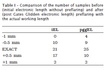
Electronic measurements were performed in both mesial root canals of mandibular molars resulting in 50 measurements before and 50 measurements after preflaring. The analysis of table I data revealed in absolute values the number of exact readings of the working length classified as negative (shorter) and positive (longer) when compared to the actual root canals length. The results revealed that there was no statistical significance (p < 0.05) between the readings before and after the root canals preflaring. Also, the results demonstrated readings closer to foraminal ending in post Gates Glidden electronic length group, in average.
Discussion
The determination of an adequate working length for chemical-mechanic preparation and subsequent obturation of root canal system is of recognized importance for endodontic treatment success 13,18. The instrumentation either up to the limit of radiographic apex or after it may irreversible compromise the endodontic treatment 14.
Several techniques have been developed aiming to facilitate odontometry during endodontic treatment. Therefore, the increasingly common introduction of electronic devices for the exact location of the foraminal ending has gained ground in the endodontists armamentarium as a valuable adjuvant in treatment success 11.
The electronic method has been studied and improved aiming to acquire accuracy, technique fastness, and measuring reliability to determine the exact location of working length, substituting or complementing the use of methods based on radiographs shots 4.
The operation of these devices depend on the dentins insulating feature to the electric current through root canal, which is markedly reduced closer to the apical constriction represented by the differences of the impedance values between the current frequencies applied within canal 15. Therefore, by performing preflaring, the dentin thickness is reduced and as a possible consequence, so is the resistance to the passage of the electric current.
Some studies evaluating the influence of the crown-down preflaring of root canal through nickel-titanium rotary instruments verified that the readings did not seem to undergo the influence of the dentin layer reduction in the location of a point close to the apical constriction and resulted in more precise readings 2,3,8,16. However, the preflaring performed by rotary files shows a more regular and uniform feature while the use of Gates Glidden burs, used in this study, may cause a greater flaring due not to observe the limit of penetration, resulting in a marked reduction of the dentin remnant, mainly in the risk zones of roots displaying an important flatness 1.
Several results have considered the electronic measurements for apical constriction between 0.5 mm and 1.0 mm 7,9. Siu et al. (2009) 19 compared three apical locators at the length in which the locator displayed 0.5 mm shorter of the apex and concluded that all locators reached high rates of reliability.
This variation is acceptable because studies on microscopy revealed that the apical constriction would be located at 1.0 mm shorter of the foraminal ending, in average 12,24. However, depending on the tolerance limit employed in each study, the results can vary within a large range, overestimating in some occasions the results obtained by apical locators 17. Heidemann et al. (2009) 6 concluded in their study that when compared to visual method at a tolerance limit of 1 mm, the electronic measurement showed the highest reliability rate than when the limit was set at 0.5 mm.
In this study, the number of precise readings increased after preflaring. The removal of the interferences within the cervical third would make easier the file to reach the canal ending and would be related to such fact. Considered the variations of +/- 0.5 mm, both measurements were capable of providing acceptable clinical results because these variations seem to be clinically irrelevant. Probably, the four measurements of 1 mm shorter of the apex should be the result of the reduction in dentins wall thickness, which caused an impedance decrease.
Conclusion
Based on the employed methodology, it can be concluded that cervical preflaring with Gates Glidden burs did not significantly influence the apical locator accuracy in determining the actual working length.
References
1. Abou-Rass M, Frank AL, Glick DH. The anticurvature filing method to prepare the curved root canal. J Am Dent Assoc. 1980;(101):792-4. [ Links ]
2. Anele JA, Tedesco M, Marques da Silva B, Baratto-Filho F, Leonardi DP, Haragushiku G et al. Análise ex vivo da influência do preparo cervical na determinação do comprimento de trabalho por três diferentes localizadores apicais eletrônicos. RSBO. 2010 Jun;7(2):139-45.
3. Bramante CM, Berbert A. A critical evaluation of some methods of determining tooth length. Oral Surg Oral Med Oral Pathol. 1974;37(3):463-73.
4. Goldberg F, Marroquín BB, Frajlich S, Dreyer C. In vitro evaluation of the ability of three apex locators to determine the working length during retreatment. J Endod. 2005:31(9):676-8.
5. Gordon MPJ, Chandler NP. Electronic apex locators. Int Endod J. 2004;37(7):425-37.
6. Heidemann R, Vailati F, Texeira CS, Oliveira CAP, Pastemak Junior B. Análise comparativa ex vivo da eficiência na odontometria de três localizadores apicais eletrônicos: Rootzx, Bingo 1020 e Ipex. RSBO. 2009:6(1):7-12.
7. Höer D, Attin T. The accuracy of electronic working length determination. Int Endod J. 2004;37:125-31.
8. Ibarrola JL, Chapman BL, Howard JH, Knowles KI, Ludlow MO. Effect of prefaring on Root ZX apex locators. J Endod. 1999;25(9):625-6.
9. Kaufman AY, Keila S, Yoshpe M. Accuracy of a new apex locator: an in vitro study. Int Endod J. 2002;35(2):186-91.
10. Katz A, Kaufman AY, Szajkis S. An in vivo model for testing the accuracy of apex locators. Revue Française DEndodontie. 1992;11:67.
11. Kobayashi C. Electronic canal root measurement. Oral Surg Oral Med Oral Pathol. 1995;79:226-31.
12. Kuttler Y. Microscopic investigation of root canals. J Am Dent Assoc. 1955;50(5):544-52.
13. Lucena-Matín C, Robles-Gijón V, Ferrer-Luque CM, Mondelo JM. In vitro evaluation of the accuracy of three electronic apex locators. J Endod. 2004 Apr;30(4):231-3.
14. Machado MEL, Pesce HF. Estudo da região apical de dentes tratados endodonticamente até o vértice radiográfico da raiz. Rev Ass Paul Cir Dent. 1981;35(6):534-7.
15. Mc Donald NJ. The electronic determination of working length. Dent Clin North American. 1992;36(2):293-307.
16. Nguyen HQ, Kaufman AY, Komorowski RC, Friedman S. Electronic length measurement using small and large files in enlarged canals. Int End J. 1996;29:359-64.
17. Pagavino G, Pace R, Bacceti T. A SEM study of in vivo accuracy of the root ZX electronic apex locator. J Endod. 1998;24:438-41.
18. Ricucci D, Langeland K. Apical limit of root canal instrumentation and obturation, part 2. A histological study. Int Endod J. 1998;31:394-8.
19. Siu C, Marshall JG, Baumgartner JC. An in vivo comparison of the root ZX II, the apex NRG XFR and mini apex locator by using rotary nickel-titanium files. J Endod. 2009 Jul;35(7):962-5.
20. Stabholz A, Rotstein I, Torabinejad M. Effect of preflaring on tactile detection of the apical constriction. J Endod. 1995;21:92-4.
21. Sunada I. New method for measuring the length of the root canal. J Dent Res. 1962;41:375-87.
22. Suzuki K. Experimental study on iontophoresis. J Jap Stomatol. 1942;16:411-7.
23. Weiger R, John C, Geigle H, Löst C. An in vitro comparison of two modern apex locators. J Endod. 1999;25(11):765-8.
24. Vertucci F, Seeling A, Gillis R. Root canal morphology of the human maxillary second pre molar. Oral Surg Oral Med Oral Pathol. 1974;38:456-64.
 Correspondence:
João Marcelo da Silva Teixeira
Rua Gonçalo Coelho, n.º 147
CEP 20751-150 – Piedade – RJ – Brasil
E-mail:joaodonto@gmail.com
Received for publication: June 14, 2011.
Accepted for publication: November 1st, 2011.
