


Serviços Personalizados
Artigo
 pdf em Inglês
 Artigo em XML
 Referências do artigo
 Enviar este artigo por email
Links relacionados
Compartilhar

 Permalink
RSBO (Online)
versão On-line ISSN 1984-5685
RSBO (Online) vol.9 no.2 Joinville Abr./Jun. 2012
Original Research Article
Bond strength of resin cements to leucite-reinforced ceramics
Rubens Nazareno Garcia I,II; Reinaldo Francisco do Nascimento III; Ana Cristina Rocha Gomes I; Marcelo Giannini IV; Luiz Carlos Machado Miguel I; Peter Clayton Moon V
I Department of Dentistry, University of the Region of Joinville – Joinville – SC – Brazil.
II School of Dentistry, University of Itajai Valley – Itajai – SC – Brazil.
III RPD Dental Laboratory – Joinville – SC – Brazil.
IV Department of Restorative Dentistry, Piracicaba Dental School, State University of Campinas – Piracicaba – SP – Brazil.
V Department of General Practice, School of Dentistry, Virginia Commonwealth University – Richmond – VA – USA.
ABSTRACT
Objective:The aim of this study was to evaluate the shear bond strength (SBS) of two resin cements to four leucite-reinforced ceramics. Material and methods: Forty ceramic blocks (4 mm wide, 14 mm length and 2 mm thick) were used and the samples abraded with aluminum oxide (90 µm). The samples were divided into eight groups (n = 5). Two resin cements (conventional RelyX ARC and self-adhesive RelyX U100 – 3M ESPE) were bonded to Creapress (CRE-Creation/Klema), Finesse All-Ceramic (FIN-Dentsply/ Ceramco), IPS Empress Esthetic (IEE-Ivoclar Vivadent) and Vita PM9 (PM9-Vita). For all groups and in each ceramic block, after application of 10% hydrofluoric acid and silanation, three Tygon tubings were positioned over the ceramics, which were filled in with the resin cements (light-cure for 40 s). The tubings were removed to expose the specimens in format of cylinders (area: 0.38 mm2) and samples were stored in relative humidity at 24±2 °C for one week. After this period, each sample was attached to testing machine and the specimens were submitted to shear bond test (applied at the base of the specimen/cement cylinder with a thin wire/0.2 mm) at speed of 0.5 mm/ min, until failure. The results were analyzed by two-wayANOVA (resin cements and ceramic systems) and Tukey test (p<0.05). Results: The means (SD) were (in MPa): ARC + CRE = 32.1±4.3; ARC + FIN = 28.3±3.7; ARC + IEE = 25.9±4.4; ARC + PM9 = 22.2±2.1; U100 + CRE = 38.0±5.2; U100 + FIN = 36.9±2.8; U100 + IEE = 38.4±2.9; U100 + PM9 = 34.3 ±7.3. U100 showed higher SBS to ceramics than ARC. U100 had higher SBS when applied on IEE ceramic than on PM9. For ARC, SBS obtained with CRE was higher than with IEE and PM9. Conclusion: RelyX U100 can provide higher SBS to leucite-reinforced ceramics than conventional resin cement. The resin cements applied on the PM9 ceramic surface resulted in lower SBS.
Keywords: ceramics; resin cements; shear strength.
Introduction
The ceramics are used to achieve esthetic dental restorations because of their superior color and translucency. Their clinical success is determined by the bond strength and bonding durability of the resin cement to tooth and ceramic 3,21.
The bonding of conventional resin cement to tooth and ceramic depends on the type of adhesive used (total etch or self-etch) and the quality of the dentin surface treatment. Self-adhesive resin cements have been developed to simplify the adhesive cementation technique because advocates no pre-treatment of tooth surfaces. These materials, so-called universal, all-purpose or multipurpose self-adhesive resin cements have been available, each purportedly bonding to enamel, dentin, amalgam, ceramic, metal- and zirconia-based restorations 14,16. They can provide fluoride ion release 1; being also an option for bonding fiber-reinforced composite posts to root canal dentin, but the conventional resin cements apparently provide higher bond strengths than self-adhesive resin cements 4.
One of these self-adhesive cements contains an organic matrix composed of multifunctional acidic methacrylates, which react with inorganic fillers that are basic in nature or with hydroxyapatite from tooth structure. The setting of the cement is based on a free radical polymerization reaction initiated by either photoactivation or a redox system 2,11.
Regarding to glass ceramic surface, the conditioning has been purposed with hydrofluoric acid followed by silanation. The acid selectively dissolves the glass matrix creating micromechanical retention, and the silanation serves for the chemical adhesion between the organic and inorganic substances, producing a strong and durable adhesion between the ceramic and the resin cement 9,19.
While many studies evaluated the bond to ceramics using traditional resin cements, few studies have shown the bonding performance and the efficiency of self-adhesive resin cements to ceramics. The aim of this study was to evaluate the shear bond strength (SBS) of two resin cements to four leucite-reinforced ceramics.
Material and methods
Forty leucite-reinforced ceramic blocks (4 mm wide, 14 mm length and 2 mm thick) were constructed in the hot pressing technique using Creapress (CRE – Creation/Klema – Batch # 8746), Finesse All-Ceramic (FIN – Dentsply/Ceramco – Batch # 2887), IPS Empress Esthetic (IEE – Ivoclar Vivadent – Batch # 0305) and Vita PM9 (PM9 – Vita – Batch # 17290). The samples were abraded with aluminum oxide (90µm / 2.5 bar / 10 mm distance) and divided into eight groups (n = 5).
For all groups and in each sample, after application of 10% hydrofluoric acid for 1 min (Condac Porcelana, FGM, Joinville, SC, Brazil – Batch # 40511 / Exp.: 05/2013), the samples were rinsed for 1 min, air-dried for 1 min, followed by the application of the silane coupling agent (Prosil, FGM, Joinville, SC, Brazil – Batch # 130411 / Exp.: 04/2012) for 1 min and then air-dried for 30 s. Next, three Tygon tubings (TYG-030, Saint-Gobain Performance Plastic, Maime Lakes, FL, USA) were positioned over the samples and filled in with the resin cements RelyX ARC or RelyX U100 and VLC for 40 s (Visible Light Cure – Led Radii-cal 1.200 mW/cm2, SDI, Bayswater, VI, Australia).
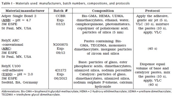
The materials and protocols are listed in the Table I and the experimental groups were: (1) ASB+ARC+CRE; (2) ASB+ARC+FIN; (3) ASB+ARC+IEE; (4) ASB+ ARC+PM9; (5) U100+CRE; (6) U100+FIN; (7) U100+IEE; (8) U100+PM9.
The tubing molds were removed to expose the specimens in format of cylinders (area: 0.38 mm2), which were stored in relative humidity at 24±2°C for one week. After this period, each sample was attached to the universal testing machine Instron (model TTC, Canton, MA, USA) and the specimens were submitted to shear bond test (applied at the base of the specimen/cement cylinder with a thin wire - 0.2 mm) at speed of 0.5 mm/min, until failure. The results were analyzed by two-way ANOVA (resin cements and ceramic systems) and Tukey test (p<0.05).
The specimens were mounted into an aluminum base, metalized with gold and examined in scanning electronic microscope (Carl Zeiss AG - EVO® 50 Series, Oberkochen, Germany). Photomicrographies of representative areas were obtained to evaluate the fracture pattern that was classified in adhesive, cohesive (either in ceramic or in cement) and/or mixed.
Results
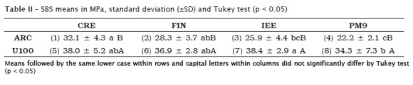
The ANOVA showed significant differences between cements (p < 0.001) and among ceramic systems (p = 0.01396). To investigate the differences between means of the ceramic systems, it was applied the Tukey test (p < 0.05). U100 showed higher SBS to ceramics than ARC. U100 had higher SBS when applied on IEE ceramic than on PM9. For ARC, SBS obtained with CRE was higher than with IEE and PM9 (table II).
Discussion
In this present study, the focus was to evaluate the bond strength of self-adhesive resin cements to leucite-reinforced ceramics, using microshear methodology proposed by Shimada et al. (2002) 25. This type of mechanical test solves problems related to tension propagations at the bonded interface in larger areas. It presents the advantage that several specimens can be obtained from one sample without cutting it, being easier and cheaper than the microtensile test 24, when the samples need to be cut to obtain the specimens.
It has been observed that shear bond testing tends to produce cohesive failures of the substrate. The improvement of the bonding properties of restorative materials have increased the bond strength and changed the failure pattern 18. This transition is most likely related to the changing stress pattern as the crack progresses across the interface. It is usually observed that as the adhesive bond increases strength and less adhesive fracture area is observed on initiation of adhesive fracture, and a bigger piece of cohesive fracture in the substrate is pulled out after the transition from adhesive to cohesive fracture occurs 29.
Resin cements have several advantages when compared to conventional powder/liquid cements: better retention, minimum solubility at oral environment, less microleakage, and acceptable biocompatibility 20. But, the conventional resin cements demand the use of either conventional or self-etching adhesive systems. The technique sensitivity and the difficulty of obtaining a hermetic sealing associated with conventional adhesives probably leads to a greater incidence of post-operative sensitivity related to indirect restorations luting procedure. Self-adhesive resin cements do not demand tooth structure pre-treatment, therefore simplifying the clinical steps during the installation procedures of the restorations. Otherwise, it is normally necessary the restoration pre-treatment 10.
In several studies testing the adhesion to ceramics, aluminum oxide sandblasting is used to increase the surface roughness, as well as to clean and to activate the surface. This method can significantly improve resin-ceramic bond strength and its durability when combined with silane or adhesive monomer-containing primers 3,28,30.
The main protocol to prepare the inner surface of ceramics for bonding is based on etching with hydrofluoric acid (HF), followed by the application of a silane coupling agent to achieve a high bond strength. Hydrofluoric acid works by creating surface pits via preferential dissolution of the glassy phase from the ceramic matrix 5,12. The removal of HF from the restoration is necessary because of its highly toxic chemical factor 17,28.
Eames et al. (1977) 8 suggested the use of a silane coupling agent for dental applications, and Roulet et al. (1995) 23 described the action mechanism that increases the wettability and forms a covalent bond with both the ceramic and the resin cement. The most commonly used silane in dentistry is 3-trimethoxysilyl propylmethacrylate diluted in a water-ethanol solution. It is marketed in a pre-hydrolysed form (one bottle) or in a form where hydrolysis can occur by mixing silane and acid (two bottles). Both types of silane coupling agents are reported to perform well, even though atmospheric moisture is unfavorable to the prehydrolyzes silanes. It activates a condensation reaction that leads to polymerized siloxanes, producing oligomers, which give the solution a white and opaque appearance 9,15.
The resin matrix of the self-adhesive consists of multifunctional acidic methacrylates. If a high content of acidic functional monomers can react with the substrate like the ceramics used in this study, and achieve enough chemical bond strength, it is possible to hypothesize that the self-adhesive resin cement RelyX U100 could be used to bond successfully to the ceramics surfaces. The leucite-reinforced ceramics showed higher SBS means (groups 5 to 8) than those observed for ARC groups (groups 1 to 4), which corroborate with De Munck et al. (2004) 6 and Lin et al. (2010) 13, that tested respectively dental substrates and ceramic. Other previous reports from Piwowarczyk et al. (2004) 22, Shimada et al. (2002) 25, Dündar et al. (2007) 7 and Shimakura et al. (2007) 26 also used SBS tests and similar methodology that can permit the positive correlation between the SBS data and the SEM analysis found in the present study.
Previous studies of Stewart et al. (2002) 27 and Fabianelli et al. (2010) 9 reported that many factors can influence the resin bond strength to tooth structure, when a resin-bonded ceramic restoration is placed, including that two interfaces need to be considered: the dentin-resin interface and the ceramic-resin interface, subject of this present study. The bond strength at these interfaces has to be optimized, as a weak interface can contribute to failure of the restoration.
For groups 1 to 4, using a combination of sandblasting, hydrofluoric acid etching and silane treatment, the mean of SBS of ASB and ARC was higher to CRE than to IEE and PM9. For groups 5 to 8, using a combination of sandblasting, hydrofluoric acid etching, silane treatment and U100, the mean of SBS was higher to IEE ceramic than on PM9.
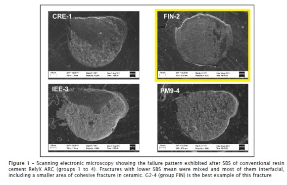
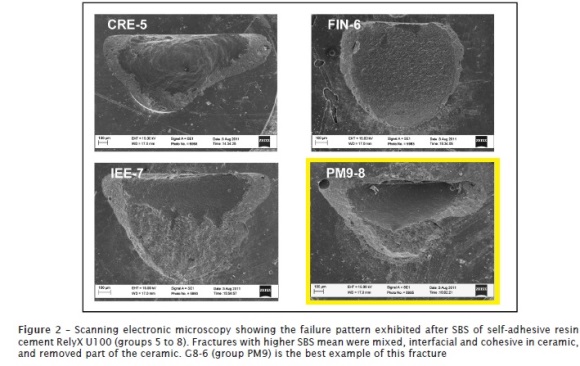
The photomicrographs of the failure pattern of the conventional resin cement RelyX ARC showed mixed fractures and most of them interfacial, including a smaller area of cohesive fracture in ceramic (figure 1). This can suggest less interaction between the cement and the ceramic surface. In the opposite side, the referent photomicrographs of RelyX U100 groups showed mixed fractures, interfacial and cohesive in ceramic, with the removal of part of the ceramic (figure 2) and they can suggest a higher interaction between the cement and the ceramic surface, as mentioned earlier and comproved by the statistical difference among the groups ARC and U100.
Conclusion
RelyX U100 can provide higher SBS to leucite-reinforced ceramics than conventional resin cement. The resin cements applied on PM9 ceramic surface resulted in lower SBS.
Acknowledgments
This study was supported by University of the Region of Joinville (UNIVILLE), Joinville, SC, Brazil (Project UNIVR, Department of Dentistry), in partnership with Virginia Commonwealth University (VCU), Richmond, VA, USA (Department of General Practice, School of Dentistry). Scanning Electron Microscopy was performed at the VCU (Department of Neurobiology & Anatomy Microscopy Facility), supported with funding from NIH-NINDS Center core grant 5P30NS047463 and NIH-NCRR grant 1S10RR022495.
References
1. Aguiar T, Pinto C, Cavalli V, Nobre-Dos-Santos M, Ambrosano G, Mathias P et al. Influence of the curing mode on fluoride ion release of self-adhesive resin luting cements in water or during pH-cycling regimen. Oper Dent. 2012 Jan-Feb;37(1):63-70. [ Links ]
2. Behr M, Rosentritt M, Regnet T, Lang R, Handel G. Marginal adaptation in dentin of a self-adhesive universal resin cement compared with well-tried systems. Dent Mater. 2004 Feb;20(2):191-7.
3. Blatz MB, Sadan A, Kern M. Resin-ceramic bonding: a review of the literature. J Prosthet Dent. 2003 Mar;89(3):268-74.
4. Calixto L, Bandeca M, Clavijo V, Andrade M, Vaz L, Campos E. Effect of resin cement system and root region on the push-out bond strength of a translucent fiber post. Oper Dent. 2012 Jan-Feb;37(1):80-6.
5. Canay S, Hersek N, Ertan A. Effect of different acid treatments on a porcelain surface. J Oral Rehabil. 2001 Jan;28(1):95-101.
6. De Munck J, Vargas M, Van Landuyt K, Hikita K, Lambrechts P, Van Meerbeek B. Bonding of an auto-adhesive luting material to enamel and dentin. Dent Mater. 2004 Dec;20(10):963-71.
7. Dündar M, Ozcan M, Gökçe B, Cömlekoglu E, Leite F, Valandro LF. Comparison of two bond strength testing methodologies for bilayered all-ceramics. Dent Mater. 2007 May;23(5):630-6.
8. Eames WB, Rogers LB, Feller PR, Price WR. Bonding agents for repairing porcelain and gold: an evaluation. Oper Dent. 1977 Summer;2(3):118-24.
9. Fabianelli A, Pollington S, Papacchini F, Goracci C, Cantoro A, Ferrari M et al. The effect of different surface treatments on bond strength between leucite reinforced feldspathic ceramic and composite resin. J Dent. 2010 Jan;38(1):39-43.
10. Garcia RN, Renzetti AGZ, Schaible BR, Frankenberger R, Lohbauer U, Miguel LCM. Bond strength of self-adhesive resin cements to deep dentin. RSBO. 2011 Oct-Dec;8(4):431-8.
11. Ibarra G, Johnson GH, Geurtsen W, Vargas MA. Microleakage of porcelain veneer restorations bonded to enamel and dentin with a new self-adhesive resin-based dental cement. Dent Mater. 2007 Feb;23(2):218-25.
12. Kato H, Matsumura H, Ide T, Atsuta M. Improved bonding of adhesive resin to sintered porcelain with the combination of acid etching and a two-liquid silane conditioner. J Oral Rehabil. 2001 Jan;28(1):102-8.
13. Lin J, Shinya A, Gomi H, Shinya A. Effect of self-adhesive resin cement and tribochemical treatment on bond strength to zirconia. Int J Oral Sci. 2010 Mar;2(1):28-34.
14. Liu Q, Meng X, Yoshida K, Luo X. Bond degradation behavior of self-adhesive cement and conventional resin cements bonded to silanized ceramic. J Prosthet Dent. 2011 Mar;105(3):177-84.
15. Matinlinna JP, Lassila LV, Ozcan M, Yli-Urpo A, Vallittu PK. An introduction to silanes and their clinical applications in dentistry. Int J Prosthodont. 2004 Mar-Apr;17(2):155-64.
16. Mazzitelli C, Monticelli F, Osorio R, Casucci A, Toledano M, Ferrari M. Effect of simulated pulpal pressure on self-adhesive cements bonding to dentin. Dent Mater. 2008 Sep;24(9):1156-63.
17. Monticelli F, Toledano M, Osorio R, Ferrari M. Effect of temperature on the silane coupling agents when bonding core resin to quartz fiber posts. Dent Mater. 2006 Nov;22(11):1024-8.
18. Moon PC, Weaver J, Brooks CN. Review of matrix metalloproteinases effect on the hybrid dentin bond layer stability and chlorhexidine clinical use to prevent bond failure. Open Dent J. 2010 Jul; 20(4):147-52.
19. Ozcan M, Valandro LF, Amaral R, Leite F, Bottino MA. Bond strength durability of a resin composite on a reinforced ceramic using various repair systems. Dent Mater. 2009 Dec;25(12):1477-83.
20. Pavan S, Santos PH, Berger S, Bedran-Russo AKB. The effect of dentin pretreatment on the microtensile bond strength of self-adhesive resin cements. J Prosthet Dent. 2010;104:258-64.
21. Peumans M, Van Meerbeek B, Lambrechts P, Vanherle G. Porcelain veneers: a review of the literature. J Dent. 2000 Mar;28(3):163-77.
22. Piwowarczyk A, Lauer HC, Sorensen JA. In vitro shear bond strength of cementing agents to fixed prosthodontic restorative materials. J Prosthet Dent. 2004 Sep;92(3):265-73.
23. Roulet JF, Söderholm KJ, Longmate J. Effects of treatment and storage conditions on ceramic/composite bond strength. J Dent Res. 1995 Jan;74(1):381-7.
24. Sano H, Shono T, Sonoda H, Takatsu T, Ciucchi B, Carvalho RM. Relation between surface area for adhesion and tensile bond strength – evaluation of a microtensile bond test. Dent Mater. 1994 Jul;10(4):236-40.
25. Shimada Y, Yamaguchi S, Tagami J. Microshear bond strength of dual-cured resin cement to glass ceramics. Dent Mater. 2002 Jul;18(5):380-8.
26. Shimakura Y, Hotta Y, Fujishima A, Kunii J, Miyazaki T, Kawawa T. Bonding strength of resin cement to silicate glass ceramics for dental CAD/CAM systems is enhanced by combination treatment of the bonding surface. Dent Mater J. 2007 Sep;26(5):713-21.
27. Stewart GP, Jain P, Hodges J. Shear bond strength of resin cements to both ceramic and dentin. J Prosthet Dent. 2002 Sep;88(3):277-84.
28. Valandro LF, Della Bona A, Antonio Bottino M, Neisser MP. The effect of ceramic surface treatment on bonding to densely sintered alumina ceramic. J Prosthet Dent. 2005 Mar;93(3):253-9.
29. Versluis A, Tantbirojn D, Douglas WH. Why do shear bond tests pull out dentin? J Dent Res. 1997 Jun;76(6):1298-307.
30. Yang B, Barloi A, Kern M. Influence of air-abrasion on zirconia ceramic bonding using an adhesive composite resin. Dent Mater. 2010 Jan;26(1):44-50.
 Correspondence:
Rubens Nazareno Garcia
Universidade da Região de Joinville – Univille – Departamento de Odontologia
Rua Paulo Malschitzki, n.° 10 – Zona Industrial – Bom Retiro
CEP 89219-710 – Joinville – SC – Brasil
E-mail:rubens.garcia@univille.net
Received for publication: November 10, 2011.
Accepted for publication: January 17, 2012.
