


Serviços Personalizados
Artigo
 pdf em Inglês
 Artigo em XML
 Referências do artigo
 Enviar este artigo por email
Links relacionados
Compartilhar

 Permalink
RSBO (Online)
versão On-line ISSN 1984-5685
RSBO (Online) vol.9 no.3 Joinville Jul./Set. 2012
Original Research Article
Effect of staining agents on color change of composites
Mateus Rodrigues Tonetto I; Carolina Santezi Neto I; Cristina Magnani Felício II; Patrícia Aleixo dos Santos Domingos II; Edson Alves de Campos I; Marcelo Ferrarezi de Andrade II
II Department of Restorative Dentistry, University Center of Araraquara – Araraquara – SP – Brazil.
ABSTRACT
Introduction: Composite resins are materials that can present color changing when exposed to pigments. Objective: The aim of this study was to evaluate, in vitro, the color changing of composites after immersion in different substances for different periods. Material and methods: Two microhybrid composite resins: Charisma (Heraeus – Kulzer) and Opallis (FGM) were used. Red wine and acai pulp were also used as immersion medium. For this study, 32 specimens with 10 mm of diameter and 2 mm of thickness were used, divided into 4 groups: Group 1 – Opallis composite immersed in red wine solution; Group 2 – Opallis composite immersed in acai berry pulp solution; Group 3 – Charisma composite immersed in red wine solution; Group 4 – Charisma composite immersed in acai berry pulp solution. The specimens were evaluated in the following time periods: T0 – baseline, T1 – 24 hours, T2 – 48 hours, T3 – 72 hours and T4 – 96 hours. For the assessment of staining, a spectrophotometer for colorimetry was used (Color Guide 45 / 0, PCB 6807 BYK-Gardner Gerestsried GmBH, Germany), and the values obtained were transferred to a computer and recorded according to CIELAB system. Results: The data were evaluated using Kruskal- Wallis non-parametric tests with the following mean values for the immersion periods of 24, 48, 72 and 96 hours, respectively: G1 – 7.35, 7.84, 9.04,10.48; G2 – 2.92, 4.15, 4.30, 4.64; G3 – 3.14, 7.35, 8.13, 8.43, G4 – 4.49, 5.99, 6.92, 6.76. Conclusion: Red wine showed a higher tendency toward altering the composite color than acai berry pulp. In addition, no significant difference was found concerning to the behavior of the two composite resins. Concerning to the immersion time periods, significant differences were only observed among the groups in the 24 hour time period.
Keywords: coloring agents; staining; composite resins.
Introduction
Dentistry has been undergoing changes and advancements which tend to develop more efficient techniques and materials for the treatment of the patients, who in turn are searching for an aesthetic and therapeutic treatment. One of the materials which most meets these requirements is composite resin. The search for the ideal composite resin has enabled a well-accepted change in the dental scenario. Because of all this development, currently, composite resins show properties very similar to those of natural teeth.
Notwithstanding, composite resins still undergo problems of resistance to degradation in the oral environment and color stability; the latter has been one of the main reasons for the replacement of the restoration 8,12.
Santos et al. 15 and Omata et al. 13 reported that color stability is a property depending on factors as the internal reactions of the material and the behavior of the patient regarding to the consumption of food containing pigments, poor oral hygiene and use of mouthrinses as well as the type of finishing and polishing procedure itself performed by the dentist. Color stability may be directly related to the different compositions of the resins regarding to the variations in the size of the particles and in matrix/inorganic load ratio 9,11,15,17.
According to Asmussem 1 and Dietschi et al. 4, there are 3 types of composite resin discoloration: 1) intrinsic discoloration, caused by the material aging itself; 2) extrinsic discoloration, caused by the accumulation of plaque and superficial pigment; 3) alteration of the surface color involving superficial degradation or mild penetration and reaction of the staining agents inside the superficial layer of composite resin (absorption). Additionally, regardless of the technique and the finishing and polishing procedures used, microleakage can occur, which can facilitate the water inlet and accelerate the phenomenon of degradation and discoloration of the material 3,8,12. The materials which show greater water absorption tend to absorb more pigments because water may conduct the staining agent toward the material, resulting in more intense staining 3,4.
The literature presents a vast number of studies reporting the influence of some beverages containing staining substances on the color stability of the aesthetic materials. Some studies 6,19 were conducted in an attempt to prove that the immersion in liquids as coffee, wine, tea, soft drinks, and mouthrinse promote a excessive staining of the restoration when the patients are frequent consumers of these beverages and found results confirming this hypothesis, with the most color alterations observed in the presence of wine and coffee. Additionally to these beverages, acai berry is a product presenting a high degree of staining of teeth at the moment of its ingestion. Notwithstanding, there are no studies in the literature evaluating its staining on composite resins.
The aim of this study was to evaluate in vitro the color stability of two different brands of composite resins after the immersion into two solutions – red wine and acai berry pulp– for different time periods.
Material and methods
Thirty-two samples were divided into four groups (n = 8) – group 1: Opallis (FGM Produtos Odontológicos, Joinville, Brazil) immersed in red wine solution; group 2: Opallis immersed in acai berry pulp solution; group 3: Charisma (Heraeus Holding GmbH, Hanau, Germany) immersed in red wine solution; group 4: Charisma immersed in acai berry pulp solution –, by employing a metallic matrix with central orifice with 10 mm of diameter and 2 mm of thickness. A colorless polyester strip and glass plate was used to support the matrix. The composite insertion was performed in a single increment. Next, another polyester strip was placed onto the resin and another glass plate was placed over it. A load of 1 kg was placed on all the set for 30 seconds, to result in a standardized smooth surface. After that period, the load and the second glass plate was removed and the material was light-cured (light-curing unit, Gnatus, frequency of 50/60 HZ, power of 15 VA, 470 nm +/- 25 nm of wavelength and 400 mw/cm² of light intensity) for 40 seconds. Following, the samples were removed from the matrix and the excesses were removed with the aid of a diamond bur (#2136FF) at high-speed without refrigeration.
The samples were stored in a incubator at 37±1°C, immersed in the immersion media, according to the groups o – acai berry pulp (Poderoso®, whole acai berry pulp, invert sugar and guarana extract) and red wine (Góes®, dry red wine, sugar and acidulant) – and the experimental times: T0 – baseline; T1 – 24 hours; T2 – 48 hours; T3 – 72 hours; T4 – 96 hours.
A spectrophotometer for colorimetry (Color Guide 45/0, PCB 6807 BYK-Gardner GmbH Gerestsried, Germany), with wavelength between 400 nm and 700 nm, with direct transmission, standard illumination D65 and calibrated over a white ceramic background.
The spectrophotometer was calibrated by the following patterns: pure white and pure black; and checked at green. Next, each sample was removed from the immersion medium, washed in tap water for 5 seconds and dried with the aid of a gauze. Just after this procedure, the device was positioned to perform the color measurement and the values were registered according to the CIELab system (Commission Internationale de lEclairage) 22, in which L* is the measurement of the luminosity, quantified with a scale ranging from zero (0) to one hundred (100), following from pure black to pure white, respectively. The measurements of a* and b* corresponded to chromaticity, in which a* is related to the axis green-red and b* to the axis blue-yellow. Therefore, the color data L (luminosity), a (deviation in the green-red axis) and b (deviation in the blue-yellow axis) are automatically obtained 18.
Following these concepts, the color difference was calculated by the equation: ΔE = [(ΔL*)2 + (Δa*)2 + (Δb*)2]½, while the luminosity alteration (ΔL*) was calculated by the formula: ΔL= L*(tx) - L*(t0), in which (tx) represents the immersion time and (t0) baseline.
The classification adopted for ΔE values were determined by the National Bureau of Standards (NBS) 10, which considers: values from 0.0 to 0.5 – extremely light change; 0.5 to 1.5 – light change; 1.5 to 3.0 – noticeable change; 3.0 to 6.0 – marked change; 6.0 to 12.0 – extremely marked change; 12.0 or more – change to another color.
Concerning to statistical analysis, ΔE evaluation among groups, at each immersion time, was performed by Kruskal-Wallis test. Color change evaluation among immersion times, for each group, was performed by Friedman tests. For both tests, the level of significance was set at 5%.
Results
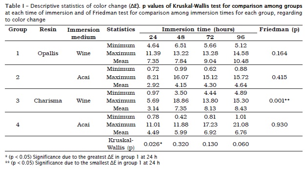
In table I are shown the means and the minimum and maximum values of the color change in the samples of Opallis and Charisma composite resins immersed in either red wine or acai berry solution, evaluated for a period of 96 hours.
Friedman test, applied separately for each group, showed a increasing of ΔE as the immersion time went by, but with only statistically significant difference for the immersion of Charisma in red wine (p < 0.05). In this case, ΔE significantly increases after 24 hours of immersion, but it maintained the value for the next hours.
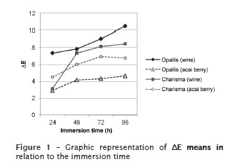
According to Kruskal-Wallis test, applied for the comparison among groups at each immersion time, there was only a statistically significant difference in the period of 24 hours (p < 0.05), caused by a greater ΔE regarding to Opallis resin immersed in red wine. Although very little statistically significant difference had occurred, we noted a tendency toward more color changing of red wine than acai berry, regardless of the resin. This fact can be visualized by the graph of the means in figure 1.
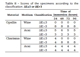
To confirm this result, the experimental data were separated into two categories: ΔE up to 3.0 (from extremely light to noticeable change according to the classification of the National Bureau of Standards) and ΔE above 3.0 (from markedly change to color change). The scores of the specimens in these categories are given in table II, confirming the tendency toward more intense color change after the immersion in red wine. After the immersion in acai berry solution, almost half of the specimens is assigned in each category, which may be the main reason that the statistical analysis have not detected significant differences.
Discussion
The present study addressed the color instability of two composite resins after the exposure to staining media coming from the diet. According to the results obtained in this study and to those of other authors, it is clear that several fluids stained not only the tooth structure but also the composite resin, so that, depending on the saturation of this staining, negative aesthetic effects are observed 1-4,7,8-13.
It is known that the etiology of the discoloration of the resins can be either intrinsic or extrinsic. Extrinsic discoloration occurs because of a process in which the staining agents coming from diet, associated to the habits, or within therapeutic agents, are in contact with the restorative material being incorporated via absorption or adsorption. The authors still affirmed that the biphasic composition of composite resins, the type of the staining agent and the duration of the contact between the agent and the material determines the staining 1,2,4,6,7.
Asmussen 1, Chan et al. 2, Dickinson et al. 3 and Dietschi 4 et al. reported that, although composite resins present some advantages over the other aesthetic materials, they evidence accentuated color change, mainly under the action of poor oral hygiene, ultraviolet rays and also by the impregnation of staining agents coming from food. Mouthrinses may also cause color changes in composite resin restorations.
The composite resins used in this study were microhybrid. Through the results obtained, it was verified statistically significant difference in the color change between them only at T1, that is, after 24 hours of immersion in wine. However, Dietschi et al. 4 observed a greater staining of Charisma than other resins evaluated; according to these authors, the susceptibility to staining of the composites is related to their composition and superficial properties.
When microfilled resin was compared to other types of resin, it is observed that they show a greater degree of staining because of their higher content of organic matrix 1,4,7,9,12-15. Concerning to the immersion medium, several solution have been used to simulate which occurs inside oral cavity, such as soft drinks, coffee, wine and nicotine 2,4,6,7,8,11-18. Currently, the consumption of acai berry has significantly increased in all regions of Brazil. Because of that, this study aimed to evaluate the color change of resins when in contact to such product and compared it to wine, which is largely used in this type of study.
The results of this research showed a wine tendency toward a greater change of resin color than acai berry, both for Charisma and Opallis resin. Villalta et al. 18 also reported that wine was the substance provoking the greatest staining in resins. Additionally to the restorative materials, it has been also noted a high degree of staining in bovine teeth after bleaching, when these were immersed in black tea, soft drinks, or wine, showing greater staining than coffee 16.
Omata et al. 13 verified that the wine solution presented the highest degree of staining, followed by tea and coffee, a result similar to that obtained by this present study.
The study of Chan et al. 2 showed that the staining of the aesthetic materials occurred more intensively between 24 hours and 7 days, becoming more intense as time goes by. Dietschi et al. 4 also verified that coffee solution provided a greater staining of the composite resin and that this staining was more accentuated in the first week of immersion. A smaller staining of resin restorations can be achieved by always performing the proper light-curing of the restorative material, associated with an efficient polishing, in addition to avoid the contact of the newly-cured restorations with staining agents concomitantly to temperature changes 4.
Conclusion
Based on the results obtained, it can be concluded:
There is a wine tendency toward more color change of the resin than acai;
There is no significant difference in the behavior of both composite resins between each other, regardless of the immersion media;
Concerning to the immersion time, significant differences only occurred among groups at 24 hour period.
References
1. Asmussen E. Factors affecting the color stability of restorative resins. Acta Odontol Scand. 1983;41:11-8. [ Links ]
2. Chan KC, Fuller JL, Hormati AA. The ability of foods to stain two composite resins. J Prosthet Dent. 1980;43:542-5.
3. Dickinson G, Leinfelder KF, Mazer R, Russel CM. Effect of surface penetrating sealant on wear of posterior composite resins. J Am Dent Assoc. 1990;121:251-5.
4. Dietschi D, Campanile G, Holz J, Meyer JM. Comparison of the color stability of ten new-generation composites: an in vitro study. Dent Mater. 1994;10:353-62.
5. Dunne SM, Davies BR, Millar BJ. A survey of the effectiveness of dental light-curing units and comparison of light testing devices. Br Dent J. 1996;180:411-6.
6. Kim J-H, Lee Y-K, Powers JM. Influence of a series of organic and chemical substances on the translucency of resin composites. J Biomed Mater Res B Appl Biomater. 2006;77(1):21-7.
7. Luce MS, Campbell CE. Stain potential of four microfilled composites. J Prosteth Dent. 1988;60:151-4.
8. Magne P, Woong-Seup S. Optical integration of incisoproximal restorations using the natural layering concept. Quintessence Int. 2008;39(8):633-43.
9. Malhotra N, Shenoy RP, Acharya S, Shenoy R, Mayya S. Effect of three indigenous food stains on resin-based, microhybrid-, and nanocomposites. J Esthet Restor Dent. 2011;23(4):250-7.
10. Mutlu-Sagesen L, Ergün G, Özkan Y, Semiz M. Color stability of a dental composite after immersion in various media. Dent Mater J. 2005;24:382-90.
11. Nasim I, Neelakantan P, Sujeer R, Subbarao CV. Color stability of microfilled, microhybrid and nanocomposite resins – An in vitro study. J Dent. 2010;38(2):e137-42.
12. Oilo G. Biodegradation of dental composites/glass-ionomer cements. Adv Dent Res. 1992;6:50-4.
13. Omata Y, Uno S, Nakaoki Y, Tanaka T, Sano H, Yoshida S et al. Staining of hybrid composites with coffee, oolong tea, or red wine. Dent Mat J. 2006;25(1):125-31.
14. Pucci CR, Lima DR, Kalczuk L, Araújo MAM, Silva RCSP. Composite resin staining with the use of bacterial plaque detector. Rev Paul Odontol. 2003;25:15-20.
15. Santos PAD, Dibb RGP, Corona SAM, Catirse ABE, Garcia PPNS. Influence of fluoride-containing solutions on the translucency of flowable composite resins. Journal of Materials Science. 2003;38(18):3765-8.
16. Téo TB, Takahashi MK, Gonzaga CC, Lopes MGK. Avaliação da alteração de cor de dentes bovinos imersos em soluções com elevado potencial de pigmentação após clareamento. RSBO. 2010;7(4):401-5.
17. Vichi A, Ferrari M, Davidson CL. Color and opacity variations in three different resin-based composite products after water aging. Dent Mater. 2004;20:530-4.
18. Villalta P, Lu H, Okte Z, Garcia-Godoy F, Powers JM. Effects of staining and bleaching on color change of dental composite resins. J Prosthet Dent. 2006;95:137-42.
19. Westland S. Review of the CIE system of colorimetry and its use in dentistry. J Esthet Restor Dent. 2003;15(1):5-12.
 Correspondence:
Mateus Rodrigues Tonetto
Rua Humaitá, n.º 1.680 – Centro
CEP 14801-903 – Araraquara – SP – Brasil
E-mail:mateus.tonetto@foar.unesp.br
Received for publication: September 28, 2011.
Accepted for publication: December 23, 2011.
