


Serviços Personalizados
Artigo
 pdf em Inglês
 Artigo em XML
 Referências do artigo
 Enviar este artigo por email
Links relacionados
Compartilhar

 Permalink
RSBO (Online)
versão On-line ISSN 1984-5685
RSBO (Online) vol.9 no.3 Joinville Jul./Set. 2012
Original Research Article
Fracture resistance, two point bending strength and morphological characteristics of pulpless teeth restored with fiber-reinforced composite posts
Betsy Kilian Martins Luiz I; Fabiana Paladini Mattei II; Karoliny Remor Moreira Francisco II;Marcelo Carvalho Chain III; Alfredo Tibúrcio Nunes Pires IV
II Federal University of Santa Catarina – Florianópolis – SC – Brazil.
III Department of Dentistry, Federal University of Santa Catarina – Florianópolis – SC – Brazil.
IV Department of Chemistry, Federal University of Santa Catarina – Florianópolis – SC – Brazil.
ABSTRACT
Introduction: Fiber-reinforced composite posts (FRC posts) have been used for tooth reinforcement after endodontic treatment. The mechanical characteristics of FRC posts can influence the clinical prognostic. Objective:The aim of this study was to evaluate the flexural strength and fracture resistance of commercially available FRC posts. Material and methods: Fourteen human single-rooted premolars with completely formed apices were selected and received endodontic treatment. The specimens were divided into two groups related to the post system: i) Group A – cylindrical-conical fiber-reinforced post (White post DC, FGM), and ii) group B – conical fiber-reinforced post (EXACTO, Angelus). The fracture resistance was evaluated and two point bending tests were carried out. The glass fiber characteristics and the tag penetration of the luting material into the radicular dentin structure were evaluated through scanning electronic microscopy in an illustrative way. One-way ANOVA and Tukey's HSD test (α = 0.05) were applied. Results: The values obtained for fracture resistance and two point bending test were, respectively, 399.29 N and 109.5 N for group A, and 386.25 N and 119.5 N for group B. No significant differences in strength values among the groups were found. Conclusion: There were no significant statistical differences between the two post groups regarding to fracture strength and two point bending strength. It can be concluded that the posts selected for this study performed satisfactorily in terms of mechanical properties so that they can be used for tooth reinforcement after endodontic treatment.
Keywords: cementation; post and core technique; resin cements.
Introduction
Crown restoration of an endodontically treated tooth often requires additional support from the root canal by means of a root canal preparation and cementation of a post and core restoration. Thus, the appropriate selection of restorative material for this procedure should consider the professionals requirements and patients preferences. The principal aims are to promote protection of the tooth support structure, obtain good marginal integrity and sufficient durability to support mastication strength and aesthetic requirements 3,9,12,13.
Due to the increasing aesthetic demands, fiber-reinforced composite (FRC) root canal posts have became more popular, offering an aesthetic alternative to the traditional metallic posts, and many types of FRC posts have been developed to restore pulpless teeth.
The first FRC posts were made of carbon/graphite fibers because of their good mechanical properties. In the fabrication of endodontic posts other types of fibers can be used, such as glass, quartz and ceramic. As fiber posts are basically composite materials, it can be assumed that their mechanical properties are improved with an increase in fiber percentage 12,15,16.
FRC posts have elastic modulus values closer to that of dentin, while metal posts are much stiffer. The flexural strength values of fiber and metal posts are reported to be four and seven times higher than those of root dentin, respectively 4,19,23. These properties of FRC posts allow teeth to flex under applied loads leading to an improved stress distribution between post and dentin. As a consequence, the risk of root fracture should be reduced, but also stress may concentrate between the cement and the endodontic post resulting in loss of adhesion 16.
Other factors that can influence the mechanical properties of FRC posts are some characteristics related to the fibers such as mean diameter, density, orientation, length and the type of matrix polymer and the strength of the interfacial bonding 15,23.
These aspects together with the large variety of FRC posts available with different shapes and diameters have led to the necessity of a systematic evaluation of their mechanical properties which can influence the clinical prognostic. This article emphasizes the importance to know the mechanical behavior of two commercially available posts and compare the obtained results with the literature. In addition, it was verified the structural characteristics of the posts and the resin-dentin inter-diffusion zones (hybrid layer) as well as resin tag formations. SEM observations can be useful in assessing the fiber/matrix relation and the fiber diameter 11.
The aim of this study was to investigate the flexural properties and fracture resistance values of different types of FRC posts. The null hypotheses tested were that there is no difference in fracture resistance and flexural strength among different types of fiber posts tested.
Material and methods
This study was approved by the Ethical Committee in Research on human beings of UFSC under protocol number #166/08 before the research execution.
Twenty-four sound single-rooted human premolars, with completely formed apices, freshly extracted for orthodontic reasons, were selected. The teeth had anatomically similar root segments, with radicular length standardized at 20 mm (± 1 mm) and with absence of visible fracture lines. All teeth were stored at room temperature in 0.1% thymol solution.
The teeth were cleaned from soft tissue and the clinical crowns were removed approximately 1.0 mm above CEJ using a diamond disc in a universal cutting machine (IsoMet 1000, Buehler Ltd, Lake Bluff, IL, USA).
Canal patency was determined by inserting a file (size 10 K-file; Dentsply Maillefer, Ballaigues, Switzerland) through the apical foramen. A step-back technique was used for canal instrumentation. The same operator instrumented all root canals with the same size (#50 file) (Dentsply Maillefer, Ballaigues, Switzerland). During instrumentation, canals were irrigated with 1 mL of 1% sodium hypochlorite. Before obturation, the root canals were dried with paper points (Dentsply Maillefer, Ballaigues, Switzerland) and filled with lateral condensation of gutta-percha (Dentsply Maillefer, Ballaigues, Switzerland) and a sealer (Cotosol, Vigodent, Rio de Janeiro, Brazil).The specimens were maintained for one week in distilled water at 37°C.
After one week, 2/3 of the gutta-percha length was removed from the cemento-enamel junction by using Gates-Glidden burs (Dentsply Maillefer, Ballaigues, Switzerland). Subsequently, the post spaces were prepared with a tapered drill specific for each of the post systems at the same length.
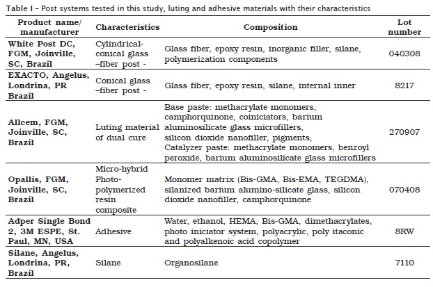
The teeth were randomly divided into 2 groups (n = 12), according to the brand of fiber-reinforced composite root canal posts: group A – cylindrical-conical fiber-reinforced post (White Post DC n. 2, FGM, Joinville, SC, Brazil); and group B – conical fiber-reinforced post (Exacto size #3, Angelus, Londrina, PR, Brazil). The posts were selected for this study based on the similarity of the cervical post diameter (1.8 ± 0.06 mm), composed of unidirectional reinforcement fibers. The materials used in this study are presented in table I.
The radicular dentin was conditioned with 37% phosphoric acid for 20 s, rinsed with water and dried. A light-cured bonding agent (Adper Single Bond 2, 3M ESPE, St. Paul, MN, USA) was applied to the walls of the post spaces, allowed to etch for 30 seconds, gently air dried and light-cured for 40 s with the light-curing unit (Curing Light 2500, 3M ESPE, St. Paul, MN, USA), with intensity of 470 mW/cm2. A dual-cured resin luting agent (AllCem, FGM, Joinville, SC, Brazil) was mixed for 20 s and placed in the post spaces using a lentulo spiral instrument (Dentsply Maillefer, Ballaigues, Switzerland). The posts were treated with silane (Angelus, Londrina, PR, Brazil) gently brushed with a microbrush point for 30 seconds and then coated with cement and slowly seated by finger pressure. Excess of cement was removed and cement was light-cured for 40 seconds with the same light-curing unit. Composite cores (shade A2, Opallis, FGM, Joinville, SC, Brazil) were built up to 5 mm height above the dentin-enamel junction by incremental technique and light-cured for 40 seconds.
Fracture strength
Before carrying out the fracture strength experiment, to simulate the periodontal ligament, the root surface of the restored tooth was coated with polyvinylsiloxane impression material (Express, 3M Espe, St. Paul, MN, USA) with a thickness of approximately 0.1 mm. Finally, the root was embedded in a clear acrylic resin block (Vipi Wave, Dental VIPI Ltda, São Paulo, Brazil) at a depth of 2 mm below the dentin-enamel junction. All specimens were stored in 100% humidity at 36°C for 24 h prior to the fracture test.
The acrylic block containing the restored tooth was tightly fixed to the inclined metal base. A 45-degree oblique load was applied to the restored teeth with a crosshead speed of 1 mm/min by means of a universal testing machine (Instron 4444, Canton, MA, USA) until fracture. Fracture loads were recorded. The vertical load was applied 2.5 mm under the incisal edge on the palatal surface of the crown.
Two point bending test
Twelve post specimens of each manufacturer were embedded in acrylic resin block (Vipi Wave, Dental VIPI Ltda, São Paulo, Brazil), with 5 mm of the cervical portion of the post exposed out of the resin and facially oriented long axes at 90o. All specimens were stored in 100% humidity at 36oC for 24 h prior to the flexural strength test.
The data were obtained in an Instron Universal Testing Machine, and the specimens were submitted to a crosshead speed of 5 mm/min with load applied 2.5 mm under the incisal edge on the post structure until fracture, and the fracture loads were recorded.
SEM evaluation
Two random specimens of each group evaluated in the bending flexural strength experiments were cross-sectioned into two halves using a diamond saw (Isomet1000, Buehler Ltd, Lake Bluff, IL, USA), and the surfaces were coated with gold to avoid a charge being induced by the electron beam and analyzed by SEM (Philips XL30, Eindhoven, Netherlands). One half of each tooth was initially demineralized in 37% phosphoric acid for 20 s, rinsed, dried, and then deproteinized in 3% sodium hypochlorite for 30 min. Resin-dentin inter-diffusion zones (hybrid layer) and resin tag formations in the cervical, middle and apical regions were evaluated.
Statistical analysis
Flexural properties and fracture strength were statistically analyzed by using one-way ANOVA and Tukeys HSD test (SPSS software, SPSS Inc., USA) to evaluate the effect of the brand of the material. Significance level was set at 0.05.
Results
Statistical analysis showed that there were no significant differences between the groups in terms of fracture strength (p = 0.153) or two point bending strength (p = 0.237).
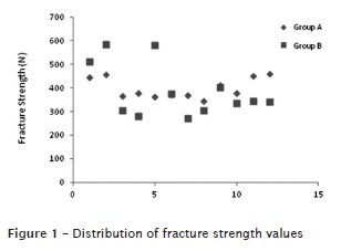
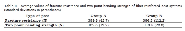
Figure 1 (a and b) shows the distribution of the results obtained in the fracture strength test for fiber-reinforced post specimens. The mean and standard deviation values of the results of the two point bending and fracture resistance tests are given in table II. The average values for fracture strength were 399.3 N (± 42.7) and 386.2 N (± 112.2) for groups A and B, respectively. For the two point bending tests, average values were 109.5 N (± 12.2) and 119.5 N (± 20) for groups A and B, respectively.
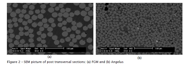
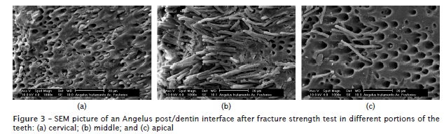
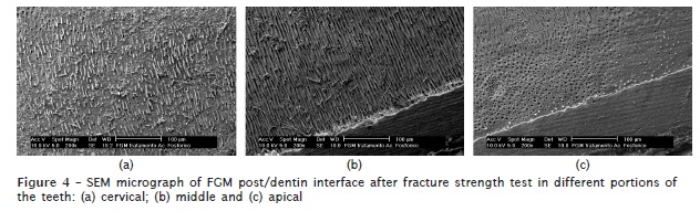
The SEM images indicated a smaller fiber diameter and higher quantity of fibers for the Angelus posts compared with the FGM posts (figure 2). The distribution of the luting material in the canal and at the interface between the luting material and the post was also evaluated through SEM. In the cervical and middle segments of radicular portion for both post groups (group B: figures 3a and b, respectively – group A: figures 4a and b, respectively), the amount of luting material tags penetration into the dentin structure was higher, when compared to the apical segment (figures 3c and 4c, for groups B and A).
Discussion
To determine the characteristics and longevity of FRC posts, simulating the clinical situation, many studies have used the mechanical properties as an evaluation parameter. In this study the tests of fracture resistance and two point bending were evaluated.
The two point bending test determines the fracture resistance of the specimen and high values indicate that a specimen is more resistant to fracture which is dependent of the specimen configuration 15,21. On the other hand, the fracture resistance test simulates the clinical condition where the post is fixed with a luting agent onto the tooth, and the axis along which the load was applied. The fiber-reinforced composite materials are anisotropic and it can be supposed that the more parallel the forces are to the long axis of the post, the higher the flexural test values will be 11,14,17. This could be observed by comparing the fracture resistance with the two point bending strength values given in table II. The values for fracture resistance were higher than those for flexural strength values, but it can be assumed that in the fracture resistance test, which simulates the clinical situation, the strength was distributed among the groups of elements: post system, tooth and the silicone materials simulating the periodontal ligament.
The results for the mechanical properties of fiber-reinforced posts, such as fracture strength, can be related to the characteristics of the fibers and matrix, the strength of the bond at the interface between these components and the geometry of the reinforcement, that is, fiber length and orientation, as well as its concentration. 25 The addition of fibers to a polymer matrix leads to a significant increase in the fracture toughness, stiffness, and fracture resistance of the material 2.
Although the fibers of both posts have different diameters and density, in this study the fracture resistance and flexural strength values were found to be similar for the two groups. On the other hand, Silva et al. 24 concluded that the different materials used in the composition of the posts and the different lengths and geometric configurations may interfere in the fracture resistance of roots. Recent reports suggested that the fiber post diameter may influence the fracture resistance values 15,23. A study reported in the literature tested the hypothesis that the fiber diameter and the surface area occupied by fibers per square millimeter of post surface are parameters related to the physical properties of a fiber post 11. Differences in the used methodology between the studies justify the different results.
Another aspect which could have led to the observed differences in the results is related to the treatment of the fibers during the manufacturing process. During the manufacturing of Angelus posts the fibers are pre-stressed under tension and then soaked in resin, which is then polymerized. During the final cure of the resin, the tension in the fibers is released and, as a result, the resin surface is placed under compression. For this reason, when the post is subjected to a flexural force, the tensile stresses which are introduced can easily be absorbed 23. On the other hand, the FGM fibers of the posts are not pre-stressed in the manufacturing process.
Another factor related to improving the strength of the fiber/matrix interface is the silanization techniques. Silanization prior to embedding in the resin matrices can particularly affect both the mechanical resistance and the structural integrity of these posts. Good interfacial bonding can lead to load transfer from the resin matrix to the fiber reinforcement 11. However, in this study, both posts contain silanized glass fibers, and thus this factor could not have influenced the results.
According to Carrilho et al. 6 the FRC posts are retained on root canal due to the strength obtained by the union between radicular dentin and post by the formation of a hybrid layer. This layer is created after acid etching dentin which removes the smear layer created during the cavity preparation. Thus, the dentin tubules are opened and the demineralized collagen fibrils are exposed. This process promotes the formation of micro channels between these fibrils which are occupied by the primer, adhesive and resin cement.
The results of SEM images of the FRC posts specimens showing the distribution of luting material tags penetration on the different radicular segments are in agreement with other studies by Bitter et al. 5, Ceballos et al. 7, Ferrari et al. 8, Grandini et al. 11, Noirrit et al. 18, Perdigão et al. 20 and Vichi et al. 26, who noted a significant difference in the length and density of tags between the crown, middle segments and the apical segment, where the tags resemble plugs blocking the dentin tubules.
The less favorable morphology of the resin tags in the apical third can probably be explained by the greater difficulty of the light beam to access this level of the root canal during the light curing, even though translucent posts are used for the cure of the luting agent so that it can be pushed out during the mechanic test. Other important aspect is the difficulty of the luting agent penetration in the apical segment, even considering that a lentulo spiral instrument will be used in the clinical situation.
Another aspect relates to the post translucence, FGM post are translucent and Angelus posts are opaque. Although it has been reported that translucent posts transmit the incident light beam to the entire length of the tooth, the polymerization degree is higher in the apical third 1,2,9,22. In this study no difference was observed between translucent and opaque posts on the micrographs.
Although there are no studies in the literature concerning to these specific post brands, the same mechanical properties have been evaluated using other post systems. For example, Garoushi et al. 10 evaluated the static load-bearing capacity and the failure mode of endodontically treated maxillary incisors. The authors reported values of 229 N and 309 N for different fiber-reinforced post systems at the failure mode, i.e. the resistance of the material to fracture.
Conclusion
The fracture resistance and two point bending strength tests are important to simulate the clinical situation and it was used to verify whether the use of posts to reinforce the dental structure can support forces and which values of maximum force it can support before fracture. From the results of the present investigation, it can be seen that there were no significant statistical differences between the two post groups in terms of fracture strength or two point bending strength. It is concluded that the posts selected for this study perform satisfactory mechanical properties so that it can be used for tooth reinforcement after endodontic treatment.
Acknowledgments
We thank the following manufacturers for the donation of materials: Angelus (Londrina, PR, Brazil) and FGM (Joinville, SC, Brazil).
References
1. Akgungor G, Akkayan B. Influence of dentin bonding agents and polymerization modes on the bond strength between translucent fiber posts and three dentin regions within a post space. J Prosthet Dent. 2006;5:145-9. [ Links ]
2. Asmussen E, Peutzfeldt A, Heitmann T. Stiffness, elastic limit, and strength of newer types of endodontic posts. J Dent. 1998;27:275-8.
3. Assif D, Bitenski A, Pilo R, Oren E. Effect of post design on resistance to fracture of endodontically treated teeth with complete crowns. J Prosthet Dent. 1993;69:36-40.
4. Baldissera P, Zicari F, Valandro LF, Scotti R. Effect of root canal treatments on quartz fiber posts bonding to root dentin. J Endod. 2006 Oct;32(10):985-8.
5. Bitter K, Paris S, Martus P, Schartner R, Kielbassa AM. A confocal laser scanning microscope investigation of different dental adhesives bonded to root canal dentin. Int End J. 2004;37:840-8.
6. Carrilho MRO, Geraldeli S, Tay F, De Goes MF, Carvalho RM, Tjäderhane L. In vivo preservation of the hybrid layer by clorhexidine. J Dent Res. 2007;86(6):529-33.
7. Ceballos L, Garrido MA, Fuentes V, Rodriguez J. Mechanical characterization of resin cements used for luting fiber posts by nanoindentation. Dent Mat. 2007;23:100-5.
8. Ferrari M, Vichi A, Grandini S. Efficacy of different adhesive techniques on bonding to root canal walls: an SEM investigation. Dent Mat. 2001;17:422-9.
9. Ferreira R, Mildemberg B, Gadotti BC, Garcia RN. Evaluation of the endodontic treatment influence on the bond strength of fiber posts reinforced by a restorative composite. RSBO. 2011 Apr-Jun;8(2):174-81.
10. Garoushi S, Vallittu PK, Lassila LVJ. Direct restoration of severely damaged incisors using short fiber-reinforced composite resin. J Dent. 2007;35:731-6.
11. Grandini S, Goracci C, Monticelli F, Franklin RT, Ferrari M. Fatigue resistance and structural characteristics of fiber posts: three-point bending test and SEM evaluation. Dent Mat. 2005;21:75-82.
12. Gutmann JL. The dentin-root complex: anatomic and biologic considerations in restoring endodontically treated teeth. J Prosthet Dent. 1992;67:458-67.
13. Ichikawa Y, Akagawa Y, Nikai H, Tsuru H. Tissue compatibility and stability of new zirconia ceramic in vivo. J Prosthet Dent. 1992;68:322-6.
14. King P, Setchell D. An in vitro evaluation of a prototype CFRC prefabricated post developed for the restoration of pulpless teeth. J Oral Rehabil. 1990;17:599-609.
15. Lassila LVJ, Tanner J, Le Bell AM, Narva K, Vallittu PK. Flexural properties of fiber reinforced root canal posts. Dent Mat. 2004;20:29-36.
16. Naumann M, Preuss A, Frankenberger R. Reinforcement effect of adhesively luted fiber reinforced composite versus titanium posts. Dent Mat. 2007;23:138-44.
17. Newman MP, Yaman P, Dennison J, Rafter M, Billy E. Fracture resistance of endodontically treated teeth restored with composite posts. J Prosthet Dent. 2003 Apr;89(4):360-7.
18. Noirrit EE, Grégoire G, Cournot M. Morphological study of fiber-reinforced post-bonding system-root dentin interface by evaluation of two bonding systems. J Dent. 2008;36:204-13.
19. Perdigão J, Gomes G, Augusto V. The effect of dowel space on the bond strengths of fiber posts. J Prosthodont. 2007 May-Jun;16(3):154-64.
20. Perdigão J, Lopes MM, Gomes G. Interfacial adaptation of adhesive materials to root canal dentin. J End. 2007;33:259-63.
21. Plotino G, Grande NM, Bedini R, Pameijer CH, Somma F. Flexural properties of endodontic posts and human root dentin. Dent Mat. 2007;23:1129-35.
22. Rathke A, Haj-Omer D, Muche R, Haller B. Effectiveness of bonding fiber posts to root canals and composite core build-ups. Eur J Oral Sci. 2009;117:604-10.
23. Seefeld F, Wenz HJ, Ludwig K, Kern M. Resistance to fracture and structural characteristics of different fiber reinforced post systems. Dent Mat. 2007;23:265-71.
24. Silva GR, Santos Filho PCF, Simamoto Júnior PC, Martins LRM, Mota AS, Soares CJ. Effect of post type and restorative techniques on the strain and fracture resistance of flared incisor roots. Braz Dent J. 2011;22(3):230-7.
25. Ulbrich NL, Franco APO, Zielak JC, Mathias AL. The stress evaluation of root posts using the finite element analysis. RSBO. 2011;8(2):189-93.
26. Vichi A, Grandini S, Davidson CL, Ferrari M. An SEM evaluation of several adhesive systems used for bonding fiber posts under clinical conditions. Dent Mat. 2002;18:495-502.
 Correspondence:
Betsy Kilian Martins Luiz
Materiais Dentários – Departamento de Odontologia – Universidade Federal de Santa Catarina
Campus Universitário – Trindade
CEP 88040-900 – Florianópolis – SC – Brasil
E-mail:betsyluiz@hotmail.com
Received for publication: October 01, 2011.
Accepted for publication: March 21, 2012.
