Serviços Personalizados
Artigo
Links relacionados
Compartilhar
RSBO (Online)
versão On-line ISSN 1984-5685
RSBO (Online) vol.9 no.3 Joinville Jul./Set. 2012
Original Research Article
Bond strength of endodontic sealers after intracanal surface pretreatment with CO2 laser
Fuad Jacob Abi Rached Junior I Aline Evangelista Souza-Gabriel I Manoel Damião Sousa-Neto II Yara Teresinha Correa Silva-Sousa I
II Department of Restorative Dentistry, University of São Paulo – Ribeirão Preto – SP – Brazil.
ABSTRACT
Introduction: The aim of this study was to evaluate in vitro the bonding of AH Plus and Endofill sealers to intraradicular canal walls after different dentin surface treatments. Material and methods: Sixty canines were sectioned obtaining dentin discs with 4-mm thickness and were embedded in acrylic resin. The canals were prepared with diamond burs. The specimens were divided into two groups (n = 30): GI – AH Plus and GII – Endofill and were subdivided into three groups based on the dentin surface treatment (n = 10): A – distilled and deionized water (control), B – 17% EDTA, C – CO2 laser with 3W in continuous mode for 10s. The specimens were submitted to push out test in a universal testing machine. Results: Tukey test revealed that the IB (AH Plus/17% EDTA) (17.59 ± 6.04) and IC (AH Plus/CO2 laser) (21.69 ± 4.93) subgroups had the highest values, which were statistically similar between each other (p > 0.05) and different from the other subgroups (p < 0.05). IIC subgroup (Endofill/CO2 laser) (7.25 ± 1.59) had intermediate values, which were statistically similar to (p > 0.05) IA subgroup (AH Plus/water) (10.99 ± 2.63), IIA subgroup (Endofill/water) (3.16 ± 0.83) and IIB subgroup (Endofill/ 17% EDTA) (5.31 ± 3.61), which had the smallest values (p < 0.05). Conclusion: The treatment of superficial intracanal dentin with CO2 laser and EDTA favored the adhesion of AH Plus and Endofill sealers.
Keywords: adhesion; sealers; Endodontics; CO2 laser.
Introduction
Different chemical solutions have been recommended for root canal instrumentation, aiming to the removal of both the debris and smear layer and root canal disinfection. Among the solutions recommended, tetra acetic acid (EDTA) is the most used for smear layer removal 4.
The smear layer is a negative factor for root canal sealing, because of the interface between the filling material and root canal walls, which reduces the bonding strength 6.
The use of laser has been a promissory alternative in endodontic therapy, by acting as an auxiliary for root canal cleaning, disinfection and removal of the smear layer 8. In vitro studies have shown the capacity of CO2 laser de of promoting root canal disinfection 9, increasing coronal dentin permeability 15, morphologically altering root dentin 1,22 and vaporizing the smear layer within intertubular dentin 22, therefore enabling a greater imbrication of the endodontic sealers to dentinal tubules 23.
Concerning to the endodontic sealers, they can be classified as: zinc oxide and eugenol-based cements, with or without medicines; calcium hydroxide-based cements; glass ionomer-based cements; and resin cements 21.
Zinc oxide and eugenol cement was introduced in 1936, by Grossman, in Endodontics to be employed in root canal obturation; in 1974, some modifications were performed in its formula, continuing until today, although it presents low biocompatibility 18, lack of adhesive properties 21 and high solubility 2.
AH Plus is a sealer based on epoxy resin with satisfactory physical-chemical properties, low solubility 3,11,16, satisfactory flowing 3,11,16, good adhesion 1,13,14 and proper biological properties 14.
Therefore, it is important to evaluate the influence of the superficial treatment of the dentin with CO2 laser on the bonding of sealers (AH Plus and Endofill) to intraradicular walls.
Material and methods
Maxillary human canines stored in 0.1% thymol solution at 9°C were washed in tap water for 24 hours to eliminate thymol residues.
Teeth were macroscopically examined and radiographed at mesiodistal direction. Inclusion criteria comprised completely formed straight roots with a single canal without calcifications or accentuated curvature. Therefore, sixty teeth were selected. Teeth were sectioned transversally 4 millimetres below the cementoenamel junction to provide 4-mm-thick dentine discs that were centred inside aluminium rings (16 mm diameter and 4 mm height) and embedded in acrylic resin. The aluminium rings containing the dentine discs were placed in a parallelometer and their coronal and apical surfaces were flattened and polished using wet 100-, 180-, 220- and 300-grit sandpapers (Bosch, São Paulo, SP, Brazil) during 15 seconds each. The root canal of each specimen was prepared using a tapered diamond bur (PM720G; KG Sorensen Ind. Com. Ltd, Barueri, São Paulo, SP, Brazil) mounted to a low-speed handpiece which was coupled to the arm of the parallelometer. This arm was lowered to a predetermined depth and a space for sealer placement was created with the following dimensions: larger diameter = 2.70 mm; smaller diameter = 2.30 mm; length = 4 mm. During preparation, the canals were irrigated with distilled water. NaOCl and EDTA solutions were prepared at a manipulation pharmacy (Fórmula & Ação, São Paulo, SP, Brazil).
Sixty specimens were randomly divided into two groups (n = 30) regarding to the sealer used: GI – AH-Plus and GII – Endofill and subdivided into three subgroups according to dentin surface treatment: A (control) – irrigation with distilled and deionized water, B – irrigation with 17% EDTA and C – surface treatment with CO2 laser (Opus Dent, Israel) with 3W in continuous mode for 10s.
Following, the specimens were placed immediately at 37ºC and 95% humidity for a period three times greater than the regular setting time of the sealer.
Subsequently, the specimens were fixed securely in a metallic apparatus by two screws at the horizontal plane. For push-out test, a stainless steel support was used to hold the samples (metallic ring + dentin cylinder) in an Instron 4444 universal testing machine (Instron Corporation, Canton, MA, USA) in such a way that the side with the smaller diameter of the root canal was faced upwards and aligned to the axis that would exert the pressure load on the sealer (apical-coronally). This method assured the alignment of the specimen in a reproducible manner, and also avoided the contact of the axis with the dentin during testing. The machine was calibrated at a constant crosshead speed of 1 mm/minute with 1.4-mm-diameter stainless steel cylindrical tip. The tensile load was applied, and the load required to cause failure at the bond interface was recorded in MPa. Data were submitted to statistical analysis by ANOVA and Tukeys test (p < 0.05).
Results
ANOVA showed statistically significant differences between the sealers, among the treatment surfaces, and among their interaction (p < 0.05).
Tukey test revealed that AH Plus sealer provided the highest mean values (16.76 ± 6.43), statistically different from those of Endofill sealer (5.24 ± 2.83) (p < 0.05).
Concerning to treatment surface, it was observed that CO2 laser promoted a higher mean value (14.47 ± 818), statistically different from that of the groups treated by EDTA (11.45 ± 7.93) and water (7.07 ± 4.42).
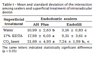
In the interaction between the endodontic sealer and the superficial treatment, it was verified that IB subgroup (AH Plus/17% EDTA) (17.59 ± 6.04) and IC subgroup (AH Plus/CO2 laser) (21.69 ± 4.93) obtained the highest mean values, statistically similar between each other (p > 0.05) and different from those of the other groups (p < 0.05). IIC subgroup (Endofill/laser CO2) (7.25 ± 1.59) presented intermediary values, statistically similar to (p > 0.05) IA subgroup (AH Plus/water) (10.99 ± 2.63) and to IIA subgroup (Endofill/water) (3.16 ± 0.83) and IIB subgroup (Endofill/17% EDTA) (5.31 ± 3.61), which obtained the smallest mean values with statistically similar values between each other (p > 0.05). Table I displays the results obtained in the different experimental groups.

Discussion
The smear layer is an amorphous structure adhered to root canal walls, produced during the biomechanical preparation. This layer is a negative factor in root canal obturation, because it is composed of dentinal debris, remnant of odontoblastic components, pulp tissue and bacterias 12, damaging the adhesion of the filling materials 20.
The removal of the smear layer can be achieved by the use of acid solutions as 17% EDTA 5-7. Sousa-Neto et al. 19 emphasized the importance of smear layer removal prior to root canal obturation, aiming to allow a higher penetration of endodontic sealers within dentinal tubules and to promote a higher mechanical imbrication. Therefore, this study evaluated the influence of the superficial treatment of the intraradicular wall with distilled and deionized water, 17% EDTA and CO2 laser on the bond strength to dentin of AH Plus and Endofill endodontic sealers.
The superficial treatment of intraradicular dentin promoted, in this present study, significantly different bond strength values of endodontic sealers to root canal walls AH Plus showed the highest bonding values when compared with Endofill, regardless of the previous superficial treatment of the dentin. Such fact can be explained because AH Plus is an epoxy resin-based sealer and it better penetrates into the micro-irregularities of root canal walls because of its flowing and high curing time 3,11,16. These properties favor a greater imbrication between the sealer and the dentin, which in addition to the cohesion among the molecules of the sealer promote a greater resistance to removal and/or displacement from the dentin surface, resulting in this present study in higher adhesion 20.
In this present study, the sealers analysed obtained an increase in the bond strength values when the dentinal surface was treated with CO2 laser. CO2 laser promotes root canal disinfection 9, increases the coronal dentinal permeability 15 and ultrastructurally alters the root dentin 1.
Additionally, this laser promotes the formations of crackers and fissures and vaporizes the smear layer of intertubular dentin 17,22. Such alterations in dentin surface were observed through scanning electronic microscopy by Trajtenberg et al. 23 and Alfredo et al. 1. According to Sousa-Neto et al. 19, smear layer removal and the presence of fissures on the dentin surface may facilitate the mechanical retention of the endodontic materials and favor the bond strength to root canal wall. Accordingly, it is believed that the ultrastructural alteration promoted by CO2 laser resulted in this present study in higher bond strength of the sealers to intraradicular wall.
Unlike to CO2 laser, 17% EDTA acts on the mineral matrix and removes the smear layer formed during biomechanical preparation, which probably enables a greater penetration of the sealers within dentinal tubules and results in an increasing of the contact surface between the sealer and the dentin 5. Therefore, EDTA action promotes values statistically similar to those treated with CO2 laser, regardless of the sealer used.
Considering the aforementioned discussion, this study encourages further studies aiming to elucidate the influence of the ultrastructural alteration of the dentin, after the superficial treatment, on the bonding of different resin-based endodontic sealers.
Conclusion
Based on the methodology employed and on the results obtained, it can be concluded that the superficial treatment of the intraradicular dentin through CO2 laser and EDTA favored the bonding of endodontic sealers.
References
1. Alfredo E, Silva SR, Ozório JE, Sousa-Neto MD, Brugnera-Júnior A, Silva-Sousa YT. Bond strength of AH Plus and Epiphany sealers on root dentine irradiated with 980 nm diode laser. Int Endod J. 2008 Sep;41(9):733-40. [ Links ]
2. Carvalho-Júnior JR, Guimarães LF, Correr-Sobrinho L, Pécora JD, Sousa-Neto MD. Evaluation of solubility, disintegration, and dimensional alterations of a glass ionomer root canal sealer. Braz Dent J. 2003 Feb;14(2):114-8.
3. Flores DS, Rached-Junior FJ, Versiani MA, Guedes DF, Sousa-Neto MD, Pécora JD. Evaluation of physicochemical properties of four root canal sealers. Int Endod J. 2011 Feb;44(2):126-35.
4. Grossman LI. Physical properties of root canal cements. J Endod. 1976 Jun;2(6):166-75.
5. Hülsmann M, Heckendorff M, Lennon A. Chelating agents in root canal treatment: mode of action and indications for their use. Int Endod J. 2003 Dec;36(12):810-30.
6. Kennedy WA, Walker WA, Gouch RW. Smear layer removal effects on apical leakage. J Endod. 1986 Jan;12(1):21-7.
7. Kouvas V, Liolios E, Vassiliadis L, Parissis-Messimeris S, Boutsioukis A. Influence of smear layer on depth of penetration of three endodontic sealers: an SEM study. Endod Dent Traumatol. 1998 Aug;14(4):191-5.
8. Lan WH, Chen KW, Jeng JH, Lin CP. A comparison of the morphological changes after Nd:YAG and CO2 laser irradiation of dentin surfaces. J Endod. 2000 Aug;26(8):450-3.
9. Le Goff A, Dautel-Morazin A, Guigand M, Vulcain JM, Bonnauremallet M. An evaluation of the CO2 laser for endodontic disinfection. J Endod. 1999 Feb;25(2):105-8.
10. Lee K, Williams MC, Camps JJ, Pashley DH. Adhesion of endodontic sealers to dentin and gutta-percha. J Endod. 2002 Oct;28(10):684-8.
11. Marin-Bauza GA, Rached-Junior FJ, Souza-Gabriel AE, Sousa-Neto MD, Miranda CE, Silva-Sousa YT. Physicochemical properties of methacrylate resin-based root canal sealers. J Endod. 2010 Sep;36(9):1531-6.
12. McComb D, Smith DC. Comparison of physical properties of polycarboxilatebased and conventional root canal sealers. J Endod. 1976;2(8):228-35.
13. Nunes VH, Silva RG, Alfredo E, Sousa-Neto MD, Silva-Sousa YT. Adhesion of Epiphany and AH Plus sealers to human root dentin treated with different solutions. Braz Dent J. 2008;19(1):46-50.
14. Onay EO, Ungor M, Ozdemir BH. In vivo evaluation of the biocompatibility of a new resin-based obturation system. Oral Surg Oral Med Oral Pathol Oral Radiol Endod. 2007;104(3):60-6.
15. Pashley EL, Homer JA, Liu M, Kim S, Pashley DH. Effects of CO2 laser energy on dentin permeability. J Endod. 1992;18(1):257-62.
16. Resende LM, Rached-Junior FJ, Versiani MA, Souza-Gabriel AE, Miranda CE, Silva-Sousa YT et al. A comparative study of physicochemical properties of AH Plus, Epiphany, and Epiphany SE root canal sealers. Int Endod J. 2009;42(9):785-93.
17. Sasaki K, Aoki A, Ichinose S, Ishikawa I. Morphological analysis of cementum and root dentin after Er:YAG laser irradiation. Lasers Surg Med. 2002;31(2):79-85.
18. Scarparo RK, Grecca FS, Fachim EV. Analysis of tissue reactions to methacrylate resin-based,epoxy resin-based and oxide-eugenol endodontic sealers. J Endod. 2009;35(2):229-32.
19. Sousa-Neto MD, Coelho FI, Marchesan MA, Alfredo E, Silva-Sousa YTC. In vitro study of the adhesion of an epoxy based sealer to human dentine submitted to irradiation with Er:YAG and Nd:YAG lasers. Int Endod J. 2005;38(12):866-70.
20. Sousa-Neto MD, Marchesan MA, Pécora JD, Brugnera A, Silva-Sousa YTC, Saquy PC. Effect of Er: YAG laser on adhesion of root canal sealers. J Endod. 2002;28(3):185-7.
21. Sousa-Neto MD, Guimarães LF, Saquy PC, Pécora JD. Effect of different grades of gum rosins and hydrogenated resins on the solubility, disintegration, and dimensional alterations of Grossman cement. J Endod. 1999;25(7):477-80.
22. Takahashi K, Kimura Y, Matsumoto K. Morphological and atomic changes after CO2 laser irradiation emotted at 9.3 µm on human dental tissues. J Clin Laser Surg. 1998;16(3):167-73.
23. Trajtenberg CP, Pereira PN, Powers JM. Resin bond strength and micromorphology of human teeth prepared with an Erbium: YAG laser. Am J Dent. 2004;17(5):331-6.
24. Vilanova WV, Carvalho-Junior JR, Alfredo E, Sousa-Neto MD, Silva-Sousa YT. Effect of intracanal irrigants on the bond strength of epoxy resin-based and methacrylate resin-based sealers to root canal walls. Int Endod J. 2012 Jan;45(1):42-8.
 Correspondence:
Aline Evangelista Souza-Gabriel
Av. Costábile Romano, n.º 2.201 – Ribeirânea
CEP 14096-900 – Ribeirão Preto – SP – Brasil
E-mail:aline.gabriel@gmail.com
Received for publication: March 12, 2012.
Accepted for publication: May 19, 2012.


