


Serviços Personalizados
Artigo
 pdf em Inglês
 Artigo em XML
 Referências do artigo
 Enviar este artigo por email
Links relacionados
Compartilhar

 Permalink
RSBO (Online)
versão On-line ISSN 1984-5685
RSBO (Online) vol.9 no.4 Joinville Out./Dez. 2012
Original Research Article
Comparison of the effectiveness of the protaper system versus hand instrumentation in endodontic retreatment: a scanning electron microscopy study
Luana Schwerz I ; Carlos Eduardo Fontana II ; Carlos Eduardo da Silveira Bueno II ; Roberta Aranha de Araújo Arruda II ; Rina Andréa Pelegrine II ; Flavia Casale Abe II ; Alexandre Sigrist De Martin II
II Department of Endodontics, São Leopoldo Mandic Center of Post-Graduation – Campinas – SP – Brazil
ABSTRACT
Introduction: Several rotary systems have been evaluated for removal of endodontic filling materials from the canal. Moreover, studies focusing on the comparison of the effectiveness of rotary systems versus hand instrumentation have yielded mixed results in terms of the efficacy and amount of time required by each technique. Objective: To compare the effectiveness of a nickel-titanium rotary system and of hand instrumentation using stainless steel files and Gates-Glidden burs in the removal of gutta-percha from root canals, as well as the time required to complete the procedure by each method. Material and methods: Forty single-rooted teeth were prepared and obturated then divided in two groups, according to the method employed for removal of the gutta-percha: ProTaper Universal rotary retreatment system (rotary instrumentation) and stainless steel hand files with Gates-Glidden burs (hand instrumentation). The time required to remove gutta-percha by each method was recorded. Roots were then sectioned lengthwise and the apical, middle, and coronal thirds were analyzed by SEM under two magnifications: x400 and x1,000. Results: Rotary instrumentation promoted better cleansing compared with hand instrumentation. The apical third was less clean than the coronal and middle thirds (Kruskal-Wallis p < 0.05). Rotary instrumentation was faster than hand instrumentation (Tukey p < 0.05). Conclusion: Although none of the methods promoted complete cleanliness of the canal walls, ProTaper Universal system showed better results and was faster than hand instrumentation.
Keywords: gutta-percha; NiTi; retreatment.
Introduction
In recent years, the great technological and scientific progress attained in the field of Endodontics has led to increase in the number of successful treatments. However, in some instances, failure associated with difficulties during the instrumentation and/or root canal filling occurs 5,16, requiring retreatment 4,6.
Endodontic therapy failure presents clinically through signs and symptoms such as pain to percussion, thermal sensitivity, recurrent abscesses, fistulas, and radiographically visible periapical lesions 6,13,18,26. In these situations, the treatment of choice is nonsurgical retreatment with the goal of eliminating the cause of failure, which is often associated with presence of bacteria within the canals 1,13. Nonsurgical endodontic retreatment is generally preferred to endodontic surgery due to the potential for post-surgical discomfort 13.
Gutta-percha, associated with endodontic sealers, is the most widely used filling material for root canals 25,25. Its removal is timeconsuming, particularly when the initial treatment involved careful condensation against the canal walls. Undoubtedly, the time spent in any clinical procedure is a relevant factor that should be taken into account when choosing between treatment options 14.
Several rotary systems have been evaluated for removal of endodontic filling materials from the canal. Moreover, studies focusing on the comparison of the effectiveness of rotary systems versus hand instrumentation 12,15,17 have yielded mixed results in terms of the efficacy and amount of time required by each technique 2,24.
The ProTaper Universal endodontic rotary instruments size D1, D2, and D3 (Dentsply-Maillefer, Ballaigues, Switzerland) have been developed specifically for retreatment. The goal of the present study was to evaluate, by means of scanning electron microscopy, the effectiveness of these instruments in the removal of root canal filling materials and compare their performance with hand instrumentation. Additionally, this study compared the amount of time required by each method to remove the filling materials from the canal.
Material and methods
This study was accepted by the Ethics Committee in Research of CPO-São Leopoldo Mandic, under protocol number #07/185 – 2007.
Preparation of the specimens
Forty single-rooted anterior teeth with completely formed roots and no evidence of either calcification or resorption were used. The teeth had their crowns removed with a diamond disc (KG Sorensen Ltda., São Paulo, Brazil) in order to standardize root length at 17 mm.
Root canal treatment
The following steps were performed by a single operator. The working length was determined by introducing a 15 K-file into the canal until its tip was visible at the apical foramen, then retracting 1mm. The root canals were instrumented by the crowndown technique up to size F2 file in the ProTaper Universal system (Maillefer Corp., Ballaigues, Switzerland) attached to an electric handpiece (XSmart – Maillefer Corp., Ballaigues, Switzerland) set at 300 rpm and 3 N/cm torque. After that, the canals were filled with size F2 gutta-percha cones (Dentsply Ind. e Com. Ltda., Petrópolis, Brazil) and AH Plus sealer cement (Dentsply Ind. e Com. Ltda., Petrópolis, Brazil), following Buchanan's continuous wave of condensation technique. Teeth were then sealed with a cotton pellet and Coltosol temporary cement (Vigodent S.A., Rio de Janeiro, Brazil) and maintained in oven at 37°C for 30 days to allow complete setting of the cement and closely simulate the clinical conditions observed during endodontic retreatments.
Retreatment techniques
The specimens were randomly divided into two groups according to the instrumentation method used for removal of the filling materials from the canal, as follows:
• Rotary instrumentation: ProTaper Universal System NiTi rotary files were attached to an X-Smart electric handpiece ( Maillefer Corp., Ballaigues, Switzerland) set at 400 rpm and 3 N/cm torque. The size D1, D 2, and D 3 retreatment files were used sequentially following the crown-down technique until the working length was reached. After that, ProTaper Universal size F1, F2, F3, and F4 files were used up to the working length at 300 rpm and 3 N/cm torque.
• Hand instrumentation: Gates-Glidden drills were attached to a conventional contra-angle (Dabi-Atlante Ltda., Ribeirão Preto, Brazil) set at approximately 10,000 rpm. Stainless steel files sizes 4, 3, and 2 (in this sequence) were used to instrument the coronal and middle thirds. The apical third was instrumented with K-Flexofiles sizes 15, 20, 25, 30, 35, and 40 (in this sequence) up to the working length.
All the hand files, as well as the rotary instruments, were discarded after five uses. At each instrument change, the canals were irrigated with 2.5% sodium hypochlorite. After instrumentation, canals were irrigated with 5 ml of 17% EDTA for 3 minutes in order to remove the smear layer, followed by a final irrigation with 2.5% sodium hypochlorite. Finally, the canals were aspirated with capillary tips (Ultradent Products Inc., USA) and dried with paper points.
After that, teeth were evaluated with the aid of an operating microscope (Opto Eletrônica S.A., São Carlos, Brazil) at x12.5 magnification, to check for remnants of gutta-percha and/or sealer. When residual gutta-percha and/or sealer were detected on the walls, the canal was reinstrumented until no remnants were visible. The time required by each technique for removal of obturating materials was recorded using a digital chronometer (Timex – USA), from the moment of introduction of the first instrument until the moment when no remnants of filling material were visualized with the aid of the operating microscope.
Evaluation
After the retreatment procedures, teeth were sectioned by carving a groove on the proximal surfaces with a diamond disc and cleaving the sections with a Le Cron carver, resulting in two hemisections for each root and exposing the lumen of the prepared canal. This allowed the visualization of the internal walls of the canal with a scanning electron microscope. The hemisection of each root that appeared most suitable for visualization with an SEM was selected and kept in an oven at 45ºC for 12 hours, in order to desiccate the specimens and allow metallization of the surfaces prior to the SEM analyses. Following that, specimens were sputter-coated using an SCD 050 apparatus (Balzers, Liechtenstein).
Analyses of the root canal surfaces were carried out using a DSM 940A scanning electron microscope (Carl Zeiss, Oberkocher, Germany) at x400 (15 kV) and x1,000 (15 kV) magnification at 1 mm, 5 mm, and 10 mm short of the working length, as described by Zmener et al14. After that, the SEM images were saved in BMP format and observed in a computer monitor by three independent observers with vast training and experience in Endodontics. Each observer attributed a score to each image, according to the guidelines described by Hülsmann and Stotz 11 for evaluation of smear layer and debris removal, where:
Score 0: No debris or only isolated small particles were present.
Score 1: Minimal debris particles were present in small clumps.
Score 2: Clumps of debris particles covered less than 50% of the canal wall.
Score 3: Clumps of debris particles covered more than 50% of the canal wall.
Score 4: Clumps of debris particles completely covered the canal wall.
Results
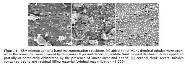
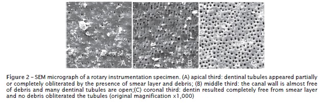
Both instrumentation methods allowed residual filling material to remain inside the root canal. Considering the results from the entire canal, the rotary instruments performed better than the hand instruments (figures 1 and 2).
The Kruskal-Wallis test showed that the true mean score for the hand instrumentation group was significantly higher than the mean score for the rotary instrumentation group (table I).

When the scores attributed to each level of the canal were compared, the Kruskal-Wallis test also indicated differences between at least two among the three thirds of the root, regardless of the technique utilized (table II).
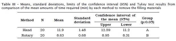
Table III shows the mean amount of time required by the two methods to remove the filling material. Tukey test showed that the rotary instruments required significantly less time than hand instruments for removal of the filling materials from the canals.
Discussion
The success of endodontic therapy is directly linked to adequate shaping of the canal during instrumentation and thorough disinfection of its walls by mechanical action of the instruments allied to an irrigation agent 13,16,18,20. Moreover, hermetic seal is known as a crucial factor for success. Unfortunately, in some instances these requirements are not fulfilled and endodontic retreatment becomes necessary 1,17.
Scanning electron microscopy has been applied to different methodologies where scores are attributed to specimens according to the degree of debris and smear layer remaining on the root canal walls after instrumentation 1,16,21. This technology allows acquisition of highresolution images of areas covered by debris and/or smear layer, as well as the identification of patent dentinal tubules. In the present study, we opted for high-resolution SEM, since all other possible techniques (including micro–computed tomography) have insufficient resolution to detect these features 19.
Several studies have shown that no endodontic instruments or instrumentation techniques are capable of achieving complete cleanliness of the root canal walls 1,2,17,23. Our results corroborated these findings: none of the instrumentation methods employed in this study was completely successful in cleaning the root canal walls.
The ProTaper Retreatment Kit (D1, D2, and D3 files) was specifically developed for retreatment of root canals. Several studies 9,23,25 have been carried out to evaluate this new system. The present work was designed with the goal of evaluating its cleansing ability and agility during endodontic retreatment.
In the present study, an operating microscope was used after reinstrumentation to visualize the degree of cleanliness of the canal walls, as described by Schirrmeister et al. 22. Even though filling material remnants were visible in some areas of the canal walls, their removal was extremely difficult, agreeing with the findings of Schirrmeister et al. 22.
Previous studies have used MEV magnifications ranging from x15 to x2,500 1,21. Lower resolutions allow visualization of great amounts of debris; however, details such as smear layer remnants or dentinal tubules require higher magnifications 26. In this study, we opted for two different magnifications, x400 and x1,000, since these are recommended in the literature for better visualization of dentinal tubules and to evaluate the presence of debris 1,21,26.
The results (scores attributed to each specimen to different thirds of the canal) were subjected to statistical analyses, where the first step was to measure the reliability of the data by applying the Kappa coefficient. Once data were deemed to be reliable, a rank-based analysis of variance was performed (ANOVA-R), showing evidence (p < 0.05) of statistically significant differences between the mean scores of each instrumentation method, and strong evidence (p < 0.01) of differences between the true scores attributed to each third of the root canal. Subsequently, the Kruskal-Wallis demonstrated that rotary instrumentation was more effective than hand instrumentation in the removal of obturating materials from the root canals.
Our results corroborate the findings from other authors 7,8,9,15,24, which also observed more favorable results with rotary instrumentation. Only two previous studies 4,10 reported better results for hand instrumentation. The statistical analyses we used also allowed comparison of the degree of cleanliness achieved in each third of the root, showing the persistence of greater amount of debris in the apical third, intermediate amounts in the middle third, and the least amount of debris at the coronal third. These findings are in agreement with some other studies 8,10,24.
Despite not playing a direct role towards the success of endodontic therapy, the time required for completion of treatment is a relevant factor in all clinical dental procedures 14 and can be considered as a measure of effectiveness of the method employed. Regarding the time required to remove obturating materials from the canal by each method evaluated in this work, the rotary instruments were statistically confirmed as faster for removing gutta-percha and sealer than the hand instruments. This observation is in agreement with results recently reported by Gu et al. 9. Furthermore, our findings agree with other authors 3,23,25 who compared the use of hand instruments and rotary instrumentation during endodontic retreatment. Only a few studies 2,3 had different outcomes, showing that hand instrumentation required less time than rotary systems.
Scanning electron microscopy is an extremely efficient means for observing the morphology of residual root filling material. Apical instrumentation with a size 40 file is probably insufficient 18 for complete removal of filling debris from the dentinal tubules. The complex morphology of the dentin surface on the root canal walls increases the risk of insufficient removal of sealer debris from the dentinal tubules.
Conclusion
Based on our results and considering the methodology used in this work, we can conclude that none of the methods evaluated promoted complete cleansing of the root canal walls. Nevertheless, the rotary retreatment system promoted better results compared with hand instrumentation.
Regarding the differences in the degree of cleanliness between the root thirds, the coronal and middle thirds were cleaner than the apical third, regardless of the technique used.
Less time was required for retreatment of the root canals using rotary instruments compared with hand instrumentation.
References
1. Ahlquist M, Henningsson O, Hultenby K, Ohlin J. The effectiveness of manual and rotary techniques in the cleaning of root canals: a scanning electron microscopy study. Int Endod J. 2001;34(7):533-7. [ Links ]
2. Barrieshi-Nussair K. Gutta-percha retreatment: effectiveness of nickel-titanium rotary instruments versus stainless steel hand files. J Endod. 2002;28(5):454-6.
3. Betti LV, Bramante CM. Quantec SC rotary instruments versus hand files for gutta-percha removal in root canal retreatment. Int Endod J. 2001;34(7):514-9.
4. Bueno CES, Delboni MG, Araújo RA, Carrara HJ, Cunha RS. Effectiveness of rotary and hand files in gutta-percha and sealer removal using chloroform or chlorhexidine gel. Braz Dent J. 2006;17(2):139-43.
5. Carvalho-Sousa B, Costa-Filho JR, Almeida-Gomes F, Ferreira CM, Gurgel-Filho ED, Albuquerque DS. Evaluation of the dentin remaining after flaring using Gates Glidden drills and Protaper rotary files. RSBO. 2011;8(2):194-9.
6. Cunha RS, De Martin AS, Barros PP, Silva FM, Jacinto RC, Bueno CS. In vitro evaluation of the cleansing working time and analysis of the amount of gutta-percha or Resilon remnants in the root canal walls after instrumentation for endodontic retreatment. J Endod. 2007;33(12):1426-8.
7. Garcia Júnior JS, Silva Neto UX, Carneiro E, Westphalen VPD, Fariniuk LF, Fidel RAS et al. Avaliação radiográfica da eficiência de diferentes instrumentos rotatórios no retratamento endodôntico. RSBO. 2008;5(2):41-9.
8. Gergi R, Sabbagh C. Effectiveness of two nickeltitanium rotary instruments and a hand file for removing gutta-percha in severely curved root canals during retreatment: an ex vivo study. Int Endod J. 2007;40(7):532-7.
9. Gu LS, Ling JK, Wei X, Huang XY. Efficacy of ProTaper Universal rotary retreatment system for gutta-percha removal from root canals. Int Endod J. 2008;41(4):288-95.
10. Hammad M, Qualtrough A, Silikas N. Threedimensional evaluation of effectiveness of hand and rotary instrumentation for retreatment of canals filled with different materials. J Endod. 2008;34(11):1370-3.
11. Hülsmann M, Stotz S. Efficacy, cleaning ability and safety of different devices for gutta-percha removal in root canal retreatment. Int Endod J. 1997 Jul;30(4):227-33.
12. Imura N, Kato AS, Hata G-I, Uemura M, Toda T, Weine F. A comparison of the relative efficacies of four hand and rotary instrumentation techniques during endodontic retreatment. Int Endod J. 2000;33(4):361-6.
13. Kvist T, Reit C. Results of endodontic retreatment: a randomized clinical study comparing surgical and nonsurgical procedures. J Endod. 1999;25(12):814-7.
14. Ladley RW, Campbell AD, Hicks ML, Li SH. Effectiveness of halothane used with ultrasonic or hand instrumentation to remove gutta-percha from the root canal. J Endod. 1991;17(5):221-4.
15. Maciel ACC, Scelza MFZ. Efficacy of automated versus hand instrumentation during root canal retreatment: an ex vivo study. Int Endod J. 2006;39(10):779-84.
16. Mayer BE, Peters OA, Barbakow F. Effects of rotary instruments and ultrasonic irrigation on debris and smear layer scores: a scanning electron microscopic study. Int Endod J. 2002;35(7):582-9.
17. Masiero AV, Barletta FB. Effectiveness of different techniques for removing gutta-percha during retreatment. Int Endod J. 2005;38(1):2-7.
18. Mickel AK, Chogle S, Liddle J, Huffaker K, Jones JJ. The role of apical size determination and enlargement in the reduction of intracanal bacteria. J Endod. 2007;33(1):21-3.
19. Pirani C, Pelliccioni GA, Marchionni S, Montebugnoli L, Piana G, Prati C. Effectiveness of three different retreatment techniques in canals filled with compacted gutta-percha or Thermafil: a scanning electron microscope study. J Endod. 2010;35(10):1433-40.
20. Pires LB, Albergaria SJ, Fagundes Tomazinho FS, Tomazinho LF. Avaliação radiográfica do desvio apical de canais radiculares curvos após emprego da instrumentação manual e rotatória. RSBO. 2009;6(3):279-85.
21. Schäfer E, Zapke K. A comparative scanning electron microscopic investigation of the efficacy of manual and automated instrumentation of root canals. J Endod. 2000;26(11):660-4.
22. Schirrmeister JF, Hermanns P, Mayer KM, Goetz F, Hellwig E. Detectability of residual Epiphany and gutta-percha after root canal retreatment using a dental operating microscope and radiographs – an ex vivo study. Int Endod J. 2006;39(7):558-65.
23. Só MV, Saran C, Magro ML, Vier-Pelisser F, Munhoz M. Efficacy of ProTaper retreatment s y s t em in root canal s f i l l ed wi th gut tapercha and two endodontic sealers. J Endod. 2008;34(10):1223-5.
24. Somma F, Cammarota G, Plotino G, Grande NM, Pameijer CH. The effectiveness of manual and mechanical instrumentation for the retreatment of three different root canals filling materials. J Endod. 2008;34(4):466-9.
25. Takahashi CM, Sanches Cunha R, De Martin AS, Fontana CE, Silveira CFM, Bueno CES. In vitro evaluation of the effectiveness of ProTaper Universal Rotary retreatment system for guttapercha removal with or without a solvent. J Endod. 2009;35(11):1580-3.
26. Zmener O, Pameijer CH, Banegas G. Effectiveness in cleaning oval-shaped root canals using Anatomic Endodontic Technology, ProFile and manual instrumentation: a scanning electron microscopic study. Int Endod J. 2005;38(6):356-63.
 Correspondence:
Carlos Eduardo Fontana
Avenida 2, n. 1.220
CEP 13500-411 – Rio Claro – SP – Brasil
E-mail: ceffontana@hotmail.com
