


Serviços Personalizados
Artigo
 pdf em Inglês
 Artigo em XML
 Referências do artigo
 Enviar este artigo por email
Links relacionados
Compartilhar

 Permalink
RSBO (Online)
versão On-line ISSN 1984-5685
RSBO (Online) vol.9 no.4 Joinville Out./Dez. 2012
Original Research Article
Comparative study among Dentistry undergraduates and Forensic Odontology postgraduate students through smile photographs for human identification
Rhonan Ferreira da Silva I,II,III ; Laíse Nascimento Correa Lima III ; Leandro Brambilla Martorell II,IV ; Mauro Machado do Prado I ; Luiz Francesquini Júnior III ; Eduardo Daruge Júnior III
II School of Dentistry, Paulista University, Campus Flamboyant – Goiânia – GO – Brazil
III School of Dentistry of Piracicaba, State University of Campinas – Piracicaba – SP – Brazil
IV Postgraduation Program in Bioethics, University of Brasília – Brasília – DF – Brazil
ABSTRACT
Introduction: The execution of forensic odontology technique for human identification depends on the existence of dental files produced ante-mortem (dental records, clinical notes, radiographs or dental casts). However, when these are not present, other sources of dental data should be searched, such as photographs of the smile. Objective: To compare the performance of undergraduates of Dentistry and postgraduate students of Forensic Odontology to execute human identification through the analysis of photographs of the smile based on decisive dental parameters. Material and methods: Forty Dentistry undergraduates of a School of Dentistry (20 presenting history of orthodontic treatment and 20 without treatment were photographed as follows: 1) extraoral photograph of posed smile, at frontal position and; 2) intraoral photograph, at frontal position. Using these 40 pairs of photographs, four tests were prepared (A, B, C and D) which were sent to both 12 undergraduates of Dentistry and 12 postgraduate students of Forensic Odontology both from another School of Dentistry. The examiners should analyze and correlate a picture randomly and previously selected (photograph of the smile or intraoral photograph) with its corresponding one (photograph of the smile or intraoral photograph), which was set in a showcase composed by 10 images, by pointing out the main criteria for reaching a final conclusion. Results: All the subjects of the research, in both groups, correctly answered to test A (analysis of a photograph of the smile in a group of 10 intraoral photographs). The tests B (analysis of an intraoral photograph in a group of 10 photographs of the smile) and D (analysis of a photograph of the smile in a group of 10 intraoral photographs) had 91.6% success among postgraduate students; and test C (analysis of an intraoral photograph in a group of 10 photographs of the smile) had 83.3% success among undergraduate students. Conclusion: Among the most relevant parameters to achieve the result of Forensic dentistry identification through the analysis of photographs of the smile, the morphology of the incisal edges of anterior teeth was the aspect most often cited by both undergraduate students (83.3%) and postgraduate students (72.9%), within the 48 tests applied to each group. Most of the Dentistry undergraduates and Forensic Odontology postgraduate students were capable of performing the human identification through the analysis of photographs of the smile, considering the wide variety of potentially demonstrable dental characteristics of the anterior teeth.
Keywords: Forensic Dentistry; human identification; smiling.
Introduction
The human identification through dental features is a task routinely executed by several Brazilian Forensic Odontology services, mainly in cases in which the fingerprint of the person to be identified could not be analyzed, such as charred, decomposing, mutilated, macerated and skeletonized individuals 20.
The importance of the Forensic Odontology is highlighted not only in single cases, but also in mass disasters, such as those reported by Schuller- Gotzburg and Suchanek 16, who demonstrated that, of the 2,679 victims of the tsunami occurred in 2004 in the city of Phuket (Thailand), 1,105 were identified exclusively through forensic odontology analysis.
Therefore, it is necessary to compare the anteand post-mortem information within a safe data source 13, which is traditionally in Dentistry achieved through dental files and records, intraoral photographs and dental casts 17,19. On the other hand, similarly to other methods of human identification, these traditional forensic odontology techniques may be inappropriate for several reasons, among which: extensive destruction of the buccomaxillofacial complex, lack of dental records obtained for clinical purpose or dental records with incomplete or irrelevant forensic information.
Because of these reasons, currently, the forensic experts acting in the practice of human identification search for information in alternative sources, such as photographs of the face 2, shootings 8 or photographs of the smile, which exhibit specific characteristics of each subject. This makes important the analysis of photographs present in family albums or produce in social events in which the victim had participated. The justification for this alternative investigation is based on the search of data on shape, dimensions and alignment of the teeth of a person, which can comprise a unique and specific set. Moreover, there is a tendency towards using digital cameras that allowed the production of either photographic shots or shootings, in which is possible to executed the image zoom or search for a central focus on the face of the subjects, more specifically the smile.
Considering that in the analysis of bodies so-called "unrecognizable" the forensic odontology techniques superposes the other methodologies – because of their lower operational cost, faster analysis and data interpretation, higher reliability of the results obtained and the presence of qualified professionals (forensic odontology experts) –, it becomes essential that new parameters be either developed or obtained, aiming to evidence and identify as unique the dental features of each individual. The literature reports three cases in which the victims were identified through using the photographs of the smile together with other forensic techniques, such as Forensic Anthropology, dental radiographs and DNA examination 18. The aforementioned photographs were obtained with relatives of the victim and produced by traditional digital cameras.
Therefore, the aim of this study was to demonstrate the importance and applicability of photographs of the smi le as informat ion source for human identification, through the comparative analysis of the performance of lastyear dental undergraduates and Forensic Dentistry postgraduates as well as to correlate the photographs of the smile to intraoral photographs and to identify which parameters would be more recurrent to establish a positive forensic dentistry identification through using photographs of the smile.
Material and methods
The research was divided into two stages: collection of data and images (intraoral and smile photographs) and analysis of the images obtained at the first stage.
Firstly, images of 40 Dentistry undergraduates of a School of Dentistry (Paulista University – Unip, GO, Brazil) were used, comprising 20 undergraduates which had been submitted to orthodontic treatment and other 20 who had not been orthodontically treated, and willing to participate in the study after reading and signing a free and clarified consent form.
Inclusion criteria comprised individuals without fixed orthodontic appliance on the labial surfaces of the anterior teeth and the possibility of visualization of the anterior teeth inside the area of exposition of the smile. Exclusion criteria were composed of smiles that did not show the incisal edge of the maxillary anterior teeth and the presence of diastemas between the maxillary central incisors.
For each individual, two types of photographs were produced: smile and intraoral, which resulted in 20 pairs of photographic images for each group (with and without orthodontic treatment).
To obtain the photograph of the smile, all face of the individual was framed in frontal norm; the individual exhibited a forced or social smile, so that the incisal edges of the anterior teeth could be seen. This photograph type was performed with tripod and conventional digital camera (Sony® CyberShot, DSC W210, 12.1 megapixels), with 3.0 megapixel of resolution. The individual was placed in a seated position on a dental stool at 1.5 m far from the lens, with the Frankfurt plan parallel to the floor. The photograph was posteriorly cut through Adobe Photoshop® software version 7.0, resulting in the image of the area between the infraorbital margin and the chin.
To obtain the intraoral photograph, the participant was photographed at lying position on a dental chair with the dental reflector turned off. The lips were manually separated, so that the incisal edges of the maxillary and mandibular teeth could be seen. At that moment, the photographic record was carried out with semiprofessional camera (Nikon® Coopix 7800, 8 megapixels), with 3.0 megapixels of resolution.
To simulate probable forensic odontology identification, the smi le photographs were considered as standard images, that is, produced during life (ante mortem), and the intraoral photographs were considered as question images, that is, they would have been produced after death (postmortem).
The second stage of the research comprised a voluntary participation of 12 last-year dental undergraduates and 12 Forensic Odontology postgraduate students after the signing and reading of the FCCF, all from the School of Dentistry of Piracicaba (State University of Campinas, FOPUnicamp). This group of 24 students must simulate the action of a forensic odontology expert in two situations, as follows: the identification of one missing person when there are 10 non identified bodies, through using the smile photograph; and the identification of one non identified body when there are 10 non identified bodies.
At this stage of the study, the participants were informed that either the smile or intraoral image that should be analysed had its correspondent pair necessarily present within the photographic set, simulating the situation which occurs in closed events, that is, when the list of missing people is known, such as in the cases of air crashes.
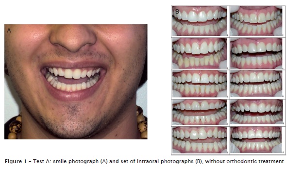
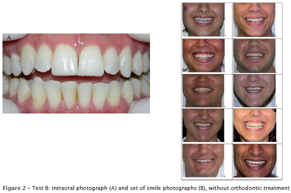
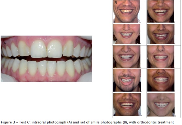
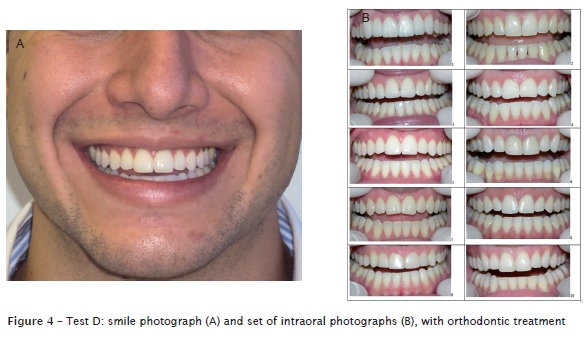
To make the analysis viable, four different types of tests were created by dividing the images as follows: A – one smile and ten intraoral images in the group without orthodontic treatment (figures 1A and 1B); B – one intraoral and tem smile images in the group without orthodontic treatment (figures 2A and 2B); C – one intraoral and ten smile images in the group submitted to orthodontic treatment (figures 3A and 3B); D – one smile and ten intraoral images in the group submitted to orthodontic treatment (figures 4A and 4B).
The images displayed in each set were numbered form 1 to 10; then a number was drawn which enabled the search for its correspondent image (intraoral or smile, according to the case), which was recorded on a answer sheet.
Therefore, each one of the 24 participants of this stage performed four tests, comprising a total time period of 20 minutes (5 minutes for each test). Following to the comparative analysis, the part icipant should mark on the answer sheet: the number of the photograph of the set correspondent to the image analysed and the type of particularity (crowding, incisal alignment, giroversions, fractures, weariness surfaces, crown morphology, etc.) which based the final conclusion, whose criteria should be visualized in both photographs (smile and intraoral).
For statistical analysis, Fisher's exact test was applied, comparing the performance of the undergraduates with that of the postgraduates in positively correlating intraoral with extraoral photographs. All analyses were performed with level of significance set at 5% (p < 0.05).
This present study was approved by the Ethical Committee in Research of the State University of Campinas under protocol number #105/2009.
Results
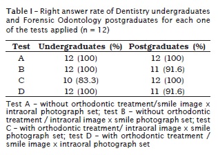
By analyzing the data within the answer sheets filled in by the undergraduates and postgraduate students, it was possible to observe that all participants were right in test A, for both groups. Tests B and D showed 91.6% of right answers among the postgraduate students, while test C showed 83.3% among the undergraduates (table I).
The most relevant parameters pointed out by both the undergraduates and postgraduate students to reach the positive identification through the smile photographs are seen in table II.
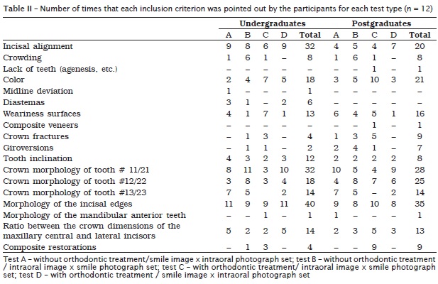
Concerning to test A, it was possible to observe that the morphology of the incisal edges (91.6%) and the incisal alignment of the anterior teeth (75%) were the parameters used by the undergraduates to reach a conclusion. On the other hand, the postgraduate students used the morphology of the crowns of the maxillary central incisors (83.3%) and the morphology of the incisal edges of the anterior teeth (75%).
The results of test B showed that the undergraduates used the parameters of the morphology of teeth #11 and #21 (91.6%) and the morphology of the incisal edges of the anterior teeth (75%), while the postgraduate students pointed out the morphology of the maxillary lateral incisors (66.6%) and the morphology of the incisal edges of the anterior teeth (66.6%).
By analyzing the results of test C, the undergraduates used the morphology of the incisal edges of the anterior teeth (75%), the color of the teeth (58.3%) and the presence of weariness surfaces (58.3%), while the postgraduates used the incisal edges morphology (83.36%) and the color of the anterior teeth (83.36%).
Finally, the result of test D demonstrated that the undergraduates employed the parameters of the morphology of the incisal edges of the anterior teeth (91.6%) and the crown morphology of the teeth #11 and #21 (83.3%). On the other hand, the crown morphology of teeth #11 and #21 (75%) and the morphology of the incisal edges of the anterior teeth (66.6%) were cited by the postgraduate students.
The analysis of the agreement data obtained by the comparison between intra- and extraoral photographs performed by the undergraduates and the postgraduate students showed that there were no statistically significant differences among the assessments, regardless of the experimental condition evaluated (p > 0.05).
Discussion
The forensic dentistry identification through photographs of the smile aims to the comparative analysis between ante mortem and postmortem features of individuals who for any reason do not have their identity established through fingerprint analysis. Therefore, the more relevant and specific are the dental particularities evidenced in an individual, the more reliable and safe will be the identification process.
In this present study, it was observed that most of the participants of the first stage presented sound anterior teeth, fact which complicating parameter because the analysis of the dental particularities sought in the images is restrained by the search for either the anatomic (shape, presence of pits and fissures), pathological (amelogenesis imperfecta), functional (weariness surfaces) or traumatic features (crown fractures). Moreover, since half of the sample had already undergone orthodontic treatment, important features such as diastemas, giroversions, crowding, and incisal misalignment, frequently present, had been corrected by the orthodontic mechanics. Therefore, it is worth highlighting that if on one hand, the identification through smile photographs is difficult because of the orthodontic treatment; on the other hand, this type of treatment makes easy the classical forensic odontology identification because of the presence of the orthodontic files of the individual, comprising radiographs, photographs and dental casts.
Concerning to the performance of the last-year undergraduates compared with that of the Forensic Odontology postgraduate students, it was possible to observe that the right answer rate for the four tests was high (95.8%); only four identification errors occurred, 2 for each one (2.1% for undergraduates and 2.1% for postgraduate students). Additionally, there were no statistically significant differences between the performances of the groups for all tests (p > 0.05), fact which demonstrated that both undergraduates and postgraduate students were technically prepared to execute human identification through the smile photographs of people who had been or had been not submitted to orthodontic treatment.
Considering to the test B (without orthodontic treatment), only one postgraduate participant did not answered right (97.9%), demonstrating that the lack of orthodontic treatment enabled the identification of important dental features that were not therapeutically corrected, such as incisal misalignments, giroversions and crowding 7.
By analyzing tests C and D (with orthodontic treatment), other features were sought in the smile photographs, such as the morphology of the central incisors, lateral incisors and canine teeth as well as the ratio among these teeth. In these two tests, three errors occurred (6.3%) within 48 identifications: two performed by undergraduates (4.2%) and one by one postgraduate student (2.1%), evidencing that the analysis of the photographs of individuals already submitted to orthodontic treatment had a higher difficulty rate than that of individuals without orthodontic treatment.
Among the main parameters used to reach the forensic odontology identification through the analysis of the smile photographs, the morphology and the alignment of the incisal edges of the anterior teeth and the morphology of the central and lateral incisors were the most cited by both the undergraduates and postgraduate students. Thus, it is emphasized the importance of morphological variations in the incisal edges of the anterior teeth, mainly in the maxillary central and lateral incisor crowns, which tend to be a single and specific set of dental features within the smile of each individual. Such morphological parameters have already been emphasized by the studies on both tooth morphology 11 and analysis of bite marks 5.
It is worth highlighting that this set can be even more specific when it is associated with one or more factors resulting from either tooth anomalies of position, shape, and development, among others, or from the action of environmental factors such as fractures, color pigment, weariness and dental treatment 10. Accordingly, to classify the maxillary central incisors regarding to their shape (square, triangle and ovoid) 11 is an important stage to start to include or exclude the individuals for the analysis of the smile photographs.
Other factor considered as relevant for most of the undergraduates to establish identification was the analysis of the incisal alignment – a line that can be drawn bordering the incisal edges of the anterior teeth. The incisal alignment or line is frequently analyzed during either the orthodontic clinical practice or the planning of aesthetical tooth interventions 3,12. Notwithstanding, this parameter may constitute an important element in evidencing a set of specific dental features, through using specific software for image editing, as Adobe Photoshop®.
Most of the studies on smile have been clinical investigations in which the physiological mechanisms of the muscles of the facial expression that allowed a smile exposure more or less pleasant to the light of the aesthetical patterns of one community or time 14,15,23. Some studies also classified the smile in forced or spontaneous according to the level of muscle contraction and the size of the area of smile exposure 1,6,12,22, which may show more or less teeth with their individual features.
In this present study, the participants of the first stage were asked to show a posed smile aiming to reproduce the type of smile that normally is produced in social events and exhibited the greater amount of teeth as possible. It is known that in some cases, the maxillary first molars can be seen in this type of smile 9,21. Consequently, the forensic odontology expert should ask the family to provide photographs in which the largest smile possible is seen, to enable the visualization of more dental features within the smile exposure.
Moreover, the older the individual, the smaller the smile area at vertical direction, as described by Desai et al. 4. Thus, the successful identification of dental features within a smile of a young individual tends to be higher than within a smile of a aged individual, fact which may contribute to make difficult the identification of older people through smile analysis. Consequently, it is important that the forensic odontology expert be aware regarding to the time (date) in which the smile photograph was taken. Ideally, the time interval between the image production and the moment of the forensic examination should be as short as possible, to avoid that the smile of the individual be modified by either several environmental factors or aesthetical dental treatment, once therapeutical dental procedures can significantly alter the smile of a person.
It is worth noting that in test C, the person to be identified showed an important feature – the absence of maxillary lateral incisors due to agenesis – posteriorly closed through orthodontic treatment and reanatomization of the canine tooth crowns. Only one postgraduate student was able to see and identify such features. Unlikely, the other participants did not identify those features even with the possibility to observe the presence of palatal cusps in the area of the maxillary canine teeth. However, this did not strongly affected the final identification for most of the participants (22 right answers for test C), indicating that the qualitative aspect of the morphology of the incisal edges and the crowns of the remnant teeth as well as the incisal alignment were the factors most used to reach the final conclusion.
The identification of the individual dental features in images produced ante- and post-mortem cannot be simply faced, because all data set should be properly described, evidenced, and discussed on the forensic report so that this document could constitute the fundamental tool for establishing a positive correlation between a missing person and a non identified body. In this sense, the forensic odontology expert is the professional who has the best post-graduation level to interpret the dental vestiges and exhibit them efficiently to the Justice.
Finally, considering the reduced field of visualization of the dental features in a smile photograph – which is basically limited to the maxillary anterior teeth -, it can be inferred that the photographs constitute an adequate information source for human identification. Therefore, the services of Forensic Odontology should perform the training of their experts and employees to search for the localization of images in family albums. Consequently, the possibility of the identification of a victim increases when traditional methods cannot be used, effectively helping the Justice in solving legal issues.
Conclusion
Considering the great amount of dental features that can be potentially found in anterior teeth (anatomic, functional, pathological, traumatic or therapeutical), the smile photographs can be considered as an adequate information source to establish a positive forensic odontology identification.
Both the last-year undergraduate and the Forensic Odontology postgraduate student demonstrated technical capacity to analyze the smile photographs aiming to human identification in a direct analysis of comparison. The parameters most cited by the undergraduates to reach the positive forensic dental identification through smile photographs were: morphology (83.3%), alignment of the incisal edges of the anterior teeth (66.6%) and morphology of the maxillary central incisors (66.6%).
For all tests performed by the postgraduates, the morphology of the incisal edges of the anterior teeth (72.9%), the morphology of the crowns of the maxillary central incisors (58.3%) and lateral incisors (52.1%) were the most cited.
References
1. Ackerman MB, Ackerman JL. Smile analysis and design in the digital era. J Clin Orthod. 2002;36(4):221-35. [ Links ]
2. Bilge Y. The identification of a dismembered human body: a multidisciplinary approach. Forensic Sci Int. 2003;137(2-3):141-6.
3. Câmara CALP. Estética em ortodontia: diagramas de referências estéticas dentárias (DRED) e faciais (DREF). R Dental Press Ortodon Ortop Facial. 2006;11(6):130-56.
4. Desai S, Upadhyay M, Nanda R. Dynamic smile analysis: changes with age. Am J Orthod Dentofacial Orthop. 2009;136:e1-10.
5. Kieser JA, Bernal V, Waddell JN, Raju S. The uniqueness of the human anterior dentition: a geometric morphometric analysis. J Forensic Sci. 2007;52(3):671-6.
6. Krishnan V, Daniel ST, Lazar D, Asok A. Characterization of posed smile by using visual analog scale, smile arc, buccal corridor measures, and modified smile index. Am J Orthod Dentofacial Orthop. 2008;133(4):515-23.
7. Mackley RJ. An evaluation of smiles before and after orthodontic treatment. Angle Orthod. 1993;63(3):183-90.
8. Marks MK, Bennett JL, Wilson OL. Digital video image capture in establishing positive identification. J Forensic Sci. 1997;42(3):492-5.
9. Maulik C, Nanda R. Dynamic smile analysis in young adults. Am J Orthod Dentofacial Orthop. 2007;132(3):307-15.
10. Menezes Filho PF, Barros CHO, Noronha JAA, Melo Junior PC, Cardoso RM. Avaliação crítica do sorriso. Int J Dent. 2006;5(1):14-9.
11. Paranhos LR, Jóias RP, Velasco LG, Bérzin F, Daruge Júnior E. Prevalence of the different maxillary central incisor shapes in individuals with natural normal occlusion. Braz J Oral Sci. 2010;9(2):104-7.
12. Ritter DE, Gandini Jr. LG, Pinto AS, Ravelli DB, Locks A. Analysis of the smile photograph. World J Orthod. 2006;7(3):279-85.
13. Rothwell BR. Principles of dental identification. Dent Clin North Am. 2001;45(2):253-70.
14. Rubin LR. The anatomy of a smile: its importance in the treatment of facial paralysis. Plast Reconstr Surg. 1974;53(4):384-7.
15. Schabel BJ, Baccetti T, Franchi L, MacNamara Jr JA. Clinical photography VS digital video clips for the assessment of smile esthetics. Angle Orthod. 2010;80(4):878-84.
16. Schuller-Gotzburg P, Suchanek J. Forensic odontologists successfully identify tsunami victims in Phuket, Thailand. Forensic Sci Int. 2007;171(2- 3):204-7.
17. Silva RF, Botelho TL, Prado FB, Kawagushi JT, Daruge Júnior, Berzin F. Human identification based on cranial computed tomography scan – a case report. Dentomaxillofac Radiol. 2011;40(4):257-61.
18. Silva RF, Pereira SDR, Prado FB, Daruge Junior E, Daruge E. Forensic odontology identification using smile photograph analysis – case reports. J Forensic Odontostomatol. 2008;26(1):12-7.
19. Silva RF, Portilho CDM, Reges RV, Leles CR, Freitas GC, Daruge Júnior E. Importância pericial dos registros odontológicos decorrentes de tratamento restaurador. R Dental Press Estét. 2007;4(4):32-8.
20. Silva RF, Prado MM, Barbieri AA, Daruge Júnior E. Utilização de registros odontológicos para identificação humana. RSBO. 2009;6(1):95-9.
21. Soares GP, Valentino TA, Lima DANL, Paulillo LAMS, Silva FAP, Lovadino JR. Esthetic analysis of the smile. Braz J Oral Sci. 2007;6(21):1313-9.
22. Tarantili VV, Halazonetis DJ, Spyropoulos MN. The spontaneous smile in dynamic motion. Am J Orthod Dentofacial Orthop. 2005;128(1):8-15.
23. Zachrisson BU. Esthetic factors involved in anterior tooth display and smile: vertical dimension. J Clin Orthod. 1998;32(7):432-45.
 Correspondence:
Rhonan Ferreira da Silva
Faculdade de Odontologia de Piracicaba – Universidade Estadual de Campinas
Avenida Limeira, n. 901 – Areião
CEP 13414-903 – Piracicaba – SP – Brasil
E-mail: rhonanfs@terra.com.br
