


Serviços Personalizados
Artigo
 pdf em Inglês
 Artigo em XML
 Referências do artigo
 Enviar este artigo por email
Links relacionados
Compartilhar

 Permalink
RSBO (Online)
versão On-line ISSN 1984-5685
RSBO (Online) vol.9 no.4 Joinville Out./Dez. 2012
Case Report Article
Anatomical variations in the permanent mandibular canine: forensic importance
Rhonan Ferreira da Silva I,II,IV ; Mauro Machado do Prado I ; Tessa de Lucena Botelho II ; Rogério Vieira Reges II ; Décio Ernesto de Azevedo Marinho III,IV
II School of Dentistry, Paulista University, Campus Flamboyant – Goiânia – GO – Brazil
III School of Medicine, Federal University of Goiás – Goiânia – GO – Brazil
IV Forensic Institute, Forensic Police of the State of Goiás – Goiânia – GO – Brazil
ABSTRACT
Introduction: The uniqueness of dental morphology plays an important role in Forensic Odontology, especially for human identifications, in which a single tooth can provide information for dental identification. Objective: To address the importance of a permanent lower canine with two roots for dental identification considering the internal and external morphology of the canine roots. Case report: To report a forensic case in which a two-rooted permanent lower canine was found in a decomposed human body. Conclusion: Although the victim was not identified by the dental parameter, it was observed that this type of morphological variation is of little incidence in some populations, therefore constituting a valuable tool for dental human identifications.
Keywords: Forensic Dentistry; anatomy; canine.
Introduction
Anatomical variation constitutes an expression that can be defined as all morphological alteration which do not determine functional impairment to the individual and that can be visualized in internal or external structures/organs 1. In Dentistry, the anatomical variations have been studied in different structures comprising the bucomaxillofacial system (teeth, bone, muscles, vessels, nerves, glands, etc.) aiming to understand which can be considered as normal or abnormal, subsidizing the correct diagnosis and treatment planing.
The dental anatomical variations, specifically, have been studied in determined dental specialties, such as Operat ive Dent istry, which seek to reconstruct the anatomy of the tooth crown as closest as the natural tooth; Endodontics, which explores the interior of the teeth aiming to enable the cleaning of the root canal system; and Bucomaxillofacial Surgery, in which the morphology and the tooth positioning directly interfere in the election/execution of the extraction technique, for instance. Determined tooth types show more anatomical variations than others, e.g., mainly the third molars which exhibited dimensional and morphological variations both in the internal and external part of the crown and root. Still in this context, the lower permanent canine teeth may also exhibit the anatomical variations. Its most expressive variations are within the internal and external part of the root. Normally, this tooth show a single root, with a single canal; however, the literature has reported single-rooted canine with two 2,6,12 or three root canals 7 and, still, canine teeth with two different roots 3,5,16.
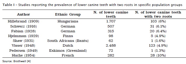
Specifically about the prevalence of lower canine teeth with two roots in determined population groups, Brothwell 4 described the presence of this anatomical variation in a Danish population living at the Neolithic age (between 2000-1500 b.c.) and in another population living at1200-1500 a.c., finding an incidence of 5.6% and 6.4%, respectively. The author also cited data of prevalence of lower canine with two roots in eight studies conducted in specific population groups, according to the table I.
Taylor 11 reported that the presence of a bifid root is not an uncommon particularity for the lower permanent canine and described the morphological variations of two roots when present in this tooth, in which bifurcation can be found at the cervical, medium, or apical thirds. The author analyzed the size ratio between the labial and lingual roots in a study of 179 lower canine teeth with two roots and observed that 65.3% exhibited proportional roots, 27.4% exhibited the labial root greater than the lingual roots and the remaining teeth (7.3%) showed the lingual root greater than the labial root.
In a posterior study, Pécora et al. 8 analyzed the internal morphology, the number of the roots and canals in 830 extracted lower canine teeth and obtained the following results: 98.3% of the teeth had a single root, 92.2% had one canal and one apical foramen, 4.9% with two canals and one apical foramen, 1.2% with two canals and two apical foramens. 1.7% of the cases showed two different roots, each one with one canal. The mean of length was 25.5 mm, ranging from 20.3 to 32.8 mm.
Sharma et al. 9 also reported an important study on the internal anatomy of 65 permanent lower canine teeth with two well-defined roots. In all cases, two root canals were found (one in each root). The lateral canals were present in 68.9% of the cases and 19.7% showed a lateral canal in the bifurcation. The external mensuration revealed a maximum and minimum length of 26.7 mm and 17.9 mm, from the cusp point to the labial root as landmark; and 27.2 mm and 17.1 mm, from the cusp point to the lingual root as landmark, respectively for both parameters. The bifurcation of the roots was more frequently present at the apical third (56.9%), followed by the medium third (40%) and less frequently at the apical third (3.1%). Proportionally, it was observed that the labial root was larger than the lingual one in 47.7% of the cases; 43.1% of the roots were proportional; and only in 9.2% of the cases, the lingual root was larger than the labial one.
Versiani et al. 15 analyzed the internal and external anatomy of 14 permanent canine teeth with two roots and two canals using a computed microtomography. The images were processed, and it was observed the root lengths, the area of bifurcation and the presence of accessory canals and others aspects related to the internal anatomy of this tooth. The area of the bifurcation was observed at the apical and medium third in 42.8 and 57.2% of the cases, respectively.
By knowing that in Forensic Dentistry the tooth anatomical variations are of great importance, because they allowed the differentiation of individuals through the specific dental characteristics which make a person unique from a forensic point of view, the aim of this study was to emphasize the forensic relevance of the anatomical variations present in the permanent lower canine through the reporting of a forensic case.
Case report
In the mid of 2011 a body in an advanced stage of decomposition was sent for forensic dental examination in the Section of Forensic Anthropology and Forensic Dent istry of the Forensic Institute in Goiânia, since there was no possibility of analyzing the fingerprints of the victim.
Aiming to facilitate the forensic dental analysis, the skull and mandible were disjointed and submitted to a cleaning process.
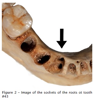
It is known that as the putrefaction process goes by, the periodontal ligament are also decomposed and the single-rooted teeth are typically displaced from their sockets, therefore occurring the socalled tooth avulsion or postmortem tooth loss. In the case presented herein, all the anterior teeth were avulsed and during their repositioning in the socket, it was possible to identify that tooth #43 had two distinct roots: labial and lingual (figure 1). By examining the sockets, this information was also confirmed and it was possible to observe two distinct sockets for the two roots in the position corresponding to tooth #43 (figure 2).
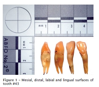
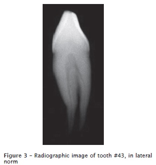
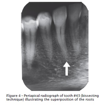
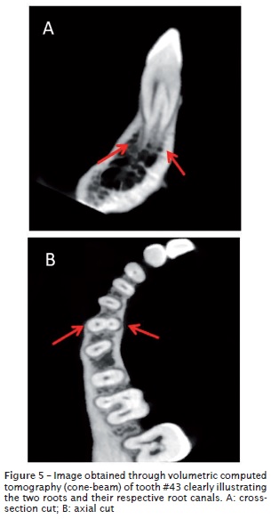
To provide a more comprehensive study of dental anatomy, conventional photographic and radiographic images of tooth #43 were obtained inside and outside of the tooth socket, as well as images through volumetric computed tomography (cone-beam). With these images it was possible to observe that: the bifurcation was located at the middle third of the root; both roots had proportional lengths, with the lingual root 1 mm smaller than the labial one; and each one had a single root canal (figure 3). The maximum length of the tooth was 25 mm. In the image obtained by the periapical radiographic examination, performing through bisecting angle technique with the tooth inside the socket, it was possible to see the superposition of the roots, which makes extremely difficult to identify accurately this dental anomaly (figure 4). On the other hand, the CT image clearly demonstrated the presence of two roots in the cross and axial sections. Thus, it was confirmed the presence of a single root canal in each root, in its entire length (figure 5).
During the direct and indirect alveolar inspection (through images) of the maxilla and mandible, it was seen that only the tooth #43 exhibited anatomical variation in the number of roots. In this context, there was the presence of a relevant dental particularity for the identification of the victim; however, it should be preferably that it had been properly registered in an imaging examination that could be confronted with the postmortem findings. As no dental files containing relevant records was sent to confrontation, the victim was later identified through a genetic evaluation.
Discussion
The forensic dental identification is a forensic method usually applied in establishing the identity of the bodies when papiloscopic analysis cannot be performed, as in the case of charred, mutilated, decomposing and skeletonized bodies 10. This technique, as well as other methods of human identification, is based on both technical and biological requirements, which are: uniqueness, immutability, permanence, classificability, and pract icality 14. Speci f ical ly concerning to uniqueness or individuality, it is essential that the dental characteristics and particularities be unique, that is, they should not be found in more than one individual. In this sense, the anatomical variations and dental anomalies which may be determined by genetic information or may arise during the various stages of development are highlighted. It differs from the anatomical variation of anomaly, because in the first case there is no impairment of the function to be performed by the structure or organ 1. Thus, the anatomical dental variations basically comprise morphological differences in the length, width, height, area or volume of the crown and root, within the parameters stipulated as "normal" for literature 4,12. Also, it can be considered as anatomical variations different locations; depths and dimensions of descriptive elements such as grooves, pits and fissures; the slope of the sides of the cusps; the shape of the incisal surface; the ratio among enamel, dentin and cementum; the dimensions of pulp chambers; and the quantity, size and morphology of the number of roots and root canals 12.
In the case presented here, it was found a permanent lower canine tooth that had two distinct and proportionate roots, each one containing a root canal, with 24 and 25 mm long. This finding agrees with the greater frequency of results obtained by Pécora et al. 8, Sharma et al. 9 and Taylor 11. The region of the bifurcation (middle third) coincided with the second more prevalent sample according to study of Sharma et al. 9 and with most of the cases observed in the study of Versiani et al. 15.
Concerning to the prevalence within the population, the incidence of lower canines with two distinct roots ranged from 1.3% to 10% 4,8,11, therefore constituting a relatively rare peculiarity of great importance to subsidize a forensic dental identification.
From a clinical point of view, the anatomical variations related to lower canine must be properly identified mainly when this tooth is involved in a diagnosis or treatment planing as endodontic, orthodontic and surgical procedures. In Endodontics, a proper exploration of the root canal system should be carefully observed by the dentist in the sense that all the roots/canals are handled by avoiding the failure in therapy performed 2,3,5-7,12,16, by the appearance or persistence of complications such as pain, infection and periapical lesion, involving the need for endodontic retreatment, change of the restoration, replacement of fixed prostheses and even tooth loss. In Orthodontics, dental movement should be performed with careful attention to the specific anatomic limits of the region, such as the mental foramen and thickness of the alveolar cortical, which may prevent complications during the execution of this therapy (bone fenestrations, compression of the neurovascular bundle, root resorption, etc.). In Buccomaxillofacial Surgery, the size and shape of the crown and roots and the number of roots associated with the tooth position in the dental arch 13 are important factors to be considered for election of the extraction technique, which can be more or less invasive (need for flap, osteotomy, and tooth sectioning).
Conclusion
In the forensic context, the forensic dentist should be aware and properly record all morphological characteristics evidenced on teeth that have any type of anatomical variation in the crown or root (root number, root length, the bifurcation region and number of canals), observed through either physical examination or various modalities of imaging tests, both in exams produced in life and in the pot-mortem, since the disclosure of dental rare anatomical features can be of extremely importance in cases of human identification.
References
1. Alves N, Cândido P. Anatomia para o curso de odontologia – geral e específica. 2. ed. São Paulo: Santos; 2009.
2. Andrei OC, Margarit R, Daguci L. Treatment of a mandibular canine abutment with two canals. Rom J Morphol Embryol. 2010;51(3):565-8.
3. Andrei OC, Margarit R, Gheorghiu IM. Endodontic treatment of a mandibular canine with two roots. Rom J Morphol Embryol. 2011;52(3):923-6.
4. Brothwell DR. Dental anthropology. Oxford: Pergamon Press; 1963.
5. D'Arcangelo C, Varvara G, De Fazio P. Root canal treatment in mandibular canines with two roots: a report of two cases. Int Endod J. 2001;34(4):331-4.
6. Ghoddusi J, Zarei M, Vatanpour M. Mandibular canine with two separated canals. N Y State Dent J. 2007;73(6):52-3.
7. Orguneser A, Kartal N. Three canals and two foramina in a mandibular canine. J Endod. 1998;24(6):444-5.
8. Pécora JD, Sousa Neto MD, Saquy PC. Internal anatomy, direction and number of roots and size of human mandibular canines. Braz Dent J. 1993;4(1):53-7.
9. Sharma R, Pécora JD, Lumley PJ, Valmsley AD. The external and internal anatomy of human mandibular canine teeth with two roots. Endod Dent Traumatol. 1998;14:88-92.
10. Silva RF, Chaves P, Paranhos LR, Lenza MA, Daruge Júnior E. Utilização de documentação ortodôntica na identificação humana. Dental Press J Orthod. 2011;16(2):52-7.
11. Taylor RMS. Variation in morphology of teeth – anthropologic and forensic aspects. Illinois: Charles C. Thomas; 1978.
12. Tiku AM, Kalaskar RR, Damle SG. An unusual presentation of all the mandibular anterior teeth with two root canals – a case report. J Indian Soc Pedod Prev Dent. 2005;23(4):204-6.
13. Xavier CRG, Dias-Ribeiro E, Ferreira-Rocha J, Duarte BG, Ferreira-Júnior O, Sant'Ana E et al. Avaliação das posições dos terceiros molares impactados de acordo com as classificações de Winter e Pell & Gregory em radiografias panorâmicas. Rev Cir Traumatol Buco-Maxilo-Fac. 2010;10(2):83-90.
14. Vanrell JP. Odontologia legal e antropologia forense. 2. ed. Rio de Janeiro: Guanabara Koogan; 2009.
15. Versiani MA, Pécora JD, Sousa-Neto MD. The anatomy of two-rooted mandibular canines determined using microcomputed tomography. Int Endod J. 2011;44(7):682-7.
16. Victorino FR, Bernardes RA, Baldi JV, Moraes IG, Bernardinelli N, Garcia RB et al. Bilateral mandibular canines with two roots and two separate canals – case report. Braz Dent J. 2009;20(1):84-6.
 Correspondence:
Rhonan Ferreira da Silva
Instituto Médico-Legal
Avenida Atílio Correa Lima, n. 1223 – Cidade Jardim
CEP 74425-030 – Goiânia – GO – Brasil
E-mail: rhonanfs@terra.com.br
