


Serviços Personalizados
Artigo
 pdf em Inglês
 Artigo em XML
 Referências do artigo
 Enviar este artigo por email
Links relacionados
Compartilhar

 Permalink
RSBO (Online)
versão On-line ISSN 1984-5685
RSBO (Online) vol.10 no.1 Joinville Jan./Mar. 2013
ORIGINAL RESEARCH ARTICLE
Analysis of apical sealing of canals irradiated with Er: YAG and Nd: YAG lasers and filled with AH Plus®
Angela Toshie ArakiI; Alexandre Gomes BezerraII; Priscila Alonso HenriquesI; Andrea Kanako Yamazaki ArasakiI; Igor ProkopowitschI; Celso Luis CaldeiraIII
IDepartment of Endodontics, Dental School, Cruzeiro do Sul University – São Paulo – SP – Brazil.
IIDepartment of Endodontics, Dental School, Santa Cecília University – Santos – SP – Brazil.
IIIDepartment of Endodontics, Dental School, University of São Paulo – São Paulo – SP – Brazil.
ABSTRACT
Introduction: Laser technology is gaining increasing importance in dental practice and also in the field of Endodontics with its ability to promote disinfection and experimentally in the preparation of root canal. The action of different types of lasers results in changes representing the increase in permeability of dentinal tissue (Er: YAG) or sometimes by a decrease in melting and recrystallization of dentin (Nd: YAG).
Objective: this study assessed through apical dye leakage, the influence of irradiation with two types of laser, regarding to the quality of apical sealing of endodontic fillings.
Material and methods: Thirty-six single-rooted teeth were used after being prepared with the ProFile system up to size #40 instrument and then divided into four experimental and two control groups. The technique used previously to the filling was as follows: G1 – not irradiated; G2 – irradiated with Er: YAG; G3 – irradiated with Nd: YAG and G4 – irradiated with Er: YAG followed by Nd: YAG. After external waterproofing and dry, the specimens were filled with a cold vertical condensation technique, using AH Plus sealer, and immediately immersed into 0.5% methylene blue solution for subsequent cleavage. The linear values of apical marginal leakage were obtained with the aid of an optical microscope connected to a computer using the Image Lab® software.
Results: Data analysis showed the non-existence of statistically significant (p = 0.05) differences between different groups.
Conclusion: It was concluded that the laser does not have influence on the apical sealing.
Keywords: erbium; neodyminum; root canal.
Introduction
The apical sealing of the canal system is crucial for endodontic therapy success. For an adequate apical sealing, in addition to other factors, the presence of smear layer and the type of the endodontic cement to be used must be considered. The smear layer is a mixture of dentin chips, decomposed organic waste, and microorganisms 2. Its removal is recommended, considering that the absence of smear layer promotes a better sealing 6.
The smear layer removal of the canal wall using citric acid or ethylenediaminetetraacetic acid (EDTA) has been recommended 16 but so far no clear evidence showed that this procedure enhances disinfection or treatment responses 10. The efficacy of sodium hypochlorite (NaOCl) as an irrigating solution was corroborated by several studies assisting in debridement and also contributing as an antimicrobial action 9,18.
Some factors have influence on the survival of microorganisms including particular ecological niches, nutrition, anaerobiosis, pH and competition with other microorganisms 7. In some situations, the persistence of signs and symptoms that hinder the completion of endodontic therapy may occur. Frequently, this is related to the presence of microorganisms maintaining an infectious process.
Aiming the solution of resistant cases, since the 1980s, the use of high energy density lasers has been proposed, whose main characteristic is the ability to reduce intracanal microbiota. Yasuda et al. 20 used Nd: YAG and Er: YAG lasers to evaluate the antimicrobial action and concluded that they are more efficient in straight canals.
Lasers may activate morphological changes in dentin when associated with microbial reduction, according to the used type and parameters. The Er: YAG laser is able to vaporize the magma present in the canal walls, exposing the dentinal tubules 13 whereas the laser Nd: YAG lasers may cause melting and recrystallization 5,8. Other authors 11 consider that the Nd: YAG and Er: YAG lasers do not remove debris and the smear layer from root canal wall.
Due to the controversy around the influence of morphological changes on the final quality of the obtained sealing, the aim of this study is to assess through dye leakage the quality of apical marginal sealing in teeth irradiated with Er: YAG and Nd: YAG lasers and filled with gutta-percha and AH Plus®.
Material and methods
Thirty-six single-rooted teeth from the Permanent Human Teeth Bank (Faculty of Dentistry, University of São Paulo) (Report nr. 143/03) were used. The selected teeth were prepared using the Profile.04 system (Maillefer, Ballaigues, Switzerland) until the instrument #40, 1 mm from the apex.
After the chemical-surgical preparation, all specimens were irrigated with 10 ml of sodium hypochlorite solution (NaOCl) at 0.5% (Fórmula e Ação, São Paulo, SP, Brazil) and later with 10 ml of EDTA-T pH 7.2 (Fórmula e Ação, São Paulo, SP, Brazil). Subsequently, canals were dried with a small caliper aspiration cannula and absorbent paper cones.
Laser irradiation and experimental groups
Experimental groups: G1 (9) – non-irradiated; G2 (9) – Er: YAG laser irradiation; G3 (9) – Nd: YAG laser irradiation; G4 (9) – Er: YAG followed by Nd: YAG Laser irradiation.
As parameters, it was used for the Nd: YAG Laser (neodymium, yttrium, aluminum, Granada) of the American Dental Technologies – Pulse Master® 1000 (GA, USA): 100 mJ, 1.5 W and 15 Hz, four times, with speed of 2mm/second in a helicoidally motion towards the cervical-apex and apical-cervical and for the Er: YAG (Erbium, Yttrium, Aluminum, Granada) of Kavo – Kavo Key® (Kavo Co., Biberach, Germany). 1 W, 100 mJ/pulse, 10 Hz, four times, with speed of 2mm/second, helicoidally motion towards cervical-apex and apical-cervical with canal filled with 0.5% NaOCl solution.
Root canal filling
After drying, the main gutta-percha point size #40 (Dentisply, Konstanz, Germany), was selected to tug fit in each root canal. Fillings of root canal were carried out using the technique of vertical condensation with AH Plus sealer®.The gutta-percha accessory points (Dentisply, Konstanz, Germany), were embedded into the endodontic sealer and taken to the open spaces in the obturation mass. This procedure was repeated until the operator was not capable to insert more gutta-percha points. All root canals obturation was executed by the same operator through vertical adaptation with Paiva's condenser size #3 (SSWhite, Rio de Janeiro, Brazil).
Evaluation of apical sealing
Immediately after filling, the access cavities were sealed with temporary cement (Cimpat®, Septodont Brasil Ltda., São Paulo, Brazil). The samples were coated externally, excluding the apical 3 mm, with two layers of ethyl cyanoacrylate (Super Bonder®, Loctite Ltd., SP, Brazil). Then, they were submerged into a 0.5% methylene blue solution, pH 7.2 (Fórmula e Ação, São Paulo, SP, Brazil), keeping the temperature at 36ºC for 48 hours.
After this period, the specimens were washed with water to remove the excess of the indicator dye and were cleaved and observed in an optical microscope. To measure the linear dye leakage, the image analysis software was used – Imagelab®. Data were then tabulated and statistically analyzed by Kruskal-Wallis (GMC 2002, Geraldo Maia Campos, FORP, SP, Brazil) with a significance level of 0.05.
Results
The values of dye leakage for each specimen and the averages for each group are shown in table I.
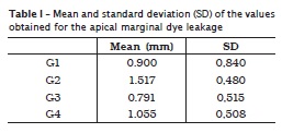
In the table I, it can be observed a higher leakage (mm) in the specimens of group 2, which used Er: YAG laser irradiation while the lowest dye leakage occurred in group 3, in the specimens which were irradiated with Nd: YAG; in the group in which the two lasers were applied, the amount of leakage was smaller than in group 2 was and higher than in group 1 (without irradiation).
The nonparametric statistical test (Kruskal-Wallis) showed no significant differences in the comparison of groups.
Discussion
The quality of the canal system filling is essential for endodontic therapy success. The evaluation of dye leakage is commonly used to detect the apical microleakage in in vitro studies, by measuring dye penetration between the walls and filling materials 19.
The EDTA has been widely used aiming to remove the smear layer because of its chelating capacity 10. Laser was selected for this study because since the 1980s its use was allowed in Endodontics for providing a high microbial reduction without causing thermal damage to the tooth structure and in the surrounding tissue 12,13.
In contrast, it is known that intracanal irradiation produces morphological changes in dentin 1,4,5 that could have an influence on the adaptation of the filling material to the root canal walls. The Er: YAG laser is absorbed by water causing micro-explosions evaporation (ablation) whereas the Nd: YAG laser is absorbed by mineral structures such as hydroxyapatite, phosphatase and carbon beginning disorder processes in crystals 3.
The wavelength variation and energy parameters used for each laser promoted different interactions with the dentin. Berkiten et al. 1 found areas of recrystallization in the canal wall after irradiation with Nd: YAG laser with 1.8 W and 2.4 W.
The mean apical marginal leakage was 0.791 mm (G3), which means that this was the group with the lowest penetration of methylene blue dye solution. This result is in accordance with the results obtained by Dederich et al. 5 on the glassy surface which can bring reduced permeability resistant to fluids when compared with non-irradiated areas of the dentin (G1).
According to Moura-Netto et al. 14, melting and crystallization of dentinal walls with tubule occlusion increase the apical sealing ability in addition to the bactericidal action of Nd: YAG laser, with the best results obtained with AH Plus sealer®.
The Er: YAG laser as opposed to the Nd: YAG laser is able to remove both the infected dentin surface and the smear layer after any type of mechanical preparation of root canal; the most exposed dentinal tubules facilitate the contact with the cement, fact which is essential for endodontic therapy success 17.
In table I, we can observe that the highest dye penetration occurred in G2, irradiated with Er: YAG. According to Pécora et al. 15 the canal instrumented with water and then irradiated with Er: YAG presented a greater increase in permeability when compared with that irrigated with NaOCl. Also, according to these authors, this is an important factor for the disinfection of the root canal system and the mechanical connection between cement and dentin.
However, the ANOVA and nonparametric statistical test (Kruskal Wallis), with 95% significance (p = 0.05) showed no significant differences in comparison between groups.
Conclusion
There is no significant interference in the quality of apical sealing in canals irradiated with Nd: YAG and Er: YAG lasers by employing AH Plus® sealer.
References
1. Berkiten M, Berkiten R, Okar I.Comparative evaluation of antibacterial effects of Nd:YAG laser irradiation in root canals and dentinal tubules. J Endod. 2000;26(5):268-70. [ Links ]
2. Brännström M, Nyborg H. Bacterial growth and pulpar chances under inlays cemented with zinc phosphate cement and Epoxylite CBA 9080. J Prost Dent. 1974,31(5):556-65. [ Links ]
3. Brugnera Junior A, Zanin F, Barbin EL, Spanó JC, Santana R, Pécora JD. Effects of Er:YAG and Nd:YAG laser irradiation on radicular dentine permeability using different irrigationg solutions. Lasers Surg Med. 2003;33(4):256-9. [ Links ]
4. Carvalho CAT, Valera MC, Gow-Soares S, Eduardo CP. Effects of Nd:YAG and Er:YAG lasers on the sealing of root canal fillings. J Clin Laser Med Surg.2002;20(4):215-9. [ Links ]
5. Dederich DN, Zakariasen KL, Tulip J. Scanning electron microscopic analysis of canal wall dentin following neodymium-yttrium-aluminum- garnet laser irradiation. J Endod. 1984;2984(9):428-31. [ Links ]
6. Economides N, Kokirikos I, Panagiotis B, Gogos C. Comparative study of apical sealing ability of a new resin-based root canal sealer. J Endod. 2004;30(6):403-5. [ Links ]
7. Estrela C, Estrela CRA, Decurio DA, Hollanda ACB, Silva JA. Antimicrobial efficacy of ozonated water, gaseous ozone, sodium hypochlorite and chlorhexidine infected human root canals. Int Endod J. 2007;40(2):85-93. [ Links ]
8. Gutknecht N, Moritz A, Conrads G, Sievert T, Lambert F. Bactericidal effect of the Nd:YAG laser in vitro root canals. J Clin Laser Med Surg. 1996;14(2):77-80. [ Links ]
9. Hardee MW, Miserendino LJ, Kos W, Walia H. Evaluation of the antibacterial of intracanal Nd:YAG laser irradiation. J Endod. 1994;20(8):377-80. [ Links ]
10. Hulsmann M, Heckendorff M, Lennon A. Chelatin agents in root canal treatment: mode of action and indications for their use. Int Endod J. 2003;36(12):810-30. [ Links ]
11. Kivanc BH, Ulusoy OI, Görgül G. Effects of Er:YAG laser and Nd:YAG laser treatment on the root canal dentin of human teeth: a SEM study. Lasers Med Sci. 2008;23(3):247-52. [ Links ]
12. Lage-Marques JL, Eduardo CP, Matsumoto K. A study on morphological changes of the root canal walls lased by pulsed Nd:YAG laser. JJEA.1995;16:64-9. [ Links ]
13. Moritz A, Schoop U, Goharkhay K, Jakolitsch S, Kluger W, Wernisch J et al. The bactericidal effect of Nd:YAG, Ho:YAG and Er:YAG laser irradiation in root canal: an in vitro comparison. J Clin Laser Med Surg. 1999;17(4):161-4. [ Links ]
14. Moura-Netto C, Carvalho CF, Moura AAM, Davidowicz H, Antoniazzi JH. Influence of Nd:YAG and diode laser irradiation on apical sealing when associated with AH Plus and endoREZ endodontic cements. Photomed Laser Surg. 2007;25(5):413-7. [ Links ]
15. Pécora JD, Brugnera Junior A, Cussiolli AL, Zanin F, Silva R. Evaluation of dentin root canal permeability after instrumentation and Er:YAG laser application. Lasers Surg Med. 2000;26(3):277-81. [ Links ]
16. Scelza M, Pierro V, Scelza P, Pereira M. Effect of three different time periods of irrigation with EDTA-T, EDTA and citric acid on smear layer removal. Oral Surg Oral Med Oral Pathol Oral Radiol Endod. 2004;98(4):499-503. [ Links ]
17. Schoop U, Moritz A, Kluger W, Patruta S, Goharkhay K, Sperr W et al. The Er:YAG laser in Endodontics: results of an in vitro study. Lasers Surg Med. 2002;30(5):360-4. [ Links ]
18. Siqueira Junior JF, Machado AG, Silveira RM, Lopes HP, Uzeda M. Evaluation of the effectiveness of sodium hypochlorite used with three irrigation methods in the elimination of Enterococcus faecalis from the root canal. In vitro. Int Endod J. 1997;30(4):279-82. [ Links ]
19. Tamse A, Katz A, Kablan F. Comparation of apical leakage shown by four different dyes with two evaluating methods. Int Endod J. 1998;31(5):333-7. [ Links ]
20. Yasuda Y, Kawamorita T, Yamaguchi H, Saito T. Bactericidal effect of Nd:YAG and Er:YAG lasers in experimentally infected curved root canals. Photomed Laser Surg. 2010;28(2):75-8. [ Links ]
 Corresponding author:
Angela Toshie Araki
Avenida Robert Kennedy, n. 2.126
CEP 09860-000 – São Bernardo do Campo – SP – Brasil
E-mail: a_araki@uol.com.br
Received for publication: March 8, 2012
Accepted for publication: September 4, 2012
