


Serviços Personalizados
Artigo
 pdf em Inglês
 Artigo em XML
 Referências do artigo
 Enviar este artigo por email
Links relacionados
Compartilhar

 Permalink
RSBO (Online)
versão On-line ISSN 1984-5685
RSBO (Online) vol.10 no.1 Joinville Jan./Mar. 2013
ORIGINAL RESEARCH ARTICLE
Histological evaluation of refinement influence in the cleaning efficacy of rotary nickel-titanium ProTaper® instruments in oval-shaped root canals
Juliana Rodrigues CachapuzI; Luciana Moura SassoneII; Rivail Antonio Sergio FidelII; Fernando Sili VilhenaI
IBrazilian Navy – Rio de Janeiro – RJ – Brasil.
IIProclin Department, School of Dentistry, Rio de Janeiro State University – Rio de Janeiro – RJ – Brasil.
ABSTRACT
Introduction and objective: The aim of this in vitro study was to evaluate the cleaning and shaping efficacy of rotary nickel-titanium ProTaper® instrumentation in oval canals and the influence of ultrasonic irrigation on the final refinement.
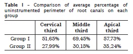
Material and methods: Twenty mandibular molars were accessed and divided in two groups. Only distal roots with oval canals were used. Group I was instrumented using only rotary nickel-titanium ProTaper® instruments. Group II received the same preparation followed by refinement with 3 minutes of ultrasonic passive irrigation. After preparation, the distal roots were sectioned for histological processing. Coronal, middle and apical thirds were analyzed. Uninstrumented perimeter in each third was measured by the software Image tool 3.0.
Results: The percentage of uninstrumented perimeter was calculated for each third of each root and average percentage was calculated for each third in both groups. Statistical analysis was performed with t-test through the software SPSS 11.0.1 for Windows. The level of significance was set at 1%. Group I showed higher percentage of uninstrumented perimeter in all thirds. Group II, which received ultrasonic irrigation showed better results in all thirds (p < 0.01). Comparing the three thirds in the same group, no statistically significant differences were found (p > 0.05).
Conclusion: Under the conditions of this ex vivo study, the three minutes use of ultrasonic irrigation after rotary instrumentation resulted in significantly more instrumented walls in the distal roots of mandibular molars.
Keywords: root canal; instrumentation; ProTaper; nickel-titanium files; refinement.
Introduction
The mechanical preparation of root canals plays a significant role in endodontic therapy 12. One of the objectives of the root canal preparation is to clean and shape the root canal system while maintaining the original configuration. It provides a progressive conical shape toward the apex, which offers a shape conducive to the placement of three dimensional filling of the root canals and all accessory canals 20.
One of the most important steps in any root canal treatment is canal preparation 17 but many methods of preparing root canals mechanically still fail to cleanse root canal systems effectively 20. Variations in the internal root canal anatomy might be present, making the current instrumentation techniques inefficient at cleaning all surfaces and irregularities within the root canal space 21 and large portions of the canal walls can remain uninstrumented, leaving organic and inorganic debris in the root canal 5. Therefore, irrigation is an essential part of a root canal treatment as it allows for cleaning beyond the root canal instruments 22.
Stainless steel files probably are still the instruments of choice by most of dentists for cleaning and shaping canals, but rotary-driven nickel-titanium files are gaining rapidly in popularity 6. A number of studies have shown that rotary nickel-titanium (Ni-Ti) instruments allow more rapid, more centered, rounder and more conservative shaping of canals than stainless steel instruments 11,19,25. However, rotary instrumentation with Ni-Ti files has limited area of action. Due to their super elasticity, it is known that they cannot be pressed against the root canal walls 5. It may result in a larger flare on one side of the canal wall due to the tendency of creating round preparations, even in oval shaped canals and a few areas may remain uninstrumented 24,25.
One approach to overcome this limitation is the use of ultrasonic irrigation of the root canal. The use of an ultrasonic irrigating needle allowed for the continual deposition and renewal of irrigating solution within the canal 13. Passive ultrasonic irrigation relies on the transmission of acoustic energy from an oscillating file to an irrigant in the root canal 22. The energy is transmitted by means of ultrasonic waves and can induce acoustic streaming and cavitation of the irrigant 1,2. As the root canal has already been shaped, the file can move freely and the irrigant can penetrate more easily into the apical part of the root canal system 14,15 and the cleaning effect will be more powerful 1,2. The cleaning efficacy of passive ultrasonic irrigation implies the effective removal of dentine debris, microorganisms (planktonic or in biofilm) and organic tissues from the root canal. Because of the active streaming of the irrigant, its potential to contact a greater surface area of the canal wall will be enhanced 22. Therefore, it improves the removal of bacteria, pulpal remaining, debris and smear layer from the root canal 7,23.
The aim of this in vitro study was to evaluate the cleaning efficacy of rotary nickel-titanium ProTaper® instrumentation in oval-shaped canals and the influence of ultrasonic irrigation on the final refinement.
Material and methods
A set of twenty permanent human mandibular molars, except for the third molars, with oval-shaped distal root canals, freshly extracted from patients, were used in this study and approval was obtained from the Ethics Committee of UERJ (University of Rio de Janeiro State). After extraction, teeth were immersed into 5.25% sodium hypochlorite (NaOCl) for 30 minutes and stored into 0.1% thymol solution. Coronal access was made and the patency of each canal was confirmed by inserting a size 10 K-file (Dentsply Maillefer Balaigues, Switzerland) through the apical foramen. The working length was defined by observing under a clinical optical microscope the tip of the file protruding through the apical foramen and subtracting 1mm from the recorded length.
Next, teeth were subdivided into two groups of 10 teeth each. The teeth were prepared by crown-down technique, using rotary instrumentation withProTaper® NiTi files (Dentsply/Maillefer, Balaigues, Switzerland) according to the recommended sequence of the manufacturers until the working length achieved a size of #30 (F3).
All teeth were instrumented using an electric endodontic engine (Endo Pro Torque, VK Driller Equipamentos Elétricos, São Paulo, Brazil). The device was adjusted to a 300-rpm constant clockwise speed and to a torque of 2 N.cm. For both groups, canals were flushed with 2mL of 2.5% sodium hypochlorite (Mil formulas- Farmácia de manipulação, Rio de Janeiro, Brazil) at each change of file and patency was verified with a size #10 K-File (Dentsply/Maillefer, Balaigues, Switzerland)
Group I was cleaned and shaped only by ProTaper® NiTi files. Group II received the same preparation of Group I, followed by ultrasonic irrigation used for final refinement. Final refinement of Group II was conducted with size #20 ultrasonic file at the ENAC ultrasonic unit (Osada Enac, Tokyo, Japan), introduced up to the working length. This file was used for 3 minutes for each tooth, comprising: 1 minute of continuous flush of ultrasonic irrigation and aspiration of distilled water; then the root canal was filled by 5.25% NaOCl and this solution was agitated with the ultrasonic file for another 1 minute; and finally, continuous flush of ultrasonic irrigation and aspiration with distilled water was carried out for more 1 minute.
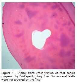
After preparation, the distal roots were cross-sectioned and only these were used for histological processing. After decalcification, the roots were sectioned with a surgical blade on their coronal, middle and apical thirds. Serial 5-μm-thick cross-sections of each third of each root were stained with hematoxylin and eosin, and examined with an optical microscope at x40 magnification (Nikon Eclipse E200, Nikon Tokyo, Japan) and images were taken with a digital camera (Nikon Coolpix 5000, Nikon, Japan).
Image analysis and processing were completed using the software Image tool 3.0 (UTHSCSA, San Antonio, Texas, USA). Through this software, total perimeter of each third of each root canal could be measured, the unistrumented perimeter in each sample of groups I and II were also analyzed. The percentage of uninstrumented perimeter was calculated for each third of each root and average percentage was calculated for each third in the two groups. Statistical analysis was performed with t-test through the software SPSS 11.0.1 for Windows. The level of significance was set at p < 0.01.
Results
Group I showed higher average percentage of uninstrumented perimeter in all thirds. Group II, which received ultrasonic irrigation showed better results in all thirds (p < 0.01) (table I). Comparing three thirds in the same group, no statistically significant difference were found (p > 0.05). Figure 1 shows that some canal walls were not touched by the rotary files.
Discussion
Chemo-mechanical preparation is the key to successful endodontic treatment. Its objective is to clean the root canal and its ramifications as thoroughly as possible, creating ideal conditions for tissue regeneration and health 5. Cleaning and shaping oval-shaped canals represent a real challenge because in most preparation techniques, the final preparation is usually round in one side of the root canal, leaving uninstrumented areas. This in vitro study investigated the ultrasonic refinement influence in the cleaning efficacy of rotary nickel-titanium ProTaper® instruments in oval root canals.
Cleaning and shaping of root canals is the single most important phase of endodontic treatment 20. It is very important to remove vital and necrotic pulp tissue, infected dentine and micro-organisms from the root canal system. The ability to achieve some of these objectives was examined in this in vitro investigation on oval shapped root canals using NiTi rotary ProTaper® instruments. Our study, used natural human teeth because their root canal anatomy is remarkably flattened and irregular 12 and may provide conditions that are close to the clinical situation 10. This aspect of human root canals may increase the difficulty of instrumentation and make it difficult to completely remove debris from isthmus areas 12.
This study showed statistically significant difference between the uninstrumented perimeters when two groups were compared. Group II, which was ultrasonically irrigated for 3 minutes showed better results. Group I, which was instrumented only by ProTaper® files, showed higher average percentage of uninstrumented perimeter in all thirds, leaving uninstrumented extensions and tending to produce round preparations on one side of the oval-shaped root canal. This is in agreement with several previous studies and showed that the rotary instrumentation had limited working area, and was not capable of reaching all root canal walls 5,7,8,18,25.
In the present study, the cleaning efficiency was histologically examined by measuring the total perimeter and the uninstrumented perimeter on the coronal, middle and apical thirds of each sample. In all samples, uninstrumented areas with remaining debris were found. This finding has also been described by other authors 4,5,7,12. However, by comparing the three thirds in the same group, no statistically significant differences were found. Foschi et al.9 found presence of several areas of dentine that were not cut and/or shaped only in the apical third when compared with coronal and middle third and suggest the use of a larger master file to remove greater portion of debris from apical third. In their study they use maxillary single canal incisors, mandibular premolars and mandibular canines, and all teeth that presented round canals.
Peters et al. 16 evaluated the performance of ProTaper® nickel-titanium instruments shaping root canal classified as "wide" and "constricted" using micro CT. In that study, "wide" canals had a significantly higher proportion of unprepared surfaces than "constricted" canals. The results suggested that these instruments may be more effective in shaping narrow canals than in wider ones. This may explain the higher average percentage of uninstrumented perimeter in all thirds in the group instrumented only by ProTaper® instruments in our study. The distal canal of mandibular molars is usually wide and oval-shaped and instruments are not able to contact all canal walls. A similar result were obtained by Grande et al. 11 that evaluated morphologic differences of two different preparation techniques in shaping oval canals. This study suggested that NiTi rotary instruments do not come in close contact with all the dentinal walls, especially at the middle third.
Although we observed high average percentages of non-instrumented perimeter, the best result was obtained with final ultrasonic irrigation. Activation of the irrigants seems to be advisable to improve chemical dissolution of residual debris and disinfection of the root canal system 3,8. Van der Sluis et al. 22, in a review of the literature article, says that passive ultrasonic irrigation relies on the transmition of acoustic energy from an oscillating file to an irrigant in the root canal. The energy is transmitted by means of ultrasonic waves and can induce acoustic streaming and cavitation of the irrigant 1,2. After the root canal has been shaped the file can move freely and the irrigant can penetrate more easily into the apical part of the root canal system 14 and the cleaning effect will be more powerful 1. In the present study the apical diameter obtained by ProTaper rotary instrumentation was size #30 and the oscilating file used with ultrasonic irrigation was size #20, consequently we can suppose that acoustic streaming phenomenon was responsible for the best results achieved in the group submitted to the ultrasound action.
Conclusion
In conclusion, under the conditions of this ex vivo study, the three minutes use of ultrasonic irrigation after rotary instrumentation resulted in significantly more instrumented walls in the distal roots of mandibular molars.
References
1. Ahmad M, Pitt Ford TR, Crum LA. Ultrasonic debridement of root canals: an insight into the mechanism involved. J Endod. 1987 Mar;13(3):93-100. [ Links ]
2. Ahmad M, Pitt Ford TR, Crum LA. Ultrasonic debridement of root canals: acoustic streaming and its possible role. J Endod. 1987 Oct;13(10):490-9. [ Links ]
3. Alves FRF, Almeida BM, Neves MAS, Moreno JO, Rocas IN, Siqueira Jr JF. Disinfecting oval-shaped root canals: effectiveness of different suplementary approaches. J Endod. 2011 Apr;37(4):496-501. [ Links ]
4. Arruda MP, Carvalho Junior JR, Miranda CES, Pascholato C, Silva SRC. Cleaning of flattened root canals with different irrigating solutions and nickel-titanium rotary instrumentation. Braz Dent J. 2009;20(4):284-9. [ Links ]
5. Barbizam JVB, Fariniuk LF, Marchesan MA, Pécora JD, Sousa-Neto MD. Effectiveness of manual and rotary instrumentation techniques for cleaning flattened root canals. J Endod. 2002 May;28(5):365-6. [ Links ]
6. Berman LH. Contemporary concepts in endodontics: 2003 and beyond. Gen Dentistry. 2003 May-Jun;224-30. [ Links ]
7. Burklein S, Hiller C, Huda M, Schafer E. Shaping ability and cleaning effectiveness of Mtwo versus coated and uncoated EasyShape instruments in severely curved root canals of extracted teeth. Int Endod J. 2011;44:447-57. [ Links ]
8. Burklein S, Hinschitza K, Dammaschke T, Schafer E. Shaping ability and cleaning effectiveness of two single-file systems in severely curved root canals of extracted teeth: Reciproc and WaveOne versus Mtwo and ProTaper. Int Endod J. 2012;45:449-61. [ Links ]
9. Foschi F, Nucci C, Montebugnoli L, Marchionni S, Breschi L, Malagnino A et al. SEM evaluation of canal wall dentine following use of Mtwo and ProTaper NiTi rotary instruments. Int Endod J. 2004;37:832-9. [ Links ]
10. Gonzáles Sánches JA, Duran-Sindreu F, de Noé S, Mercadé M, Roig M. Centering ability and apical transportation after overinstrumentation with ProTaper Universal and ProFile Vortex instruments. Int Endod J. 2012;45:542-51. [ Links ]
11. Grande NM, Plotino G, Butti A, Messina F, Pameijer CH, Somma F. Cross-sectional analysis of root canals prepared with NiTi rotary instruments and stainless steel reciprocating files. Oral Surg Oral Med Oral Pathol Oral Radiol Endod. 2007 Jan;103(1):120-6. [ Links ]
12. Grecca FS, Garcia RB, Bramante CM, Moraes IG, Bernardineli N. A quantitative analysis of rotary, ultrasonic and manual techniques to treat proximally flattened root canals. J Appl Oral Sci. 2007;15(2):89-93. [ Links ]
13. Gutarts R, Nusstein J, Reader A, Beck M. In vivo debridement efficacy of ultrasonic irrigation following hand-rotary instrumentation in human mandibular molars. J Endod. 2005 Mar;31(3):166-70. [ Links ]
14. Krell KV, Johnson RJ, Madison S. Irrigation patterns during ultrasonic canal instrumentation. Part I: K-type files. J Endod. 1988 Feb;14(2):65-8. [ Links ]
15. Krell KV, Johnson RJ. Irrigation patterns of ultrasonic endodontic files. Part II: Diamond-coated files. J Endod. 1988 Nov;14(11):535-7. [ Links ]
16. Peters OA, Peters CI, Schonenberger K, Barbakow F. ProTaper rotary root canal perparatio: effects of canal anatomy on final shape analysed by micro CT. Int Endod J. 2003;36:86-92. [ Links ]
17. Peters OA. Current challenges and concepts in the preparation of root canal systems: a review. J Endod. 2004 Aug;30(8):559-67. [ Links ]
18. Rasquin LC, Carvalho FB, Lima RKP. In vitro evaluation of root canal preparation using oscillatory and rotary systems in flattened root canals. J Appl Oral Sci. 2007;15(1):65-9. [ Links ]
19. Schäfer E, Zapke K. A comparative scanning electron microscopic investigation of the efficacy of manual and automated instrumentation of root canals. J Endod. 2000 Nov; 26(11):660-4. [ Links ]
20. Schilder H. Cleaning and shaping the root canal. Dent Clin North Am. 1974 Apr;18(2):269-96. [ Links ]
21. Siqueira JF, Araújo MC, Garcia PF, Fraga RC, Dantas CJ. Histological evaluation of the effectiveness of five instrumentation techniques for cleaning the apical third of root canals. J Endod. 1997 Aug;23(8):499-502. [ Links ]
22. van der Sluis LWM, Versluis M, Wu MK, Wesselink PR. Passive irrigation of the root canal: a review of the literature. Int Endod J. 2007;40:415-26. [ Links ]
23. van der Sluis LWM, Vogels MPJM, Verhaagen B, Macedo R, Wesselink PR. Study on the influence of refreshment/activation cycles and irrigants on mechanical cleaning efficiency during ultrasonic activation of the irrigant. J Endod. 2010 Apr;36(4):737-40. [ Links ]
24. Weiger R, ElAyouti A, Löst C. Efficiency of hand and rotary instruments in shaping oval root canals. J Endod. 2002 Aug;28(8):580-3. [ Links ]
25. Wu MK, Wesselink PR. A primary observation on the preparation and obturation of oval canals. Int Endod J. 2001;34:137-41. [ Links ]
 Corresponding author:
Luciana Moura Sassone
Boulevard Vinte e Oito de Setembro, n. 157 – Vila Isabel
CEP 20551-030 – Rio de Janeiro – RJ – Brasil
E-mail: lsassone@uerj.br
Received for publication: August 2, 2012
Accepted for publication: September 11, 2012
