


Serviços Personalizados
Artigo
 pdf em Inglês
 Artigo em XML
 Referências do artigo
 Enviar este artigo por email
Links relacionados
Compartilhar

 Permalink
RSBO (Online)
versão On-line ISSN 1984-5685
RSBO (Online) vol.10 no.1 Joinville Jan./Mar. 2013
ORIGINAL RESEARCH ARTICLE
Evaluation of apically extruded debris during endodontic retreatment
Marcelo de Morais VitorianoII; Bernardo Almeida AguiarI; Iuri Lopes MesquitaI; Cláudio Maniglia-FerreiraI; Fábio Almeida GomesI; Roberto Alves dos SantosIII; Marco Antonio Hungaro DuarteII
IDepartment of Dentistry, University of Fortaleza – Fortaleza – CE – Brazil.
IIDepartment of Endodontics, School of Dentistry of Bauru, University of São Paulo – Bauru – SP – Brazil.
IIIDepartment of Endodontics, School of Dentistry of Pernambuco, University of Pernambuco – Camaragibe – PE – Brazil.
ABSTRACT
Introduction: A growing interest to preserve teeth into the mouth by patients resulted in the increasing number of endodontic retreatments, and when these happen, many different types of irritants are extruded through the foramen.
Objective: This study analyzed in vitro the amount of debris extruded through the foramen using four instrumentation techniques during endodontic retreatment.
Material and methods: Forty mesial-buccal roots of first molars were selected, instrumented with anatomical diameter up to size #30 ISO file and then obturated with gutta-percha and grossman sealer by lateral condensation. After, they were separated and randomly allocated into four groups with 10 teeth each for the endodontic retreatment procedure: G1 – conventional technique + solvent, G2 – conventional technique without solvent, G3 – ProTaper retreatment + solvent, G4 – ProTaper retreatment without solvent. In all groups, gutta-percha in the coronal portion was removed by using size 1-3 Gates Glidden drills. All teeth were irrigated with distilled water. The debris extruded through the foramen were collected and weighed by an analytical balance.
Results: Group 4 had the lowest average for material extrusion through the foramen followed by groups 2, 3 and 1. When Tukey test for statistical analysis was applied, no significant difference among groups were found (p = 0.5664).
Conclusion: We conclude that all instrumentation techniques used in this study produced debris which goes beyond the foramen.
Keywords: oral health; Endodontics; retreatment.
Introduction
Currently, it is believed that the endodontic treatment success is directly associated with several factors relating to each other as links in a chain; if one of these links is broken, treatment success probability decreases significantly. Among these factors, it can be cited the accurate diagnosis, the maintenance of an aseptic chain, the knowledge on tooth morphology, the proper chemical-mechanical preparation, the tridimensional obturation of the root canal system, proservation, and when necessary, the use of intracanal medications. All these factors converge to a crucial point: the root canal system contamination, which should be prevented in biopulpectomy cases and eliminated in necropulpectomy cases. Because of the endodontic treatment failure and the increasing interest in maintaining the teeth by the patients, the number of endodontic retreatments has increased 11. Although shaping advancements and microbial control have allowed endodontic therapy improvements, commonly adverse situations occur, not consistent with tissue repair. These failures may occur because of several reasons: from diagnosis errors, lack of operative care, incorrect pulp cavity access, not detected root canals, to endodontic obturations without proper sealing 13. Thus, endodontic retreatment may be necessary aiming to the re-instrumentation and antisepsis of the root canal system, as well as to obtain the adequate shape favoring a new compact and tridimensional obturation to reverse the failures of prior therapies 15.
When the endodontic retreatment is performed, various irritants may be extruded through the foramen. Filling materials, necrotic tissues, bacterias or irrigants may be undesirably introduced into the periapical tissues. The extrusion of these materials may potentially cause damage, such as: post-operative pain, flare-ups, foreign-body reaction, and even failure in the lesion repair 16.
Currently, root canal preparation with motor-driven nickel-titanium instruments has become frequent. More recently, the modern design of the instruments with non-cutting tips, different cross-sections, and several tapers has been developed to offer a safer and faster procedure 1.
All instrumentation technique, regardless of the material used, performed either only within the root canal or surpassing the apical foramen, causes debris extrusion 3. However, the amount of apically extruded debris may vary according to the technique used. Consequently, proper retreatment techniques should be selected to remove as much previous filling material as possible, with the minimum debris extrusion for the periapical tissues 12.
The aim of this study was to compare the amount of debris extruded through the apical foramen during endodontic retreatment. Two conventional hand-instrumentation techniques l (k-files – with or without solvent), and two rotary-instrumentation techniques (ProTaper – with or without solvent) were used.
Material and methods
Previously to the beginning of the procedures, the project was submitted and approved by the Ethical Committee in Research of the University of Fortaleza under protocol number #324/2011.
Obtainment and selection of the specimens
Forty mesial-buccal roots of human lower molars with curvature lower than 30º were selected. This degree of curvature was determined by the technique developed by Schneider, in 1971, in which a radiograph is taken of each tooth. On the radiograph two lines were drawn: one on the long axis of the tooth and the other from the apex to the point in which the root canal begins its deviation. The acute angle determined by the two lines is measured with the aid of a protractor.
All teeth had their extractions properly indicated. Exclusion criteria comprised teeth with incomplete rhizogenesis, internal and/or external resorptions, fracture lines and dilacerated roots. After the extraction, the teeth were stored into 10% formalin because this solution is capable of promoting a cellular fixation and antisepsis.
Preparation of the samples
After the root selection, the root canals were negotiated with the aid of a size #10 K file (Dentsply Maillefer, Ballaigues, Switzerland), and preflared with the aid of a size #20 K file up to all the length of the roots. Following, sizes # 4, 3, 2 Gates Glidden drills (Dentsply Maillefer, Ballaigues, Switzerland), in decreasing order, were used to prepare the cervical and medium thirds of the roots. A size #10 K file was used at every GG drills change to maintain the root patency. After the cervical and medium thirds preparation, a size #20 K type was introduced into the canal up to be seen in the apical foramen. This length was recorded and the working length was determined at 1 mm short of this measurement. Apical preparation was initiated with size #25 Flexofile file at the working lenght; apical stop was constructed with size #30-06 K3 file (SybronEndo, Kerr, Mexico), driven at a velocity of 250 rpm, at 0.8N torque, mounted into a rotary contra-angle handpiece (Endo – mate DT, NSK). This step aimed to standardize all root canals. During all preparation, 2 ml of 2.5% sodium hypochlorite (NaOCl) (Biodinâmica Quím. e Farm. Ltda., Ibiporã, Brazil) was used as irrigant at every instrument change. At the ending of the instrumentation, 1 ml of 17% EDTA (Biodinâmica Quím. e Farm. Ltda., Ibiporã, Brazil) was utilized for 5 minutes, with agitation to remove the smear layer. Following, the roots were washed with 5 ml of NaOCl and dried with size #30 absorbent paper points (Dentsply®).
Obturation of the root canals
The obturation was performed by lateral condensation technique and a Medium sized master cone (size 30/.06 taper) was selected (Odous Industrial e Comercial Ltda., Belo Horizonte, MG, Brazil).
After the ending of the obturation, radiographs were taken to evaluate the obturation quality. Next, the specimens were stored into humid conditions to simulate the oral environment.
Division of the experimental groups
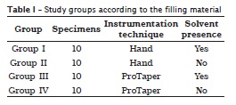
The specimens were randomly selected into four experimental groups. In each group, the endodontic retreatment was executed through a different endodontic retreatment technique, according to table I.
Platform for sample fixation
According to the methodology employed by Er et al. 3, a platform was used to fix the specimens and to measure the debris extruded.
This structure was composed of 10 ml glass flasks (Wheaton do Brasil S.A., São Bernardo do Campo, SP, Brazil) previously washed, dried and weighed in high precision scale (Balança analítica Marte – SHIMADZU AY220, Santa Rita do Sapucaí, MG, Brazil), with a standard deviation of 0.01 mg, and rubber caps with 20 mm of diameter (Adnaloy Artefatos de Borracha Ltda., São Paulo, SP, Brazil). To prevent false positive results, an arithmetic mean was calculated by weighing the flasks three times to avoid the distortion as much as possible.
The rubber caps were perforated at their center and the roots were inserted under pressure through this orifice, so that 1 mm was left above the rubber cap level. This positioning allowed and made the fixation and sealing of the root through the rubber easy. For this purpose, two layers of cyanoacrylate ester (Loctite Henkel, São Paulo, SP, Brazil) were applied.
After that, the rubber cap together with the root was properly positioned into the flask so that the apical portion of the root stayed inside the flask, therefore enabling the collection of the debris extruded through the foramen during the retreatment. Following, a needle (BD Precision Glide, 20x0.55 mm – 24G 3/4) was inserted through the rubber cap to balance the air pressure between the interior of flask an the external environment.
Then, the roots underwent the endodontic retreatment with two techniques. After that, the flasks containing the debris were sealed and stored into a greenhouse (Biomatec 36º Aparelhos Científicos Ltda., Porto Alegre – RS, Brazil) for one week. After that, a new weighing was performed similarly to the first one. The initial value was subtracted from the final value to obtain the weigh in grams of the amount of debris extruded through the apical foramens in the different techniques of endodontic retreatment.
Retreatment of the root canals
With the roots properly placed into the platform, the root canals underwent retreatment according to the technique previously established in table I.
Firstly, all groups had their cervical and medium third prepared with sizes 2, 3 and 4 GG drills.
• Group 1: The technique used followed the principle of reverse enlargement with K files (Dentsply – Maillefer®), up to reach the working length; at every instrument change, distilled water and eucalyptol (Biodinâmica, Ibiporã, PR, Brazil) was used alternately. The procedure was finished when the working lenght was reached, the apical stop was enlarged up to size #40 file with the insertion of the memory instrument (size #20 file) at 1 mm beyond the real root length at every instrument change;
• Group 2: Instrumentation was performed similarly to group 1, but without the use of eucalyptol;
• Group 3: The technique used was proposed by the manufacturer. Initially, size D1 and D2 instruments were used to remove the filling material of the cervical and medium thirds of the root. For the apical third, size D3 was used; at every instrument change, distilled water and eucalyptol (Biodinâmica, Ibiporã, PR, Brazil) was used alternately;
• Group 4: Instrumentation was performed similarly to group 3, but without the use of eucalyptol.
After the retreatment procedure, the radiographs were again taken to verify the amount of remnant material within root canals.
Results
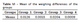
Forty teeth were submitted to an experiment evaluating the degree of extrusion of the filling during endodontic retreatment. The data obtained, that is, the difference in the initial and final weighing of the groups was statistically analyzed by Tukey test. All groups showed debris extrusion through the apical foramen.
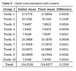
In group 1 (n = 10), in which hand instrumentation was used with the aid of a solvent, the extruded material weighing mean value was 0.0126 g. This group showed a weighing discrepancy in one of the specimens when compared with the others (table II).
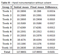
In group 2 (n = 10), in which hand instrumentation was used without solvent, the extruded material weighing mean value was 0.0015 g (table III).
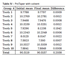
In group 3 (n = 10), in which rotary ProTaper instruments were employed with solvent, the extruded material weighing mean value was 0.0204 g. This group also showed a weighing discrepancy in one of the specimens when compared with the others (table IV).
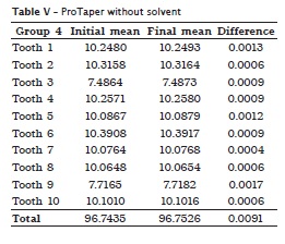
In group 4 (n = 10), in which rotary ProTaper instruments were employed without solvent, the extruded material weighing mean value was 0.0009 g (table V).
Notwithstanding, group 4 showed the smallest extrusion mean, followed by groups 2, 1 and 3. However, when data was submitted to the statistical analysis by Tukey test, there were no statistically significant differences among groups (p = 0.5664) (table VI).
Discussion
Root canal retreatment is a difficult current endodontic therapy modality 14. The performance of such procedure demands the search for a fast, safe and efficient technique, aiming to the comfort of both the patient and the clinician 2.
Regardless of the technique employed, there would be apical extrusion of the debris 3. The extrusion of filling material remnants, which are generally contaminated, through the apical foramen may cause periapical inflammation, therefore resulting in post-operative pain or even interfering in the tissue repair 9.
Based on this aforementioned discussion and on other authors 6, the aim of this study was to evaluate the degree of debris extrusion through apical foramen during the endodontic retreatment by using different techniques with or without using gutta-percha solvents.
In this study, similarly to another one 4, the use of extracted human teeth was opted, aiming to the better simulation of the clinical conditions in which the endodontic treatment is performed. The use of extracted teeth is largely reported and accepted in studies on Endodontics; however, it shows several variables 12. After obturation, the teeth were stored under humid conditions to maintain the experimental situation as closer to the reality as possible. All procedures, both for the preparation and the filling material removal, were performed by a single examiner. This corroborates other authors who think that this fact is of valuable importance for the results 7.
In this present study, hand and rotary instrumentation techniques were used. The hand technique comprised the use of K files; rotary technique comprised the use of ProTaper instruments; this latter, similarly to the study of Gu et al. 5, obtained an efficient result and a smaller operative time.
The spirals of the ProTaper universal file for retreatment can remove greater amounts of gutta-percha, while hand instruments remove such material by small increments 6. This performance is probably attributed to the singular design of the D1, D2 and D3 instruments, which exhibit three sizes and progressive tapers in addition to their convex triangular cross-section, decreasing the contact with the dentine walls.
Rotary ProTaper universal instrument for retreatment extrudes a significantly smaller amount of debris during endodontic retreatment than the conventional techniques 6, which was not in agreement with this present study. At thetime the techniques were tested, there were no statistically significant differences in the amount of debris extruded in this present study. In other words, conventional hand instrumentation technique eliminated through the apical foramen virtually the same amount of debris compared with rotary ProTaper instrumentation technique 8.
When the solvent was added, there were also no statistically significant differences regarding the amount of debris extruded; however, it is worth noting that one specimen of each technique exhibited a nonstandard extrusion. Accordingly, in the hand technique with the solvent, the extrusion mean value was 0.0126g, while the nonstandard specimen showed an extrusion value of 0.1139g, approximately nine times greater.
This fact also occurred when ProTaper instrument was used with the solvent. While the extrusion mean value of this group was 0.0204g, one specimen exhibited a value of 0.1942 g, approximately nine times greater.
Although there were no statistically significant differences among the groups with or without solvent, the fact that this product transforms the gutta-percha into a pasty mass would suggest a greater difficulty in the filling material removal 10.
One of the limiting factors of this study was the fact that the gutta-percha removed from root canals was new and did not undergo aging process, becoming dehydrated and rigid. Dehydrated and rigid gutta-percha promotes a greater wearing of the instruments during the attempt of its removal. Based on the results of this present study itself, it is not possible to recommend an endodontic retreatment technique with smaller extrusion of debris amount.
Conclusion
According to the methodology and the results of this study, it can be concluded that:
• All the techniques employed produced debris which extruded through the apical foramen, regardless of the use of solvent;
• The smallest amount of extrusion was obtained in the group 4 (ProTaper without solvent), followed by groups 2 (hand technique without solvent), 1 (hand technique with solvent) and 3 (ProTaper without solvent). However, there were no statistically significant differences (P = 0.5664);
• The groups using the rotary ProTaper system exhibited similar extrusion results, but with a significantly smaller time for the filling material removal process.
References
1. Bergmans L, Van Cleynenbreugel J, Wevers M, Lambrechts P. Mechanical root canal preparation with NiTi rotary instruments: rationale, performance and safety. Status report for the American Journal of Dentistry. American Journal of Dentistry. 2001;14:324-33. [ Links ]
2. Bramante CM, Freitas CVJ. Retratamento endodôntico: estudo comparativo entre técnica manual, ultra-som e canal finder. Rev Odontol Univ São Paulo. 1998;12(1):13-7. [ Links ]
3. Er K, Sumer Z, Akpinar KE. Apical extrusion of intracanal bactéria following use of two engine-driven intrumentation techniques. Int Endod J. 2005;38:871-6. [ Links ]
4. Garcia Jr JS, Silva Neto UX, Carneiro E, Westphalen VPD, Fariniuk LF, Fidel RAS et al. Avaliação radiográfica da eficiência de diferentes instrumentos rotatórios no retratamento endodôntico. RSBO. 2008;5(2):41-8. [ Links ]
5. Gu LS, Ling JQ, Wei X, Huang WY. Efficacy of ProTaper Universal rotator retreatment system for gutta-percha removal from root canals. Int Endod J. 2008:41(4):288-95. [ Links ]
6. Huang X, Ling J, Wei X, Gu L. Quantitative evaluation of debris extruded apically by using protaper universal tulsa rotary system in endodontic retreatment. J Endod. 2007;33(9):1102-5. [ Links ]
7. Imura N, Zuolo ML, Ferreira MO, Novo NF. Effectiveness of the canal finder and hand instrumentation in removal of guta-percha root fillings during root canal. Int Endod J. 1996;29(6):382-6. [ Links ]
8. Imura N, Kato AS, Hata GI, Uemura M, Toda T, Weine F. A comparison of the relative efficacies of four hand and rotary instrumentation techniques during endodontic retreatment. Int Endod J. 2000;33(4):361-6. [ Links ]
9. Lopes HP, Siqueira Jr JF. Endodontia – biologia e técnica. Rio de Janeiro: Medsi; 1999. p. 650.
10. Mandel E, Friedman S. Endodontic retreatment: a rational approach to root canal reinstrumentation. J Endod. 1992;18(11):565-9. [ Links ]
11. Paik S, Sechrist C, Torabinejad M. Levels of evidence for the outcome of endodontic retratament. J Endod. 2004;30:745-50. [ Links ]
12. Reddy SA, Hick ML. Apical extrusion of debris using two hand and two rotary instrumentation technique. J Endod. 1998;24:180-3. [ Links ]
13. Santos M, Aun CE. Análise comparativa in vitro da eficiência na desobstrução dos canais radiculares entre as técnicas manual e sônica. Rev Assoc Paul Cir Dent. 1992;46(1):685-8. [ Links ]
14. Stabholz A, Friedman S. Endodontic retreatment – case selection and technique. Part 2: treatment planning for retreatment. J Endod. 1998;14(12):607-14.
15. Stabholtz A, Walton RE. Avaliação do sucesso e do insucesso. In: Walton RE, Torabinejad M. Princípios e práticas em endodontia. São Paulo: Santos; 1997. p. 324-35. [ Links ]
16. Tinaz AC, Alacam T, Uzun O, Maden M, Kayaoglu G. The effect of disruption of apical constriction on periapiacal extrusion. J Endod. 2005;31:533-5. [ Links ]
 Corresponding author:
Marcelo de Morais Vitoriano
Al. Octávio Pinheiro Brisola, 9-75 – Vila Universitária
CEP 17012-901 – Bauru – SP – Brasil
E-mail: marcelovitoriano@gmail.com
Received for publication: June 12, 2012
Accepted for publication: July 10, 2012
