


Serviços Personalizados
Artigo
 pdf em Inglês
 Artigo em XML
 Referências do artigo
 Enviar este artigo por email
Links relacionados
Compartilhar

 Permalink
RSBO (Online)
versão On-line ISSN 1984-5685
RSBO (Online) vol.10 no.2 Joinville Abr./Jun. 2013
ORIGINAL RESEARCH ARTICLE
Comparative evaluation of radiopacity of MTA Fillapex® endodontic sealer through a digital radiograph system
Silvya Aparecida Vanso BicheriI; Fausto Rodrigo VictorinoI
I Department of Dentistry, University Center of Maringá – Maringá – PR – Brazil
ABSTRACT
Introduction: The radiograph represents one of the evaluation tools of the endodontic procedure. Therefore, radiopacity is an essential property of filling materials.
Objective: To evaluate the MTA Fillapex® radiopacity, as compared to that of Sealer26®, Sealapex® and AH Plus®.
Material and methods: Five specimens were prepared from each endodontic sealer, which were then positioned in the digital sensor (Kodak RVG 6100 – Digital Radiography System) and radiographed with a Kodak 2200 Intraoral Device Rx System, operating at 70 Kvp, 10 mV, with an exposition time of 0.071 tenth of second and a distance of 20 cm from the sensor. The radiopacity was assessed using the software Image Tool 3.0, which determines gray shades ranging from 0 to 255 pixels. For statistical analysis, ANOVA test was used, followed by Tukey's test at a significance level of 5%.
Results: The MTA Fillapex® presented the lowest radiopacity between all tested sealers, while AH Plus® was the most radiopaque one. Sealapex® and Sealer 26® showed intermediate radiopacity and statistically did not differ from each other.
Conclusion: MTA Fillapex® presented intermediate radiopacity. When compared with AH Plus®, Sealer 26® and Sealapex® endodontic sealers, it exhibited the smallest result.
Keywords: root canal obturation; digital dental radiograph; Endodontics.
Introduction
Root canal obturation is the endodontic treatment phase where the goal is the sealing of the root canal system to prevent a possible recontamination of the root canal 9. The evaluation of the filling procedure is performed through radiographic image. Thus, the radiograph is an indispensable tool for the evaluation of the endodontic treatment executed. The radiopacity is a physical property required by the endodontic sealers which enables to assess the contrast of the tooth structure on a radiograph 15 as well as the filling of the root canal system. Gutta-percha points in association with endodontic sealers are the most accepted approach for the root canal filling 14. The radiopacity of the endodontic sealer highly influences on the radiographic image of the filling 8.
To analyze the radiopacity, the specification n. 57 of the American Dental Association (ADA) 1 suggests an evaluation of the optical density in radiographic films through photodensitometer. Notwithstanding, the sealer radiopacity should be compared with that of a device with aluminum steps, so called penetrometer. The results are equivalent to millimeters of aluminum. The digital radiograph has been much utilized for the analysis of the radiopacity of the sealers 14,16, because it is a technique of fast visualization dispensing the chemical processing, which avoids errors of this phase, reduces the time of exposure to radiation and still enables a better visualization of the radiographic contrast.
Recently launched into the Brazilian market, MTA Fillapex® (Angelus, Londrina, PR, Brazil) is a Mineral Trioxide Aggregate (MTA)-based endodontic sealer, with little studies on it. For this reason, the aim of this present study was to assess the level of radiopacity of MTA Fillapex® sealer and compare it to that of AH Plus® (Dentsply, DeTrey, Konstanz, Germany), Sealer 26® (Dentsply, Petrópolis, RJ, Brazil) and Sealapex® (Sybron Kerr, Washington, USA) sealers through digital radiograph.
Material and methods
The sealers were divided into four groups: group I – MTA Fillapex® (Ângelus, Londrina, PR, Brazil), group II – AH Plus® (Dentsply, DeTrey, Konstanz, Germany), group III – Sealer 26® (Dentsply, Petrópolis, RJ, Brazil) and group IV – Sealapex® (Sybron Kerr, Washington, USA). Five samples were constructed for each sealer with aluminum rings of 10 mm of internal diameter and 1.5 mm of thickness. The rings were placed onto a thin lamina of cellophane over a glass plate. The sealers were proportionated and mixed according to the manufacturer's instructions and the rings were filled up to their upper border, with the aid of a dental vibrator. Additionally, a mass of 100 g was placed over the set. The samples were stored in an incubator at 37ºC.
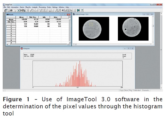
Elapsed three times the setting time of each material, the samples were removed from the incubator and the aluminum rings. Then, they were positioned on a sensor (Kodak RVG 6100 – Digital Radiography System – Kodak, Rochester, New York, USA) and radiographed with an x-ray device (Kodak 2200 Intraoral X – Ray System), operating at 70 Kvp, 10 mV, with exposure time of 0.071 tenth of second, at a distance of 20 cm from the sensor. On the radiographic images, the measurements of the optical density or gray shades of the sealers were executed on ImageTool 3.0 software (UTHSCSA, San Antonio, Texas) (figure 1). To measure the gray shades, which varied from 0 to 255 pixels, the function "histogram" was employed by using a standardized area of 20 x 19 pixels always equally positioned for all images. For the statistical analysis of the results, ANOVA and Tukey test was applied with level of significance 5%.
Results
Table I shows the mean radiopacity values in pixels and percentage for the endodontic sealers, using as reference the minimum and maximum pixel values of the histogram.
The results demonstrated that AH Plus® was the most radiopaque sealer (169.7 pixels), equivalent to 66.5% of the total of 255 pixels, while Sealapex® and Sealer 26® sealers exhibited intermediate radiopacity and did not statistically differ between each other, 60% (153.7 pixels) and 58.1% (148.3 pixels), respectively. MTA Fillapex® sealer significantly differed from the other sealers, presenting a radiopacity of 49.8% (127 pixels).

Discussion
A great technological advancement of Radiology is the use of the digitized image, a technique of fast visualization which has made easy the work of endodontists because it enables the storage of the images in the computer and to perform adjustments in the brightness and contrast, therefore improving the evaluation of the procedures executed and careful interpretation, such as anatomic details, accessory root canals, precise measurements, periapical and bone alterations 4,5,10,12. Additionally, it does not employ the chemical processing it avoids the errors inherent to this phase; also, it reduces the time of exposure to radiation decreasing the risks of effects deleterious to patients 7. Because the digital radiograph is a more sensible method than the conventional radiograph film, requires less radiation 11 and has been increasingly used, this present study opted to evaluate MTA Fillapex® radiopacity through this method.
In this present study, the digital radiographic images of the endodontic sealers were evaluated through the histogram analysis of the gray shades, ranging from 0-255 pixels, in which 0 is black color and 255 is the white color. Therefore, the greater the pixel value, the more radiopaque is the sealer. To obtain radiopacity values of the sealers in percentage, 255 pixels were considered as 100%.
Considering the results, AH Plus® was the most radiopaque sealer (66.5%), which corroborates the results of Sydney et al. 16, Aznar et al. 2 and Vidotto et al. 18. These authors demonstrated that the resin sealers are characterized by the highest radiopacity than that of the calcium hydroxide- and MTA-based sealers. Sealapex® and Sealer 26® sealers did not show statistically significant differences between each other, but a significantly smaller difference than AH Plus® and greater than MTA Fillapex®. Sealer 26® is a sealer containing calcium hydroxide and having good biological and physical-chemical characteristics. It shows a higher biocompatibility than that of the other sealers; however, it exhibits low radiopacity according the studies of Tanomaru-Filho et al. 17 and Rosa et al. 14.
The Sealapex® is a calcium hydroxide-based sealer and it is characterized by exhibiting good biocompatibility, control of the inflammatory root resorption and due its high pH shows excellent bacterial action 3. However, studies demonstrated that Sealapex® radiopacity is also smaller than that of the other endodontic sealers 2,8.
MTA Fillapex® exhibits the smallest radiopacity value (127 pixels), which represents 49.8% of the total of the histogram. A very similar result was found by Vidotto et al. 18, who obtained a value of 125.6 pixels. In the study of these authors, MTA Fillapex® exhibited the best performance because the sealers such as Acroseal®, Endomethasone® and Roekoseal® showed radiopacity smaller than 120 pixels.
In this present study, the smallest radiopacity of o MTA Fillapex® than that of the other sealers is related to its composition, once it only contains bismuth oxide as radiopacifier. Materials with this component have obtained low radiopacity values, as demonstrated by Rasimick et al. 13. On the other hand, AH Plus® sealer has zirconium oxide as radiopacifying agent and Sealer 26® and Sealapex® sealers has bismuth trioxide and titanium oxide.
Conclusion
According to the results found, MTA Fillapex® has an intermediate radiopacity (49.8%) and, when it is compared with AH Plus®, Sealer 26® and Sealapex® sealers, it obtained the smallest result.
References
1. American National Standard / American Dental Association – ANSI/ADA. Specification n. 57: Endodontic sealing materials. Modified adoption of ISO 6876:2000. Dental root canal sealing materials. Chicago; 2000.
2. Aznar FDC, Bueno CES, Nishiyama CK, Martin AS. Radiopacidade de sete cimentos endodônticos avaliada através de radiografia digital. RGO. 2010;58(2):181-4. [ Links ]
3. Bezzera da Silva LA, Leonardo MR, Da Silva RS, Assed S, Guimarães LFL. Calcium hydroxide root canal sealers: evaluation of pH, calcium ion concentration and conductivity. Int Endod J. 1997;30:205-9. [ Links ]
4. Botelho TL, Mendonça EF, Cardoso LLM. Contribuição da radiografia digital na clínica odontológica. Robrac. 2003;12(33):55-9. [ Links ]
5. Coclete GA, Tavano O, Pavan AJ. Comparação das densidades ótica e radiográfica, analisadas pelo fotodensiômetro MRA e pelo sistema digital Digora. Rev Odontol UNESP. 2003;32(1):93-8. [ Links ]
6. Costa RF, Scelza MFZ, Costa AJO. Radiopacidade de cimentos endodônticos: avaliação pela intensidade de pixel. J Bras Clín Odontol Integr. 2002;6(32):137-9. [ Links ]
7. Cruz GA, Morais LC, Médici Filho E, Castilho JCM. Utilização de radiografia digital em Odontologia. Rev ABO Nac. 2004:12(5):283-6. [ Links ]
8. Ferreira FBA, Silva e Souza PAR, Vale MS, Tavano O. Radiopacidade de cimentos endodônticos avaliados pelo sistema de radiografia digital. Rev Fac Odontol Bauru. 1999;7(1/2):55-60. [ Links ]
9. Kopper PMP, Santos RB, Viegas APK, Reis Só MV, Grecca FS, Figueiredo JAP. Estudo do selamento dos canais radiculares obturados com AH Plus® ou Endofill®, com e sem cimento nos cones acessórios. Rev Fac Odontol Univ Passo Fundo. 2007;12(1):52-5. [ Links ]
10. Kreich EM, Leal GA, Slusarz PAA, Santini RM. Imagem digital na Odontologia. Ciênc Biol Saúde. 2005;11(3/4):53-61. [ Links ]
11. Parks ET, Williamson GF. Digital Radiology: an overview. The Journal of Contemporary Dental Practice. 2002;3(4):1-13. [ Links ]
12. Pasquali AAG, Matson MR, Raitz R. Estudo comparativo da densidade radiográfica de cimentos resinosos. Rev Odontol Univ São Paulo. 2009;21(3):239-43. [ Links ]
13. Rasimick BJ, Shah RP, Musikant BL, Deutsch AS. Radiopacity of endodontic materials on film and a digital sensor. J Endod. 2007;33(9):1098-101. [ Links ]
14. Rosa RA, Bier CAS, Pereira CC, Só MVR, Wolle CFB. Simulation of soft and hard tissues and its effects on radiopacity of root canal sealers. Rev Odonto Ciênc. 2011;26(4):326-30. [ Links ]
15. Salzedas LMP, Louzada MJQ, Oliveira Filho AB. Radiopacity of restorative materials using digital imagens. J Appl Oral Sci. 2006;14(2):147-52. [ Links ]
16. Sydney GB, Ferreira M, Leonardi DP, Deonizio MDA, Batista A. Análise da radiopacidade de cimentos endodônticos por meio de radiografia digital. Rev Odonto Ciênc. 2008;23(4):338-41. [ Links ]
17. Tanomaru-Filho M, Jorge EG, Tanomaru JMG, Gonçalves M. Radiopacity evaluation of new root canal filling materials by digitalization of images. J Endod. 2007;33(3):249-51. [ Links ]
18. Vidotto APM, Cunha RS, Zeferino EG, Rocha DGP, Martin AS, Bueno CES. Comparison of MTA Fillapex radiopacity with five root canal sealers. RSBO. 2011;8(4):404-9. [ Links ]
 Corresponding author:
Fausto Rodrigo Victorino
Rua Formosa, n. 489 – Centro
CEP 86990-000 – Marialva – PR – Brasil
E-mail: frvictorino@ig.com.br
Received for publication: November 28, 2012
Accepted for publication: December 17, 2012
