


Serviços Personalizados
Artigo
 pdf em Inglês
 Artigo em XML
 Referências do artigo
 Enviar este artigo por email
Links relacionados
Compartilhar

 Permalink
RSBO (Online)
versão On-line ISSN 1984-5685
RSBO (Online) vol.10 no.2 Joinville Abr./Jun. 2013
LITERATURE REVIEW ARTICLE
Gubernacular cord and canal – does these anatomical structures play a role in dental eruption?
Danielly Cunha Araújo FerreiraI; Ana Caroline FumesI; Alberto ConsolaroII; Paulo Nelson-FilhoI; Alexandra Mussolino de QueirozI; Andiara De RossiI
I Department of Child Clinics, Preventive and Social Dentistry, School of Dentistry of Ribeirão Preto, University of São Paulo – Ribeirão Preto – SP – Brazil.
II Department of Pathology, School of Dentistry of Bauru, University of São Paulo – Bauru – SP – Brazil.
ABSTRACT
Introduction: The gubernacular cord is an original structure of the dental lamina, which undergoes apoptosis and their remnants were organized in the form of epithelial islets and strands that lined up, leaving the reduced epithelium of the enamel organ towards the oral mucosa. This structure is located within the gubernacular canal, which can be identified as a small opening in the alveolar region of the lingual or palatal surface of the deciduous teeth.
Objective: To conceptualize, identify and assess the possible contribution of the gubernacular cord and canal in the process of tooth eruption.
Literature review: A review of literature on Pubmed, Medline and Bireme databases, without datum restriction. Little amount of scientific articles were found, and only 14 studies were identified. The authors addressed the matter succinctly, with little information about these structures, which can play an important role in the process of tooth eruption.
Conclusion: The gubernacular cord and canal are anatomical structures located in the alveolar bone crest of the maxilla or mandible, behind the deciduous teeth. These structures appear to exhibit the ability to aid the eruption path of the permanent teeth successors. Despite being a relevant subject, few professionals know this structure and its possible role in the process of tooth eruption.
Keywords: tooth eruption; primary dentition; permanent dentition.
Introduction
Tooth eruption is a physiologic process in which a tooth undergoing formation migrates from its development site within the alveolar processes to its functional positions inside the oral cavity 18. During this process, the participation of different anatomic structures, cells, chemical and molecular mediators occurs and although there have been a great advancement in the scientific knowledge regarding this issue, still today the cellular, molecular and anatomic mechanisms involved in tooth eruption process are not fully understood 17,28,29. Aiming to explain the tooth eruption process, countless theories have been proposed over the years, such as the theory of root growth 18, alveolar bone growth 4,20, pulpal growth 25, in addition to the theory of the combination of genetic factors 26 and the follicular theory 6,15, which has been the most accepted nowadays.
The follicular theory postulates that the dental follicle is capable of inducing, guiding and coordinating the bone resorption above the crown of a teeth undergoing formation and bone apposition below this same crown, which enables that during the intraosseous eruptive phase of the tooth eruption process the formation of a eruptive path occur and the tooth undergoing formation be passively conducted through this path 6,15.
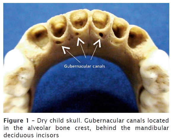
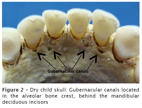
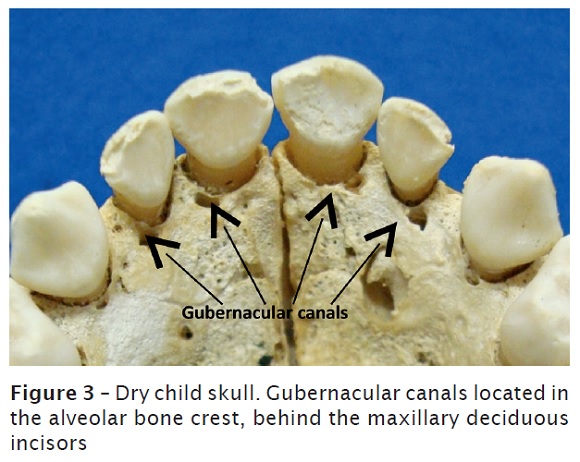
The gubernacular cord is a structure composed of conjunctive tissue which link the tooth follicle to the overlying gingiva, showing the function of guiding or directing the course of the tooth eruption. The formation of this structure starts from the remnants cells of the tooth lamina which are organized as a fibrous cord leaving the reduced epithelium of the enamel organ towards the oral mucosa 9,10. This structure is located in the alveolar ridge behind the deciduous tooth 7,12. In this cord, there is the presence of countless chemical mediators, including EGF (epithelial growth factor), a substance secreted by the epithelial cells having the capacity of stimulating the formation of clasts and consequently a bone resorption, making impossible the filling of the cord area by alveolar bone, always leaving a space surrounding this cord, so-called the gubernacular canal (figures 1 to 3) 8.9. Bothe the gubernacular canal and cord are structures very little studied on the scientific literature; however they seem to play some role in the process of tooth eruption.
The aim of this study was to conceptualize, identify and assess the possible contribution of the gubernacular cord and canal in the process of tooth eruption.
Material and methods
A bibliographic searching was conducted on the following databases Pubmed, Bireme, Medline, Google and Central Library of the University of São Paulo – Campus of Ribeirão Preto, without datum restriction, through using the terms: gubernacular cord, gubernacular canal, gubernaculum dentis, deciduous dentition, permanent dentition and tooth eruption. Only 14 scientific studies were found in all literature researched. Of these studies, ten were written in English, one in Portuguese, one in Japanese, on in Italian, and one in France.
Literature review
The first reference to the gubernacular cord and canal occurred in 1778, by an English called John Hunter, which described these structures after observing a connection between the bone ridge of the tooth in development and the gingiva, without little scientific proof 11. Almost one century later, this observation was supported by microscopic studies conducted by the France histologist Louis-Charles Malassez, in 1887. He described the existence of remnants of the tooth lamina (epithelial cells) and longitudinal fibers that were within a canal connecting the gingiva with the permanent tooth bud 14.
The gubernacular canal, which enables the continuity of the bone ridge of permanent incisors, canines and premolars with the tissue of the overlying gingiva itself, it is filled by the gubernacular cord, which is composed by a fibrous conjunctive tissue containing peripheral nerves, blood and lymphatic vessels, as well as epithelial cells or cell aggregates coming from the fragmentation of the tooth lamina. Thus, the gubernacular cord is the connection between the conjunctive tissue of the ridge with the oral mucosa 22.
During the pre-eruptive phase of tooth eruption, the tooth buds have special relationship with the growing jaws. At this phase, the gubernacular canal does not undergo perceptive alterations in shape or size 5. During the eruptive phase, according to the successor tooth moves towards the axial direction to the oral cavity, the gubernacular canal is widened by the local osteoclastic activity, with the aim of accommodating the crown of the permanent tooth undergoing eruption 7. The tooth moves towards the mucosa, the pericoronary follicle incorporates within its conjunctive tissue the islets and cords of epithelial cells from the gubernacular cord, progressively increasing the epithelial component at this area 9.
The role of the gubernacular cord in the eruption process was investigated Cahill and Marks 6, who observed that the surgical section of this structure in dogs did not alter the eruption rates and the final position of the tooth. A next study, in which the section of the gubernacular cord and the surgical removal of the crown parts of the tooth follicle were executed, enabled to observe the interruption in the tooth eruption process, demonstrating that the tooth follicle has a fundamental role in the physiologic movement of the tooth towards the oral cavity and that if the gubernacular cord has some role in the process of tooth eruption, this is not relevant for the occurrence of the process 5.
According to Hodson 10, the gubernacular cord was not described in association with the deciduous dentition, only with the permanent dentition with the deciduous predecessor. However, according to Scott 27, the permanent molar teeth, which did not have a deciduous predecessor, also have their follicles connected to the oral mucosa by gubernacular cords, so-called by the author "molar gubernacular cords".
Philipsen 23 theorized that the gubernacular cord could have implication in the development of the adenomatoid odontogenic tumor (AOT), because this contains remnants of the tooth lamina. These epithelial remnants are very closer to the crown of the permanent tooth and they can move during the tooth eruption process along with the gubernacular canal and induce AOT formation, because among the many hypotheses for the pathogenesis of this tumor are the remnants of the tooth lamina.
Discussion
Despite the first reference to the gubernacular cord and canal have been shown in 1778 by John Hunter, 234 years ago, the existence and function of this structure are still controversial and questioned. According to Hodson 10, these structures are present only in association with the permanent teeth with deciduous predecessor, fact corroborates by Cahill et al. 5 and Philipsen and Reichart 22. However, Scott 27 suggested that the permanent teeth without deciduous predecessor also displayed gubernacular cord and canal, connecting the bud to the oral mucosa.
Notably, the description of the gubernacular cord and canal was only recently incorporated to the pattern of the American texts 3. The explanation could be based on the fact that, by the omission of this gubernacular canal, the existing theories on the pressure for the eruption recommended by some professor and researchers influenced by the wrong concept that the successor tooth would be stuck by a complete bone coating, could be more easily accepted 5. Some authors 5,10,27 believe that the gubernacular canal and cord have a role in the direction of the teeth, making the eruption easier; while others 6 claimed that these structures do not exert any type of interference. According to Philipsen et al. 23, the gubernacular cord and canal can influence on AOT development, because AOT in about 80% of the cases is located at the region of the permanent incisors and canines, where the gubernacular cord and canal is present 21,24. Some authors 13,19,30 sympathize with this hypothesis, because rarely AOT is found in association with the deciduous teeth, which could be justified by the absence of the gubernacular cord in the deciduous dentition, according to Hodson 10. Ide et al. 12 questioned that sometimes (4% of the pericoronary lesions) AOT occurs in permanent molars, which did not have according Hodson 10, the gubernacular cord and canal. Notwithstanding, it valid to remember that Scott 27 reported the presence of the "molar gubernacular cords", which could explain the occurrence of this tumor type in these teeth.
Until now, the ability of the dentist to interfere or treat an included or impacted tooth due to eruption failure is limited to surgical or mechanical procedures, such as: the extraction of a deciduous tooth, surgical removal of the alveolar bone, the aid of the mucosal penetration through gingival incision and orthodontic tooth traction 2. Perhaps by knowing deeply the role of the structures such the gubernacular cord and canal and its possible role in tooth eruption process, one can develop in the future mechanisms that allow the interference in the eruptive process due to these structures.
Conclusion
The gubernacular cord is the structure coming from the tooth lamina that after undergoing apoptosis has its remnants organized in islets and epithelial cords that lined up leaving the reduced epithelium of the enamel organ towards the oral mucosa. The gubernacular cord in located within a alveolar bone scaffold, so-called the gubernacular canal.
The existence of both the gubernacular canal and cord is proved in the permanent dentition with deciduous predecessor; however, their existence is not still proved in the deciduous dentition. Concerning to the permanent dentition without deciduous predecessor, some authors defend their existence and others do not.
After the careful analysis of the studies, it is observed that still there are divergences on the function of the gubernacular cord and canal in tooth eruption process.
Although the tooth eruption process is a very researched issue in the literature, the role of the gubernacular cord and canal in tooth eruption is still very obscure, and further studies are necessary to clarify its real function. Therefore, it is necessary to deepen the knowledge on this issue, which can interfere on the treatment options for impacted or included teeth as well as on the guided direction of tooth eruption.
References
1. Andreasen JO, Andreasen FM. Textbook and color atlas of traumatic injuries to the teeth. 4. ed. Copenhagen: Munksgaard; 2001. [ Links ]
2. Assed S. Odontopediatria – bases científicas para a prática clínica. 1. ed. São Paulo: Artes Médicas; 2005.
3. Bhaskar SN. Histologia e embriologia oral de Orban. 10. ed. São Paulo: Artes Médicas; 1986. [ Links ]
4. Brash JC. Growth of the alveolar bone and its relation to the movements of teeth, including eruption. Int J Orthod. 1928;14:196-223. [ Links ]
5. Cahill DR, Marks Jr SC, Wise GE, Gorski JP. A review and comparison of tooth eruption systemsused in experimentation – a new proposal on tooth eruption. In: Davidovitch Z (Ed.). Biological mechanisms of tooth eruption and root resorption. Alabama: EBSCO Media; 1988.
6. Cahill DR, Marks Jr SC. Tooth eruption: evidence for the central role of the dental follicle. J Oral Pathol. 1980;9:189-200. [ Links ]
7. Cahill DR. Histological changes in the bony crypt and gubernacular canal of erupting permanent premolars during deciduous premolar exfoliation in beagles. J Dent Res. 1974;53:786. [ Links ]
8. Consolaro A. Reabsorções dentárias nas especialidades clínicas. 3. ed. Dental Press; 2012. [ Links ]
9. Consolaro A. Tracionamento ortodôntico: possíveis consequências nos caninos superiores e dentes adjacentes (parte 1). Dental Press J Orthod. 2010 Jul-Aug;15(4):15-23. [ Links ]
10. Hodson JJ. The gubernaculum dentis. Dent Practit. 1971 Aug;21(12):423-8. [ Links ]
11. Hunter J. The natural history of human teeth. 2. ed. Londres; 1778. [ Links ]
12. Ide F, Mishima K, Kikuchi K, Horie N, Yamachika S, Satomura K et al. Development and growth of adenomatoid odontogenic tumor related to formation and eruption of teeth. Head and Neck Pathol. 2011;5:123-32. [ Links ]
13. Kearns GJ, Smith R. Adenomatoid odontogenic tumour: an unusual cause of gingival swelling in a 3-year-old patient. Br Dent J. 1996;181:380-2. [ Links ]
14. Malassez ML. Sur la structure du gubernaculum dentis et la theorie paradentaire. Compte Soc Biol. 1887. [ Links ]
15. Marks Jr SC, Cahill DR. Experimental study in the dog of the non-active role of the tooth in the eruptive process. Arch Oral Biol. 1984;29:311-22. [ Links ]
16. Marks Jr SC, Cahill DR. Regional control by the dental follicle of alterations in alveolar bone metabolism during tooth eruption. J Oral Pathol. 1987;16:164-9. [ Links ]
17. Marks Jr SC, Schroeder HE. Tooth eruption: theories and facts. Anat Rec. 1996;245:374-93. [ Links ]
18. Massler M, Shour I. Studies in tooth development: theories of eruption. American Journal of Orthodontics and Oral Surgery. 1941;27:552-76. [ Links ]
19. Mizukoshi T, Ojima I, Mizuno Y. A case of adenomatoid odontogenic tumor in a 3-year-old boy. Jpn J Stomatol Soc. 1980;29:605-6. [ Links ]
20. O'Brien C, Bhaskar SN, Brodie AG. Eruptive mechanism and movement in the first molar of the rat. J Dent Res. 1958;37:467-84. [ Links ]
21. Philipsen HP, Reichart PA, Siar CH. An updated clinical and epidemiological profile of the adenomatoid odontogenic tumour: a collaborative retrospective study. J Oral Pathol Med. 2007;36:383-93. [ Links ]
22. Philipsen HP, Reichart PA. The development and fate of epithelial residues after completion of the human odontogenesis with special reference to the origins of epithelial odontogenic neoplasms, hamartomas and cysts. Oral Biosci Med. 2004;3:171-9. [ Links ]
23. Philipsen HP, Samman N, Ormiston IW, Wu PC, Reichart PA. Variants of the adenomatoid odontogenic tumor with a note on tumor origin. J Oral Pathol Med. 1992;21:348-52. [ Links ]
24. Rick GM. Adenomatoid odontogenic tumor. Oral Maxillofac Surg Clin N Am. 2004;16:333-54. [ Links ]
25. Sandy JR. Tooth eruption and orthodontic movement. Br Dent J. 1992;172:141-9. [ Links ]
26. Sauk JJ. Genetic disorders involving tooth eruption anomalies. In: Davidovitch Z (Ed.). Biological mechanism of tooth eruption and root resorption. Alabama: EBSCO Media; 1988. [ Links ]
27. Scott JH. The development and function of the dental follicle. Br Dent J. 1948 Nov 5;85(9):193-9. [ Links ]
28. Sutton PR. Tooth eruption and migration theories: can they account for the resence of a 13,000 – year-old mesiodens in the vault of the palate? Oral Surg Oral Med Oral Pathol. 1985;59:252-5.
29. Ten Cate AR. Erupção dentária. In: Bhaskar SN. Histologia e embriologia oral de Orban. 8. ed. Santa Maria: Artes Médicas; 1978. [ Links ]
30. Unal T, Cetingul E, Gunbay T. Peripheral adenomatoid odontogenic tumor: birth of a term. J Clin Pediatr Dent. 1995;19:139-42. [ Links ]
 Corresponding author:
Danielly Cunha Araújo Ferreira
Departamento de Clínica Infantil, Odontologia Preventiva e Social
Faculdade de Odontologia de Ribeirão Preto – USP
Av. do Café, s/n.
CEP 14040-903 – Ribeirão Preto – SP – Brasil
E-mail: daniellycaf@hotmail.com
Received for publication: May 22, 2012
Accepted for publication: November 5, 2012
