Serviços Personalizados
Artigo
Links relacionados
Compartilhar
RSBO (Online)
versão On-line ISSN 1984-5685
RSBO (Online) vol.10 no.3 Joinville Jul./Set. 2013
ORIGINAL RESEARCH ARTICLE
Analysis of apical fitting of .06 and .02 tapered gutta-percha master cones in root canals shaped with ProTaper rotary system
Manoel Eduardo de Lima MachadoI; Cleber Keiti NabeshimaI; Guilherme Henrique Rosa MartinsI; Maria Leticia Borges BrittoII
IDepartment of Operative Dentistry, School of Dentistry, University of São Paulo – São Paulo – SP – Brazil.
IIDepartment of Endodontics, Cruzeiro do Sul University – São Paulo – SP – Brazil.
ABSTRACT
Introduction: The fitting of gutta-percha master cone is important for the obturation step. The modified single-cone technique using larger taper has provided better filling of gutta-percha than the original single-cone technique.
Objective: The aim of this study was to verify if either .02 or .06 tapered gutta-percha master cone would better fit into the working length of teeth shaped using ProTaper rotary system.
Material and methods: Thirty distobuccal root canals of mandibular molars were shaped using F2 ProTaper, and size 40, 35, 30 and 25 0.6 or 0.2 tapered gutta-percha cones were tested. The best fitting into the working length was recorded. The data were gathered and compared with size 25 by using Fisher's exact test.
Results: There was no statistically significant difference between groups (p = 0.4915). Sizes 30 and 35 were the most used.
Conclusion: It can be concluded that both .02 and .06 tapered gutta-percha master cones showed best fitting in sizes larger than 0.25 mm in root canals shaped with ProTaper F2.
Keywords: Endodontics; root canal preparation; gutta-percha.
Introduction
Root canal obturation aims at the sealing of the canal therefore hindering the communication of the oral cavity with the periapical structures and vice versa. Therefore, it is of extreme importance the adaptation of the master gutta-percha cone to root canal walls, mainly at apical level. Previous studies have demonstrated this difficulty even when there is the fitting of the cone into the working lenght 2,16.
The rotary systems results in more conical and uniform preparations 12, enabling the execution of single-cone technique developed for this purpose, with easy and fast execution 4,5.
The ProTaper system is the most used worldwide and has its gutta-percha cone according to the taper characteristics of its instruments to enable the execution of single-cone technique 14. However, studies have demonstrated that ProTaper single-cone technique can show post-obturation leakages 3,6,11,17,19. Accordingly, Machado 8 affirmed that automated systems resulted in larger preparations and advocates the use of modified single-cone technique with 0.6 tapered cones and diameters larger than the last size of the rotary instrument used in preparations with ProTaper system, searching for the best fitting of the single cone at the apical third.
Notwithstanding, studies are still lacking to confirm which diameter would be ideal to be used in most part of the cases in the modified technique. Thus, considering that all modification must be analyzed on several technical and clinical aspects, the aim of this study was to verify if either .02 or .06 tapered gutta-percha master cone would better fit into the working length of teeth shaped using ProTaper rotary system.
Material and methods
After the approval of the Ethical Committee in Research of the School of Dentistry of the University of São Paulo, thirty distobuccal roots of mandibular molars with straight canals and similar morphology were selected and checked by periapical radiographs. All specimens were standardized at 12 mm of length.
All canals were prepared according the technique of Machado et al. 9. Briefly, the cervical and medium third were prepared with size 1, 2 and 3 Gates glidden burs (Dentsply Maillefer, Ballaigues, VD, Switzerland), followed by SX, S2 and F1 ProTaper rotary instruments (Dentsply Maillefer, Ballaigues, VD, Switzerland) through hand insertion towards apical direction and motor driven at 350 rpm with torque 3 and brushing movements applied to all canal walls. The working length was established at 1 mm short of the apical foramen and the apical third was firstly prepared with size 15 and 20 K flexofile (Dentsply Maillefer, Ballaigues, VD, Switzerland), followed by S1, S2, F1 and F2 rotary instruments, as aforementioned described. All root canal preparations were executed under copious irrigation with 20 ml of 1% sodium hypochlorite (Fórmula e Ação, São Paulo, SP, Brazil), associated with light Endo PTC (Fórmula e Ação, São Paulo, SP, Brazil), followed by final irrigation with 5 ml of 1% sodium hypochlorite, 5 ml of 17% EDTA (Fórmula e Ação, São Paulo, SP, Brazil), and 5 ml of 1% sodium hypochlorite.
Next, with a wet root canal, the specimens were numbered and the fitting of 0.6 tapered cone (Dentsply Maillefer, Ballaigues, VD, Switzerland) was executed. Then, all data were recorded and the canals were again irrigated with 1% sodium hypochlorite and the fitting of 0.2 tapered cone (Dentsply Maillefer, Ballaigues, VD, Switzerland) was randomly performed not to influence on the results. The following sequence of diameters was used for all specimens: 40 – 35 – 30 – 25. The selected gutta-percha cone was the first one which best fitted into the working length, followed by confirmation through periapical radiograph. The data were gathered and compared with size 25 (which corresponds to F2 file) by using Fisher's exact test with level of significance of 5%.
Results
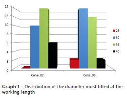
Only in two samples of group 1 size 25 fitted into the working length, the other samples of group 1 and all samples of group 2 fitted at larger diameters.
The comparison between groups did not show statistically significant differences (p = 0.4915). Sizes 30 and 35 were the most used (graph 1).

Discussion
The searching for simplicity and rapidity is constant in all technical procedures in all research areas. In Endodontics, this is clear both for the preparation – in which automated techniques are used –, and for obturation exemplified by single-cone technique. However, these objectives should be also associated with quality, once they can define either the treatment success or failure. Concerning to root canal obturation, several techniques have been proposed and the presence of the solid filling material such as gutta-percha is of extremely importance because endodontic cements can be solubilized resulting in spaces which allow bacterial penetration towards inside or outside the root canal 7,15.
The ProTaper system has seemed to be faster than the other rotary systems 13. It presents specific cones aiming to single-cone obturation. Notwithstanding, the modification of this technique proposed by the manufacturer allows the use of .06 tapered cones with larger apical diameters 8. The results found in this present study showed the positive results towards the modification because only two of the 60 samples tested (30 per group) exhibited apical diameter of 0.25 mm corresponding to F2 ProTaper, fact that confirms the hypothesis that preparations finished with F2 instruments result in diameters larger than 25. This finding can justify the high rate of leakage found by prior studies in literature with obturations executed by the original technique proposed by the manufacturer 3,6,11,17,19.
Concerning to apical fitting, a study conducted by van Zyl et al. 18 exhibited that customized gutta-percha cones (modification of the original standardization), resulted in less empty spaces at the apical third. Accordingly, other studies showed the compatibility of greater filling of root canal by gutta-percha through the modified technique. The quantification of the filling material in mandibular molars prepared with ProTaper instruments and filled with .06 tapered gutta-percha single cone showed lesser amount of cement at the apical third than those prepared by hand instrumentation and filled with lateral condensation 10. Similar results were seen when mandibular pre-molars were prepared with ProTaper and obturated either by single-cone technique proposed by the manufacturer or by modified .06 tapered gutta-percha, resulting in greater gutta-percha filling when .06 tapered gutta-percha cone was used 1.
Clinically, F2 cone seems to be well fitted to the working lenght, but this could have occurred because the cone had fitted at the other thirds, giving the false impression of apical fitting. Therefore, .02 tapered gutta-percha cones were used in this present study as a control group. There were no statistically significant differences between .02 and .06 tapered gutta-percha cones. However, the latter avoids the use of great amount of secondary gutta-percha points for root canal filling, therefore characterizing the single-cone technique as faster than the lateral condensation by using .02 tapered gutta-percha cone 4.
Based on the aforementioned discussion and the results of this study, the modified technique seemed to reach its goals. However, further studies are necessary to confirm its efficacy within biological and microbiological thresholds of Endodontics.
Conclusion
Both .02 and .06 tapered gutta-percha showed better fitting in diameters larger than 0.25 mm in root canals prepared and finished with F2 ProTaper.
References
1. Araquam KR, Britto MLB, Nabeshima CK. Comparison of two single-cone obturation techniques. ENDO (Lond Engl). 2011 May-Aug;5(2):133-7. [ Links ]
2. Carvalho RLS, Pinheiro JT, Couto GBL, Silva ACC. Avaliação da área de adaptação do cone principal de guta-percha após seu travamento. Estudo in vitro. Odontol Clín-Científ. 2006 Jul-Sep;5(3):225-30. [ Links ]
3. Damasceno JLN, Silva PG, Queiroz ACFS, Oliveira PTV, Pereira KFS. Estudo comparativo do selamento apical em canais radiculares obturados pelas técnicas cone único ProTaper e termoplástica sistema TC. RGO. 2008 Oct-Dec;56(4):417-22. [ Links ]
4. Gordon MPJ, Love RM, Chandler NP. An evaluation of .06 tapered gutta-percha cones for filling of .06 taper prepared curved root canals. Int Endod J. 2005 Feb;38:87-96. [ Links ]
5. Hörsted-Bindslev P, Andersen MA, Jensen MF, Nilsson JH, Wenzel A. Quality of molar root canal fillings performed with the lateral compaction and the single-cone technique. Endod J. 2007 Apr;33(4):468-71. [ Links ]
6. Inan U, Aydin C, Tunca YM, Basak F. In vitro evaluation of matched-taper single-cone obturation with a fluid filtration method. J Can Dent Assoc. 2009 Mar;75(2):123-6. [ Links ]
7. Kontakiotis EG, Wu M-K, Wesselink PR. Effect of sealer thickness on long-term sealing ability: a 2-year follow-up study. Int Endod J. 1997 Sep;30:307-12. [ Links ]
8. Machado MEL. Endodontia da biologia à técnica. 1. ed. São Paulo: Santos; 2007. [ Links ]
9. Machado MEL, Sapial LAB, Cai S, Martins GHR, Nabeshima CK. Comparison of two rotary systems in root canal preparation regarding disinfection. J Endod. 2010 Jul;36(7):1238-40. [ Links ]
10. Machado MEL, Shin RCF, Zólio AA, Pallotta RC, Nabeshima CK. Confronto tra la quantità di sigillante nell'otturazione canalare con l'uso di strumentazione e tecniche d'otturazione diverse. Il Dent Mod. 2010 Sep;28:50-6. [ Links ]
11. Mahera F, Economides N, Gogos N, Beltes P. Fluid-transport evaluation of lateral condensation, ProTaper gutta-percha and warm vertical condensation obturation techniques. Aust Endod J. 2009 Dec;35(3):169-73. [ Links ]
12. Moore J, Fitz-Walter P, Parashos P. A micro-computed tomographic evaluation of apical root canal preparation using three instrumentation techniques. Int Endod J. 2009 Dec;42:1057-64. [ Links ]
13. Paqué F, Musch U, Hülsmann M. Comparison of root canal preparation using RaCe and ProTaper rotary Ni-Ti instruments. Int Endod J. 2005 Jan;38:8-16. [ Links ]
14. Pereira AC, Nishiyama CK, Castro Pinto L. Single-cone obturation technique: a literature review. RSBO. 2012 Oct-Dec;9(4):442-7. [ Links ]
15. Peters DD. Two-year in vitro solubility evaluation of four gutta-percha sealer obturation techniques. J Endod. 1986 Apr;12:139-45. [ Links ]
16. Souza RA, Andrade SM, Bahia A. Avaliação da interferência do travamento do cone principal de guta-percha no selamento apical. JBE. 2003 Apr-Jun;4(12):119-21. [ Links ]
17. Taşdemir T, Er K, Yildirim T, Buruk K, Çelik D, Cora S et al. Comparison of the sealing ability of three filling techniques in canals shaped with two different rotary systems: a bacterial leakage study. Oral Surg Oral Med Oral Pathol Oral Radiol Endod. 2009 Sep;108(3):e129-34. [ Links ]
18. van Zyl SP, Gulabivala K, Ng Y-L. Effect of customization of master gutta-percha cone on apical control of root filling using different techniques: an ex vivo study. Int Endod J. 2005 Sep;38:658-66. [ Links ]
19. Yücel AÇ, Çiftçi A. Effects of different root canal obturation techniques on bacterial penetration. Oral Surg Oral Med Oral Pathol Oral Radiol Endod. 2006 Oct;102(4):e88-92. [ Links ]
 Corresponding author:
Cleber Keiti Nabeshima
Av. Amador Bueno da Veiga, n. 1.340 – Penha
CEP 03636-100 – São Paulo – SP – Brasil
E-mail: cleberkn@hotmail.com
Received for publication: January 20, 2013
Accepted for publication: February 22, 2013


