


Serviços Personalizados
Artigo
 pdf em Inglês
 Artigo em XML
 Referências do artigo
 Enviar este artigo por email
Links relacionados
Compartilhar

 Permalink
RSBO (Online)
versão On-line ISSN 1984-5685
RSBO (Online) vol.10 no.3 Joinville Jul./Set. 2013
ORIGINAL RESEARCH ARTICLE
Evaluation of the effectiveness of manual and rotary techniques in the desobturation and reinstrumentation of root canal
Érica Pozo MautoneI; Elias Pandonor Motcy de OliveiraI; Simone Viegas da Silva BonattoI; Tiago André Fontoura de MeloI
IDepartment of Dentistry, Lutheran University of Brazil – Canoas – RS – Brazil.
ABSTRACT
Introduction and Objective: This study aimed to evaluate the ability of desobturation and reinstrumentation of root canal with manual and rotary techniques.
Material and methods: Sixty single-rooted incisors were selected, prepared with ProTaper® rotary system and obturated with gutta-percha and AH Plus®, using the lateral condensation technique. The teeth were restored with Cimpat® and were kept for 90 days at 37°C and 100% humidity. After this period, the teeth were divided into three groups according to the technique of desobturation and reinstrumentation. In group A, D1, D2 and D3 instruments of ProTaper® system for desobturation were used associated with F4 instrument of the same system for reinstrumentation. In group B, K type manuals instruments were employed in the desobturation and reinstrumentation. The teeth of group C (control) were not treated. After the desobturation and reinstrumentation steps of groups A and B, the teeth were radiographed at mesiodistal and labial-lingual incidences. The images were evaluated by three examiners previously trained and calibrated.
Results and Conclusion: The data were statistically analyzed and showed no significant differences in the amount of the remaining filling material tested by both techniques, except from the medium third, at mesiodistal direction, in which the rotary technique was more effective. There was no difference in the cleaning of the canal walls after the desobturation and reinstrumentation. The apical third showed the largest amount of remaining material, regardless of the technique used.
Keywords: Endodontics; retreatment; instrumentation.
Introduction
The success rate of the endodontic retreatment still shows a great variability 26,28. According to Paik et al. 21, the success rate of the retreatment has been related in the literature between 40 and 100%.
This variation in treatment success, on the other hand, is related to several factors, such as: periapical condition 15, quality of coronal restoration 20 and ability of removing all endodontic filling material 7.
Currently, a variety of techniques and devices is employed and studied for the desobturation of root canals: use of hand endodontic instruments, heat instruments, sonic and ultrasonic devices, laser and rotary instruments 2,4,5,8,10,16. Notwithstanding, to date, none of these resources is totally efficient in removing the filling material 1,9,13,19.
Schwerz et al. 25 compared the effectiveness of NiTi rotary system, Gates-Glidden burs and the use of hand instruments in the desobturation of root canals. The authors concluded that the ProTaper® retreatment system was faster and more effective in the gutta-percha removal than hand technique with Flexofile instruments and Gates-Glidden burs.
On the other hand, Queiróz et al. 22 analyzed the ProTaper® rotary system and K type hand instruments in the endodontic desobturation and they could verify through diaphanization that there were no differences between the two techniques for the removal of the filling material.
In 2013, Klalilak et al. 17 evaluated the efficacy of the ProTaper® rotary system and H-File® hand instrument in the desobturation of root canals. They observed that the use of the rotary system was more efficient and faster in removing the filling material.
Thus, the aim of this study was to evaluate through radiographs the ability of desobturation and reinstrumentation of the root canal by using hand and rotary techniques.
Material and methods
This study was approved by the Ethical Committee in Research in Human Beigns and Aninals of the Lutheran University of Brazil (under protocol number #2007 427H).
Sixty single-rooted mandibular incisors were used. After the surgical access, the working lenght was determined and standardized at 1 mm short of the foramen opening.
Next, the roots were individually immersed into blocks of chemically-activated acrylic resin (Clássico, Brazil), aiming to standardize the radiographic shots, so that the teeth were kept at the same position.
Root canal preparation was executed through the rotary technique at crown-down technique with the ProTaper® system (Dentsply/Maillefer, Ballaigues, Switzerland). The instruments were motor driven (Endo Pro Torque - VK Driller Equipamentos Elétricos, Brasil) at a speed of 250 rpm and torque of 2 N/cm2. The memory instrument was standardized at size 3 Finishing File (F3).
At each instrument change, the root canals were irrigated with 2 ml of 1% sodium hypochlorite (Farmácia Escola da Ulbra – Campus Canoas/RS, Brazil). In the ending of the preparation the canals were irrigated with 17% EDTA (Iodontosul – Industrial Odontológica do Sul Ltda., Porto Alegre, Brazil) for 3 minutes, followed by 1% sodium hypochlorite.
Next, the root canals were dried with size F3 paper points (Dentsply/Maillefer, Ballaigues, Switzerland), and the gutta-percha master cone was determined by size F3 instrument (Dentsply/Maillefer, Ballaigues, Switzerland).
During the obturation process, AH Plus® endodontic cement (Dentsply/Maillefer, Ballaigues, Switzerland) was used together with size B7 accessory gutta-percha points (Tanari®, Manaus, Amazonas, Brazil), following the manufacturer's instructions. The accessory points were also involved into endodontic cement and inserted intothe open spaces. This was repeated until the size B spacer (Dentsply/Maillefer, Ballaigues, Switzerland) could not be inserted anymore. The obturation technique used was lateral condensation and vertical adaptation with the aid of size 3 Paiva condenser (SSWhite, Rio de Janeiro, Brazil).
After the ending of the endodontic treatment, the teeth were sealed with temporary restorative material (Cimpat® -Septodont Brasil Ltda., São Paulo, Brazil).
Following, the teeth were kept into flasks with distilled water (Iodontosul – Industrial Odontológica do Sul Ltda., Porto Alegre, Brazil) taken into an incubator (Fabbe-Primar Equipamentos e Serviços Ltda., São Paulo, Brazil) at 37°C, with 100% humidity, for 90 days.
Elapsed this time, the teeth were randomly divided into three experimental groups (table I), to be submitted to the procedures of desobturation and reinstrumentation.
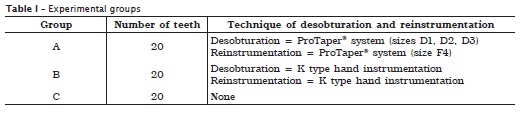
In group A, the desobturation of the root canal was executed with ProTaper® reinstrumentation system coupled with a contra-angle handpiece (Dabi Atlante, Ribeirão Preto, São Paulo, Brazil) and motor driven (Endo Pro Torque®), at a constant speed of 300 rpm and torque of 2 N/cm² 6.
The enlargement movements with constant rotation were performed in the following sequence: size D1, D2 and D3 instruments onto the cervical, medium and apical thirds, respectively. During the reinstrumentation, the size File 4 (F4) instrument of ProTaper® Finishing system was used at the working length.
In group B, root canal desobturation was executed with K type hand instruments. The procedure was completed when the size 30 K file reached the working length and no material remnants were seen. Next, the canals were re-instrumented with sizes 35 and 40 endodontic instruments through sharpening movements, at the working length.
Both in groups A and B, at every instrument change, root canal was irrigated with 2 ml of 1% sodium hypochlorite. Eucalyptol (Iodontosul – Industrial Odontológica do Sul Ltda., Porto Alegre, Brazil) was used as solvent agent during the desobturation procedures. During all procedures, both hand and rotary instruments were used at most five times and then discarded 24.
All study steps were executed by a single operator who was Specialist in Endodontics.
To assess the condition of the root canal walls regarding to the presence or absence of filling material remnants, a digital radiographic shot was executed after the desobturation and reinstrumentation procedures at two incidences: mesiodistal and labial-lingual, enabling the assessment of the labial and proximal views of each sample.
The specimens were placed into a radiographic platform and the Charge-Coupled Device (CCD) sensor (Cygnus Ray - Progeny Dental, Buffalo Grove, Illinois USA) was positioned parallely to the specimens (labial and mesial surface), at vertical position. The cylinder of TimeX device (70 kV, 7 mA, Gnatus, Riberão Preto, SP, Brazil) was positioned in order to enable that the central ray perpendicularly reached the CCD sensor, with focal distance of 28 cm and 1.5 second of exposure.
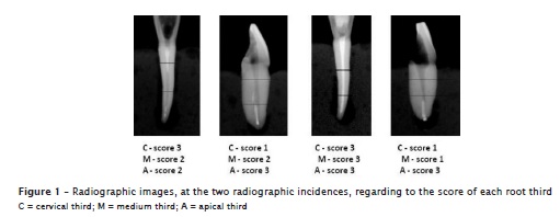
The radiographic images obtained were divided into three thirds (cervical, medium and apical) and assessed by three examiners, specialists in Endodontics, which were previously trained and calibrated.
The examiners scored the images regarding to the amount of remaining material, as follows (figure 1):
– Score 1 = lack of filling material;
– Score 2 = presence of up to 50% of filling material;
– Score 3 = presence of more than 50% of filling material.
The results were submitted to statistical analysis through Mann-Whitney and Friedman non-parametric tests followed by the test of multiple comparisons with level of significance of 5%.
Results
By applying Kendall coefficient of concordance, it was observed a good inter-examiner agreement whose index was of 0.860 for the apical third, 0.825 for the medium third, and 0.883 for cervical third.
Because in group C (control) the teeth were not desobturated and reprepared, the radiographic examination showed 100% of filling material presence. Due to that reason, there was no need to execute the comparative statistical analysis with the two other groups.
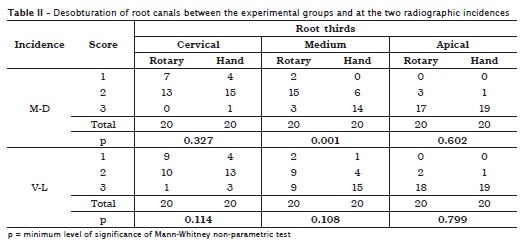
Aiming to compare the desobturation of the canal between the experimental groups, Mann-Whitney non parametric test was used and showed that there were no statistical significant differences between groups, except for the medium third where the rotary system exhibited the lowest scores (table II).
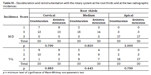
By comparing the rotary group after the desobturation and reinstrumentation procedure, no significant differences were found after Mann-Whitney non-parametric test (table III).
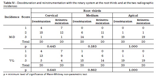
In the comparison of hand group between desobturation and reinstrumentation there were no statistical significant differences after Mann-Whitney non-parametric test (table IV).
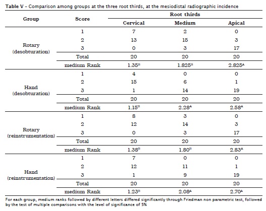
When the comparison among the three root canal thirds was performed through Friedman non-parametric test, followed by the Test of multiple comparisons with level of significance of 5%, it was verified that at the mesiodistal direction in the rotary groups (desobturation and reinstrumentation) at the apical third, the scores attributed were significantly higher than those of the other thirds. In the hand groups (desobturation and reinstrumentation) the scores attributed to the apical thirds were significantly higher than those attributed to the cervical third, but without significant differences from those of the medium third (table V).
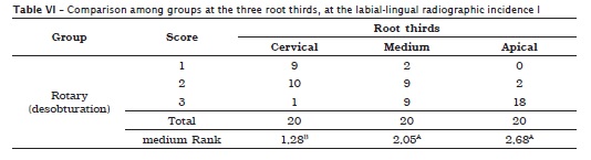
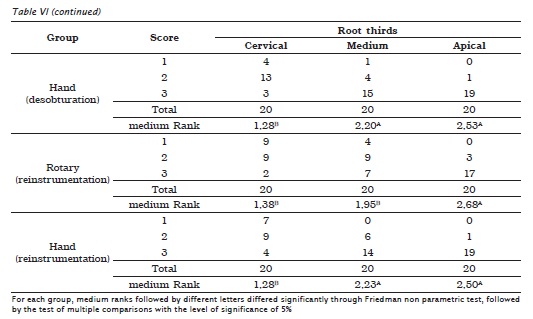
In the comparison between the experimental groups at the three root thirds, at the labial-lingual direction by applying Friedman non parametric test, followed by the test of multiple comparisons with the level of significance of 5%, it was observed that in the rotary group (desobturation) and hand group (desobturation and reinstrumentation) the scores attributed to the apical third were significantly higher than those given to the cervical third, but without statistically significant differences from those of the medium third. For the rotary group (reinstrumentation), the scores attributed to the apical third were significantly higher than those of apical and medium thirds (table VI).
Discussion
The removal of a preexisting filling material within root canals is of fundamental importance to obtain success in endodontic retreatment 22. For this reason, there is a constant searching for new techniques that make easy the removal of this filling material. The rotary instruments are a reality today in Endodontics and its use during endodontic retreatment has been searched by diverse researches 5,19,29.
The ProTaper® system has been already studied by several authors 12-14,23, who evidenced its good performance in the desobturation of the root canals. Notwithstanding, likely other rotary systems, this system was not capable of removing completely the filling material. Other studies 11,22 also observed this same research.
Concerning to the efficacy of the techniques tested, the results did not show statistic differences in the amount of remaining material after the desobturation and reinstrumentation. These findings are in agreement with other studies previously conducted by Schirmeister et al. 24, Gergi and Sabbagh 12 and Barletta et al. 3.
A better cleaning of the root canal walls was obtained with the rotary technique than with the hand technique, only at the medium third, at the mesiodistal direction. This probably occurred because of the design of the ProTaper® instruments, which exhibited a greater taper than standardized hand files, enabling a greater contact of the instrument with the root canal walls during its action.
Marques da Silva et al. 18 did not find statistically significant difference regarding the amount of remaining filling material after retreatment when ProTaper® retreatment system, D-Race® system and MTwo® retreatment instruments were compared, even when additional instruments had been employed for reinstrumentation.
In relation to the analysis of the removal of the filling material among the root thirds, it could be observed that the apical third was the region that showed the greatest amount of remaining filling material, regardless of the operative technique used. Other studies 22,25,27 also verified this same situation. Only in the study conducted by Garcia Júnior et al. 11 the apical third exhibited the smallest amount of remaining filling material, followed by medium and cervical third.
Based on the results of this study, further studies are necessary because an efficient and safe technique has not been found for the total removal of the filling material.
Conclusion
According to the results obtained, it can be concluded that:
• Hand and rotary techniques were not capable of removing completely the filling material inside root canals. The rotary technique was most efficient only at the medium third, at mesiodistal direction, without statistically significant difference between the two techniques at the other thirds and directions;
• Concerning to the cleaning of the root canal walls after desobturation and reinstrumentation, there were not statistically significant differences, regardless of the technique used;
• The apical third showed the greatest amount of filling material after the desobturation and reinstrumentation of the root canals in both techniques tested.
References
1. Baratto-Filho F, Ferreira EL, Fariniuk LF. Efficiency of the 0.04 taper ProFile during the re-treatment of gutta-percha-filled root canals. Int Endod J. 2002;35(8):651-4. [ Links ]
2. Barletta FB, Lagranha SB. Analisis comparativo in vitro de diferentes tecnicas de desobturación de conductos radiculares. Endodoncia. 2002;20(6):189-96. [ Links ]
3. Barletta FB, Sousa Reis M, Wagner M, Borges JC, Dall'Agnol C. Computed tomography assessment of three techniques for removal of filling material. Aust Endod J. 2008;34(3):101-5. [ Links ]
4. Barrieshi-Nusair KM. Gutta-percha retreatment: effectiveness of nickel-titanium rotary instruments versus stainless steel hand files. J Endod. 2002;28(6):454-6. [ Links ]
5. Betti LV, Bramante CM. Quantec SC rotary instruments versus hand files for gutta-percha removal in root canal retreatment. Int Endod J. 2001;34(7):514-9. [ Links ]
6. Carvalho Maciel AC, Zaccaro Scelza MF. Efficacy of automated versus hand instrumentation during root canal retreatment: an ex vivo study. Int Endod J. 2006;39(10):779-84. [ Links ]
7. Ersev H, Yilmaz B, Dinçol ME, Dağlaroğlu R. The efficacy of ProTaper Universal rotary retreatment instrumentation to remove single gutta-percha cones cemented with several endodontic sealers. Int Endod J. 2012;45(8):756-62. [ Links ]
8. Farge P, Nahas P, Bonin P. In vitro study of a Nd:YAP laser in endodontic retreatment. J Endod. 1998;24(5):359-63. [ Links ]
9. Fariniuk LF, Westphalen VP, Silva Neto UX, Carneiro E, Baratto-Filho F, Fidel SR et al. Efficacy of five rotary systems versus manual instrumentation during endodontic retreatment. Braz Dent J. 2011;22(4):294-8. [ Links ]
10. Ferreira JJ, Rhodes JS, Ford TR. The efficacy of gutta-percha removal using ProFiles. Int Endod J. 2001;34(4):267-74. [ Links ]
11. Garcia Júnior JS, Silva Neto UX, Carneiro E, Westphalen VPD, Fariniuk LF, Fidel RAS et al. Avaliação radiográfica da eficiência de diferentes instrumentos rotatórios no retratamento endodôntico. RSBO. 2008;5(2):41-9. [ Links ]
12. Gergi R, Sabbagh C. Effectiveness of two nickel-titanium rotary instruments and a hand file for removing gutta-percha in severely curved root canals during retreatment: an ex vivo study. Int Endod J. 2007;40(7):532-7. [ Links ]
13. Huang X, Ling J, Wei X, Gu L. Quantitative evaluation of debris extruded apically by using ProTaper Universal Tulsa rotary system in endodontic retreatment. J Endod. 2007;33(9):1102-5. [ Links ]
14. Hülsmann M, Bluhm V. Efficacy, cleaning ability and safety of different rotary NiTi instruments in root canal retreatment. Int Endod J. 2004;37(7):468-76. [ Links ]
15. Imura N, Pinheiro ET, Gomes BP, Zaia AA, Ferraz CC, Souza Filho FJ. The outcome of endodontic treatment: a retrospective study of 2000 cases performed by a specialist. J Endod. 2007;33(11):1278-82. [ Links ]
16. Imura N, Kato AS, Hata GI, Uemura M, Toda T, Weine F. A comparison of the relative efficacies of four hand and rotary instrumentation techniques during endodontic retreatment. Int Endod J. 2000;33(4):361-6. [ Links ]
17. Khalilak Z, Vatanpour M, Dadresanfar B, Moshkelgosha P, Nourbakhsh H. In vitro comparison of gutta-percha removal with H-File and ProTaper with or without chloroform. Iran Endod J. 2013;8(1):6-9. [ Links ]
18. Marques da Silva B, Baratto-Filho F, Leonardi DP, Henrique Borges A, Volpato L, Barletta FB. Effectiveness of ProTaper, D-RaCe, and Mtwo retreatment files with and without supplementary instruments in the removal of root canal filling material. Int Endod J. 2012;45(10):927-32. [ Links ]
19. Masiero AV, Barletta FB. Effectiveness of different techniques for removing gutta-percha during retreatment. Int Endod J. 2005;38(1):2-7. [ Links ]
20. Ng YL, Mann V, Gulabivala K. Outcome of secondary root canal treatment: a systematic review of the literature. Int Endod J. 2008;41(12):1026-46. [ Links ]
21. Paik S, Sechrist C, Torabinejad M. Levels of evidence for the outcome of endodontic retreatment. J Endod. 2004;30(11):745-50. [ Links ]
22. Queiróz MLP, Oliveira EPM, Melo TAF, Mautone EP, Colpo A. Analysis of two different endodontic desobturation techniques through clearing teeth technique. RSBO. 2012;9(1):44-9. [ Links ]
23. Saad AY, Al-Hadlaq SM, Al-Katheeri NH. Efficacy of two rotary NiTi instruments in the removal of gutta-percha during root canal retreatment. J Endod. 2007;33(1):38-41. [ Links ]
24. Schirmeister JF, Wrbas KT, Schneider FH, Altenburger MJ, Hellwig E. Effectiveness of a hand file and three nickel-titanium rotary instruments for removing gutta-percha in curved root canals during retreatment. Oral Surg Oral Med Oral Pathol Oral Radiol Endod. 2006;101(4):542-7. [ Links ]
25. Schwerz L, Fontana CE, Bueno CES, Arruda RAA, Pelegrine RA, Abe FC et al. Comparison of the effectiveness of the protaper system versus hand instrumentation in endodontic retreatment: a scanning electron microscopy study. RSBO. 2012;9(4):368-74. [ Links ]
26. Sjogren U, Hagglund B, Sundqvist G, Wing K. Factors affecting the long-term results of endodontic treatment. J Endod. 1990;16(10):498-504. [ Links ]
27. Somma F, Cammarota G, Plotino G, Grande NM, Pameijer CH. The effectiveness of manual and mechanical instrumentation for the retreatment of three different root canal filling materials. J Endod. 2008;34(4):466-9. [ Links ]
28. Swartz DB, Skidmore AE, Griffin Jr. JA. Twenty years of endodontic success and failure. J Endod. 1983;9(5):198-202. [ Links ]
29. Zmener O, Pameijer CH, Banegas G. Retreatment efficacy of hand versus automated instrumentation in oval-shaped root canals: an ex vivo study. Int Endod J. 2006;39(7):521-6. [ Links ]
 Corresponding author:
Érica Pozo Mautone
Rua Baronesa do Gravataí, n. 594 – Menino Deus
CEP 90160-070 – Porto Alegre – RS – Brasilsil
E-mail: epozo@terra.com.br
Received for publication: February 21, 2013
Accepted for publication: April 22, 2013
