


Serviços Personalizados
Artigo
 pdf em Inglês
 Artigo em XML
 Referências do artigo
 Enviar este artigo por email
Links relacionados
Compartilhar

 Permalink
RSBO (Online)
versão On-line ISSN 1984-5685
RSBO (Online) vol.10 no.3 Joinville Jul./Set. 2013
CASE REPORT ARTICLE
Surgical treatment of transmigration of mandibular canine
Ana Maria Estivalete MarchionattiI; Vinícius Felipe WandscherI; Felipe Wehner FloresI; Jorge Abel FloresII
IDepartment of Restorative Dentistry, Federal University of Santa Maria – Santa Maria – RS – Brazil.
IIDepartment of Stomatology, Federal University of Santa Maria – Santa Maria – RS – Brazil.
ABSTRACT
Introduction: The occurrence of canine impaction is not a rare phenomenon, but transmigration of the tooth across the midline is a less common event. The finding is normally asymptomatic and the etiologic factors involved in the transmigration process are still unclear.
Objective: To present a clinical case of surgical treatment of a transmigrated mandibular canine.
Case report: A 17-year-old male patient presented to the dental clinic to remove an unerupted canine. Clinical and radiological examinations led to the diagnosis of a transmigrated canine in the mental region. Surgical removal was the treatment of choice and further radiographs were needed to complement the clinical exam and to determine the location of the tooth precisely for the surgical procedure. Postoperative period was uneventful. Canine transmigration is a rare finding and symptoms are usually absent.
Conclusion: An early detection is important to plan the treatment and mainly to avoid future complications.
Keywords: tooth, impacted; surgery, oral; diagnosis.
Introduction
The occurrence of impacted canines is not uncommon in Odontology, and cases of mandibular unerupted canines are less frequent than maxillary ones 9. Recurrently, intraosseous teeth move to a local distant from its origin, but they usually remain in the same side of the arch 6. A rarer phenomenon is the migration of an impacted canine across the midline without pathological influences. This event is known as the term "transmigration" 9 and happens almost exclusively in the mandible 2. It is difficult to determine its incidence, but it ranges approximately from 0.33% 5 to 0.48% 2. Although usually asymptomatic 2, the situation represents functional, aesthetics, orthodontics and surgical problems 13.
Although etiology of this anomaly is not completely defined, many factors have been suggested to explain the condition. Premature loss of deciduous canine, interferences that delay canine eruption and heredity can be causes of transmigration 13. Surgical removal, orthodontic alignment and transplantation are some of the available treatment options.
It is proposed that to be considered transmigrated, canines should cross the midline at least half of its length 8. However, it has been suggested that the tendency to migrate across the midline is more important than the distance traveled. Moreover, the distance coursed depends on the stage of transmigration when the condition is diagnosed 9.
This article aims to report a case of surgical treatment of a transmigrated mandibular canine.
Case report
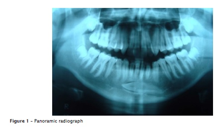
A male patient, 17 years old was referred by an orthodontist in order to remove a left canine impacted in the mandible. The patient reported that the tooth was asymptomatic. During intraoral physical examination, it was detected absence of tooth #33. Panoramic radiograph revealed that the mentioned tooth was transmigrated horizontally below the root apices of anterior teeth and both first premolars (figure 1).
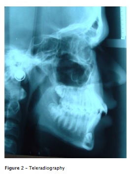
The treatment consisted of surgical removal of the transmigrated canine. For improved treatment planning, a teleradiography was requested to analyze the precise location of the dental element (figure 2).
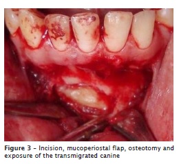
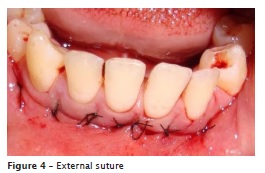
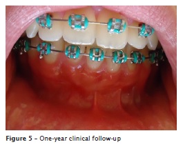
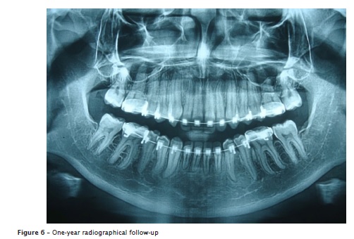
After the bilateral anesthesia of mental nerves (2% mepivacaine with 1:10,0000 epinephrine), a straight mucoperiosteal incision was made on the buccal vestibule below anterior teeth and an osteotomy was performed with round burs (figure 3). The canine was extracted in one piece using curved elevators, then the pericoronal flap was curetted and the area was irrigated with saline solution. Simple interrupted stitches were made internally with absorbable suture 4-0 and, externally, with nylon 4-0 (figure 4). Sutures were removed after 10 days and the postoperative period was uneventful. Follow-up was made during one year after surgery (figures 5 and 6).
Discussion
Canine transmigration is an unusual movement of the tooth that happens almost exclusively in the mandible, although few cases have been reported in the maxillary arch 2.
The finding is generally unilateral 8 and the left side is mostly involved than the right side 5. Females are more affected than males in a ratio of 1.6:1 12. In the present study, the patient was male and the left canine was unilaterally affected.
Transmigration of mandibular canines has not a definite etiology. Some authors have attempted to explain the possible origin of the phenomenon by genetics, trauma, premature loss or retention of the deciduous canine, the long path of canine germ, excessively long crown and agenesis of lateral incisors that may deviate the guide of eruption 2,9. Evidences suggest that the bud usually develops in its normal position and then migrates to an abnormal location 9. Regional disturbances in the dental follicle may cause defective osteoclastic function and form an abnormal eruption pathway 10. According to Stafne and Giblisico 15, the migration of unerupted teeth is possible due to rich blood circulation and active alveolar bone formation in the stage of development of the tooth apex. In the case presented in this report, the cause of transmigration is unclear, since the patient did not relate early loss or retention of deciduous teeth nor trauma.
Clinically, the presence of the primary canine and volume in the symphyseal region may suggest an impacted or transmigrated canine 2. The only sign suggesting the canine transmigration in the case presented in this article was absence of the permanent canine. Radiographs are important auxiliary examinations to determine the diagnosis 1, considering that most cases are asymptomatic. There are cases that periapical radiographs fail to detect the canine due to its position 5 and occlusal, panoramic or teleradiographies are more suitable to confirm the clinical diagnosis 4,7. Early detection is important for a correct planning and avoiding complications, such as the resorption of the roots of adjacent teeth and pathological lesions 7.
Treatment options proposed for transmigrated canines are surgical exposure with orthodontic alignment, transplantation and extraction 2,3,6,7,14. Regular follow-up is an alternative when there are no symptoms and anatomical structures could be damaged during interventions 7. Inthese patients, periodical radiographs must be taken in order to avoid future complications 5,6. Orthodontic alignment was excluded since the unfavorable position of the canine would make it impossible to bring it to its normal place. Also transplantation was not possible because there was not enough space in the arch and the apex was closed, with reduced probability of revascularization 11. Surgical removal was the treatment of choice, in agreement with various studies 4,7,12,13.
When surgical treatment is proposed, care must be taken regarding innervation. Besides performing a nerve block on the migrated side, local anesthesia must also be performed on the side that the tooth belongs, as the canine maintains nerve supply from the original side 6,7,9.
Conclusion
Transmigration of mandibular canine is a rare phenomenon and usually asymptomatic. Early detection by means of clinical and radiographic exam has primary importance for improved planning and selection of the most adequate treatment.
References
1. Abuabara A, Cruz GV, Nóbrega MJ. Casual disclosure of an enlargement of the sella túrcica during orthodontic treatment planning. RSBO. 2010 Oct-Dec;7(4):499-501. [ Links ]
2. Aktan AM, Kara S, Akgünlü F, Malkoç S. The incidence of canine transmigration and tooth impaction in a Turkish subpopulation. Eur J Orthod. 2010 Oct;32(5):575-81. [ Links ]
3. Al-Waheidi AMH. Transmigration of unerupted mandibular canines: a literature review and a report of five cases. Quintessence Int. 1996 Jan;27(1):27-31. [ Links ]
4. Alaejos-Algarra C, Berini-Aytes L, Gay-Escoda C. Transmigration of mandibular canines: report of six cases and review of the literature. Quintessence Int. 1998 Jun;29(6):3995-8. [ Links ]
5. Buyukkurt MC, Aras MH, Caglaroglu M, Gungormus M. Transmigrant mandibular canines. J Oral Maxillofac Surg. 2007 Oct;65(10):2025-9. [ Links ]
6. Camilleri S, Scerri E. Transmigration of mandibular canines – a review if the literature and a report of five cases. Angle Orthod. 2003 Dec;73(6):752-62.
7. González-Sánchez MA, Berini-Aytés L, Gay-Escoda C. Transmigrant impacted mandibular canines: a retrospective study of 15 cases. J Am Dent Assoc. 2007 Nov;138(11):1450-5. [ Links ]
8. Javid B. Transmigration of impacted mandibular cuspids. Int J Oral Surg. 1985 Dec;14(6):547-9. [ Links ]
9. Joshi MR. Transmigrant mandibular canines: a record of 28 cases and a retrospective review of the literature. Angle Orthod. 2001 Feb;71(1):12-22. [ Links ]
10. Marks SC Jr, Schroeder HE. Tooth eruption: theories and facts. Anat Rec. 1996 Jun;245(2):374-93. [ Links ]
11. Pacini NM, Nery DTF, Carvalho DR, Junior NL, Miranda AF, Macedo SB. Dental autotransplant: case report. RSBO. 2012 Jan-Mar:9(1):108-13. [ Links ]
12. Peck S. On the phenomenon of intraosseous migration of nonerupting teeth. Am J Orthod Dentofacial Orthop. 1998 May;113(5):515-7. [ Links ]
13. Pippi R, Kaitsas R. Mandibular canine transmigration: aethio-pathogenetic aspects and six new reported cases. Oral Surgery. 2008 Jun;1(2):78-83. [ Links ]
14. Rebellato J, Schabel B. Treatment of a patient with an impacted transmigrant mandibular canine and a palatally impacted maxillary canine. Angle Orthod. 2003 Jun;73(3):328-36. [ Links ]
15. Stafne EC, Gibilisco JA. Oral roentgenographic diagnosis. Philadelphia: WB Saunders; 1975. [ Links ]
 Corresponding author:
Ana Maria Estivalete Marchionatti
Rua Floriano Peixoto, n. 1.184
CEP 97015-372 – Santa Maria – RS – Brasil
E-mail: anamarchionatti@hotmail.com
Received for publication: September 10, 2012
Accepted for publication: April 16, 2013
