


Serviços Personalizados
Artigo
 pdf em Inglês
 Artigo em XML
 Referências do artigo
 Enviar este artigo por email
Links relacionados
Compartilhar

 Permalink
RSBO (Online)
versão On-line ISSN 1984-5685
RSBO (Online) vol.10 no.4 Joinville Out./Dez. 2013
ORIGINAL RESEARCH ARTICLE
Analysis of remaining tissue after using LA AXXESS® drill in flaring of cervical mesial canals of mandibular molars
Elias Pandonor Motcy de Oliveira I; Alexandre Azevedo Salles I; Vânia Regina Camargo Fontanella I; Tiago André Fontoura de Melo II; Amanda Erthal II; Tâmara Bacci II
I Department of Dentistry, ULBRA Dental School – Canoas – RS – Brazil
II Department of Dentistry, São Leopoldo Mandic / SOBRACURSOS – Porto Alegre – RS – Brazil
ABSTRACT
Introduction and Objective: The objective of this study was to analyze, in vitro, the remaining tissue at the risk zone in the mesial canals of the mandibular molars after cervical flaring using LA AXXESS® drill. Material and methods: Thirty molars were randomly divided into two experimental groups. Twenty teeth received cervical flaring using LA AXXESS® drill (group A), and ten were kept as control group (group B) without undergoing any kind of flaring. The teeth had their mesial roots cross-sectioned at a 3.5 mm standardized height apical to the cementum junction. Specimens were scanned and analyzed by a professional previously trained and calibrated. In order to measure the distances of the remaining tissue in relation to the furca, a Photoshop® program ruler version 7.0 was used. Results and Conclusion: It was possible, through statistical analysis using Student's t-test for paired and independent samples with a 5% significance level, to verify that there was no difference regarding the remaining tissue at the risk zone between the group that had cervical flaring and the control group.
Keywords: endodontics; dental instruments; root canal flaring; risk zone.
Introduction
A priori, the enlargement of the root canal cervical third, comprising the area where the highest dentin apposition occurs resulting in its narrower portion, is a caution to be taken in order to obtain good chemical-mechanical flaring.
The concept of cervical anticurvature wear, introduced by Abou-Rass et al. 1 in 1980, claims the elimination of interferences regarding the middle and coronal thirds of the root canal. Due to the anatomical complexity of the tooth morphology, the cervical flaring, when done, enables the endodontic instrument to have a more rectilinear access to the apical third of the root canal, thus reducing the possibility of accidents while handling chemical-mechanical flaring, such as: ledge formation, apical transport, perforations and endodontic instrument fracture 17.
In addition, some precautions must be taken during cervical flaring, especially in the lower molar group as the region next to the distal walls of the mesial roots is not very thick, there might be at greater risk of accidents.
The cervical wear and anticurvature technique are performed using rotary instruments, and the Gates Glidden burs are the most commonly used in the past decades. However, disadvantages, such as the possibility of fracture and pronounced wear at the risk zone, may result in root perforation 9,10. Thus, due to technological advances and in order to decrease the risk of such accidents, countless studies have been carried out with different drills and instruments.
Coutinho-Filho et al. 5 compared the amount of remaining cervical dentine at 3 mm below mesial roots bifurcation of lower molars, using orifice shapers and size #2, #3 and #4 Gates Glidden drills. The authors have not found any statistical differences between the two systems, although the Gates Glidden drills resulted in a greater wearing area.
However, Skeldton-Macedo et al. 15 compared different ways of preparing the entrance of the mesial-buccal canals of the maxillary molar: Gates Glidden, Largo, Peeso drills and endodontic instruments associated with Gates Glidden. The results have shown that Largo and Peeso drills resulted in greater weariness when compared with the other groups.
In that same year Garala et al. 8, carried out a study where they analyzed the minimum width of the dentinal remnants of the distal wall of the mesial canals of the mandibular molar, after cervical flaring, using nickel-titanium Profile® and Hero 642® rotary instruments. Although the authors pointed out that the width of the root canal wall before the use of instruments can be considered as a determining factor for the results after flaring, they reached the conclusion that there was no significant statistical difference between the two systems tested regarding how much the width of the remaining tissue was affected.
Despite of the countless studies approaching the different techniques and instruments, whether manual, rotary, or both, used in cervical flaring, the LA AXXESS® drills have recently been launched in the dental market with the objective of removing the interferences at the root canal cervical third, consequently reducing the degree of curvature and offering greater security during the endodontic treatment 13.
Thus, the objective of this study was to analyze, in vitro, the remaining tissue of the lower molar mesial canals when submitted to cervical flaring using a LA AXXESS® drill.
Material and methods
The present study was approved by the Ethical Committee in Human and Animal Research of Ulbra under protocol number #2009-427H. The mesial roots of mandibular molars were used with 18 and 22 mm long.
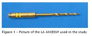
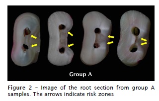
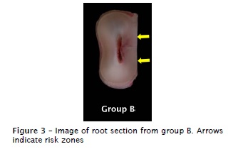
The teeth were randomly distributed into two experimental groups. Group A, comprised 20 teeth, where cervical flaring was carried out using a LA AXXESS® drill (SymbroEndo, Glendora, USA) size 20 taper 0.6 (figure 1). Group B, was composed of 10 teeth, as control for tissue wear, where cervical flaring was not performed, and an anatomical mean was obtained in relation to the security zone.
In group A, the LA AXXESS® drill was attached to a counter-angle handpiece (Dabi Atlante, Ribeirão Preto, São Paulo, Brazil) and introduced at low speed into the interior of the mesial canals at cervical level, under irrigation with sodium 1% hypochlorite solution. A single operator applied light apical pressure towards the anticurvature by means of intermittent movements of introduction and traction at the mesial-buccal and mesial-lingual canals for 5 seconds, measured by a digital chronometer Oregon SI210.
After finishing the cervical flaring the teeth were measured from the enamel-dentine junction to the pulp; a mean was obtained from these measurements defining the height of 3.5 mm to execute the root sectioning. This cut was done by means of a diamond disk attached to a straight handpiece according to the indication previously marked on each root surface.
After the cuts, the root discs obtained were fixed on a wax slide used to enable the image digitalization.
Initially, all root sections were digitalized using an Epson Perfection V500 (Epson, California, USA) scanner with a slide adapter at 300 dpis and 8 bits mode. The scanned images in different modes – anatomical (figure 2) and surgical (figure 3), were randomly codified and distributed into a temporary file and given to a calibrated radiology teacher, expert in this type of analysis. The images were sent to the Photoshop® program, version 7.0, where, by means of the ruler tool, canal measurements up to the thinnest possible measurement at the furca in each section were obtained.
After the measurements were obtained, they were submitted to statistical analysis using the Student's t-test for independent samples at 5% significance level.
Results
Through the results obtained in the Student's t-test for independent samples at 5% significance level, it was possible to verify that there was no significant difference for the values found between the experimental groups with respect to cervical flaring (table I).
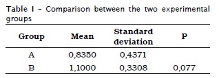
It was verified that the average amount of dental structure at the risk zone, was above the minimum reported as security measure in the literature searched (> 0.2 mm) (table II).

Discussion
Although great advances are available through the development of new rotary instruments for the cervical flaring of root canals, some risks of damage at the dental wall risk zones still remain.
With the emergence of a new rotary instrument in the dental market – the LA AXXESS® drill – for the flaring of the root canal and because of the lack of studies regarding its use, especially in flat and curved roots, as well as the risk of perforations and excessive wear, to carry out this study was considered as important.
The choice for the mandibular molars and their mesial roots was due to their flatness feature; they were also used in other studies such as Busquim and Santos 4 and Spazzin et al. 16.
Several methodologies were used to evaluate the thickness of the root canal walls, such as radiographic method 12, CT scan 2 and diaphanisation 18. For the purpose of this study, transversal cuts along the axis of the root were adopted and then the analysis of the sectioned surfaces were performed, similar to that conducted by Ferreira et al. 7 and Santiago et al. 14
Although there are studies, such as Berutti and Fedon 3 and Ferreira et al. (2002) 7, who measured the thickness of the dentin and cementum at a 1.5 mm distance below the bifurcation, in this present study, root sections were obtained at 3.5 mm apically to the enamel-cementum junction and perpendicular along the root axis. Such measurement is similar to the study of Coutinho-Filho et al. 5, which standardized it at 3 mm. According to Fariniuk et al. 6 and Wu et al. 19, the risk zone for root perforation is localized at this distance.
The results obtained from the present study have shown that the use of LA AXXESS® drills – according to the manufacturer's recommendations – proved to be safe, differently from the result found by the study of Santiago et al. 14, where no significant statistical difference was found between the dentinal wear of the risk zone of lower molar mesial roots when Gates Glidden and LA AXXESS® had been used.
In the study carried out be Spazzin et al. 16 the cervical flarings were executed using both Gates Glidden and LA AXXESS® drills, the presence of root alterations at apical level were also found.
However, in the study of Pecora et al. 13, where the influence of different cervical reamers was evaluated when determining the anatomic apical diameter, the authors could reach the conclusion that the LA AXXESS® drills allowed for more accuracy than other instruments among which, the Gates Glidden drills.
At the same time, the minimum amount of remaining tissue, after the flaring of the mechanical-chemical interventions in this region, must be pointed out. Lim and Stock 11 showed that in the presence of cementum, dentine thickness of 0.2 to 0.3 would be clinically sufficient to resist the possibility of perforation or root fracture during canal filling.
Although the average thickness recorded in this experiment was 0.965 mm for MB canal and 0.705 mm for ML canal, above the minimum previously described in the literature, the anatomical variables should not be underestimated, especially in relation to the concavity present along the root, the presence of secondary dentin, the different levels and positions of the curvature area, as well as the flatness in the proximo-proximal direction.
It must be still pointed out that, although the flarings have been performed by a single operator one must not dismiss the pressure variable exerted on the drills, justified by the different cervical anatomies as well as the oscillation in the air pressure of the dental compressor.
Although it was not the objective of this study because of the lack of research study using these drills, it must be highlighted that due to their resistance and taper 13, no fracture or perforation case was reported during the experimental period, different from that reported by Ferreira et al. 7 in the use of Gates Glidden drills.
Having in mind that the pertinent literature lacks further studies in this area, new investigations must be carried out in order to ratify the results found in the present study.
Conclusion
According to the results found, it can be observed that:
– There was no significant statistical differences between the remaining tissue after the use, or not, of the LA AXXESS® drill;
– The average thickness of the remaining tooth wall in relation to the risk zone was above (MB = 0.965 mm / ML = 0.705 mm) the minimum reported as safe measure in the literature consulted (> 0.2 mm).
References
1. Abou-Rass M, Frank AL, Glick DH. The anticurvature filing method to prepare the curved root canal. J Am Dent Assoc. 1980;101(5):792-4. [ Links ]
2. Bergmans L, Van Cleynenbreugel J, Beullens M, Wevers M, Van Meerbeek B, Lambrechts P. Progressive versus constant tapered shaft design using NiTi rotary instruments. Int Endod J. 2003;36(4):288-95.
3. Berutti E, Fedon G. Thickness of cementum/dentin in mesial roots of mandibular first molars. J Endod. 1992;18(11):545-8.
4. Busquim SS, Santos M. Cervical shaping in curved root canals: comparison of the efficiency of two endodontic instruments. Pesqui Odontol Bras. 2002;16(4):327-31.
5. Coutinho-Filho T, De Deus G, Pinto TG, Gurgel-Filho ED, Maniglia-Ferreira C. A computer evaluation of the dentin remaining after cervical preparation in curved canals: gates-glidden drills vs. orifice shaper. Bras J Oral Sci. 2002;1(3):116-20.
6 . Fariniuk LF, Sassone LM, Baratto-Filho S, Fidel RAS. Avaliação da zona de perigo após a modelagem do canal radicular utilizando Profile série 29 .04 e .06 em raízes mesiais de molares inferiores. JBE. 2004;5(18):222-6.
7. Ferreira EL, Fariniuk LF, Ambrosio AR, Gabardo MCL. Avaliação da técnica da força balanceada na zona de perigo de molares inferiores. Rev Fac Odontol Bauru. 2002;10(4):239-44.
8. Garala M, Kuttler S, Hardigan P, Steiner-Carmi R, Dorn S. A comparison of the minimum canal wall thickness remaining following preparation using two nickel-titanium rotary systems. Int Endod J. 2003;36(9):636-42.
9. Kessler JR, Peters DD, Lorton L. Comparison of the relative risk of molar root perforations using various endodontic instrumentation techniques. J Endod. 1983;9(10):439-47.
10. Lopes HP, Elias CN, Costa Filho AS. Estudo das superfícies da fratura das brocas Gates Glidden. Rev Paul Odontol. 1992;14(6):34-6.
11. Lim SS, Stock CJ. The risk of perforation in the curved canal: anticurvature filing compared with the stepback technique. Int Endod J. 1987;20(1):33-9.
12. Palo RM, Paradella TC, Valera MC, Araújo MAM. Avaliação do desgaste das paredes internas de canais radiculares de molares. Rev Assoc Paul Cir Dent. 2006;60(5):375-8.
13. Pecora JD, Capelli A, Guerisoli DM, Spano JC, Estrela C. Influence of cervical preflaring on apical file size determination. Int Endod J. 2005;38(7):430-5.
14. Santiago CN, Camões ICG, Gomes CC, Freitas LF, Souza AT, Sambati S. Avaliação "in vitro" da espessura dentinária da zona de risco de molares inferiores, após o uso de Gates Glidden e La Axxess. Rev Odontol UNICID. 2010;22(1):6-11.
15. Skelton-Macedo MC, Cardoso RJA, Bombana AC. Avaliação do desgaste do terço cervical dos canais mésio-vestibulares de molares superiores submetidos a diferentes procedimentos de preparo de suas entradas. JBE. 2003;4(15):317-23.
16. Spazzin WO, Spazzin AO, Cecchin D, Mesquita MF, Magro ML, Barbizam JVB. Efeitos do preparo cervical com brocas Gates-Glidden e LA Axxess no desvio apical após preparo biomecânico de canais radiculares. RFO. 2008;13(1):39-42.
17. Torabinejad M. Passive step-back technique. Oral Surg Oral Med Oral Pathol. 1994;77(4):398-401.
18. Wasti F, Shearer AC, Wilson NH. Root canal systems of the mandibular and maxillary first permanent molar teeth of south Asian Pakistanis. Int Endod J. 2001;34(4):263-6.
19. Wu MK, van der Sluis LW, Wesselink PR. The risk of furcal perforation in mandibular molars using Gates-Glidden drills with anticurvature pressure. Oral Surg Oral Med Oral Pathol Oral Radiol Endod. 2005;99(3):378-82.
 Corresponding author:
Elias Pandonor Motcy de Oliveira
Rua Gonçalves Dias, n. 606, apto. 1.003 – Menino Deus
CEP 90130-060 – Porto Alegre – RS – Brasil
E-mail: eliaspmo@uol.com.br
Received for publication: January 25, 2013
Accepted for publication: February 22, 2013
