


Serviços Personalizados
Artigo
 pdf em Inglês
 Artigo em XML
 Referências do artigo
 Enviar este artigo por email
Links relacionados
Compartilhar

 Permalink
RSBO (Online)
versão On-line ISSN 1984-5685
RSBO (Online) vol.10 no.4 Joinville Out./Dez. 2013
ORIGINAL RESEARCH ARTICLE
Effect of substrate and adhesive system type on composite resin restorations
Clarissa Cabral Trinta I; José Ferreira Costa I; Rosana Costa Casanovas de Carvalho I; Sandra Augusta de Moura Leite I; Daniele Meira Conde I; Adriana Silva de Carvalho II
I Department of Dentistry I, Federal University of Maranhão – São Luís – MA – Brazil
II Department of Dentistry, São Leopoldo Mandic University – Campinas – SP – Brazil
ABSTRACT
Introduction: Although it is possible to obtain a reliable bond between enamel and composite resin, the ideal bonding method of composite resin to dentin still needs to be developed. Variables such as the type of adhesive system used and type of dentin substrate can influence on the adhesion. Objective: The purpose of this study was to compare the shear bond strength of composite resin restorations to human and bovine dentin using three adhesive systems. Material and methods: Fifteen human third molars sectioned into two halves and 30 bovine incisors were cut into blocks (4x4mm), embedded in acrylic resin and ground flat to expose the dentin. The specimens were randomly divided into six groups (n = 10): group 1 – human dentin using Scotchbond Multi-Purpose; group 2 – human dentin using Adper Single Bond 2; group 3 – human dentin using Adper Prompt L-Pop; group 4 – bovine dentin using Scotchbond Multi-Purpose; group 5 – bovine dentin using Adper Single Bond 2; group 6 – bovine dentin using Adper Prompt L-Pop. After composite resin restoration procedure, the specimens were stored into distilled water for 24h at 37ºC and then submitted to the shear test using a universal testing machine. The failure patterns were examined microscopically and classified as adhesive, cohesive in resin, cohesive in dentin or both, and mixed. The ANOVA (two-way) and Tukey's post hoc were used. Chi-square test for independence was used for analysis of failure mode. The significance level was set at 5%. Results: A significant difference in shear bond strength was observed among adhesive systems (p = 0.031), with higher values for one-bottle adhesive (8.87±2.72) and lower for self-etching (6.38±3.15), and between the two types of substrate (p = 0.018), with higher values for human dentin. However, there was no significant difference for the adhesive system/substrate interaction (p = 0.11). Adhesive failure was the predominant failure mode for all adhesive systems and for the two substrates. Conclusion: Shear bond strength was different between human and bovine substrates and for the adhesive system used.
Keywords: dental adhesives; dentin; shear strength.
Introduction
Although it is possible to obtain a reliable bond between enamel and composite resin, the ideal bonding method of composite resin to dentin still needs to be developed. There is consensus that the difficulty in obtaining a reliable bond in composite resin restorations ending in dentin is related to the complexity of this substrate, which is heterogeneous, hydrophilic and physiologically dynamic, a fact interfering with the effectiveness of adhesive systems 7,14,18. Despite phosphoric acid has been intensely used to etch the dental substrates for bonding, self-etching adhesives are considered as alternative methods to prepare the tooth for restorative procedures 15,25. Self-etching adhesives systems have been shown to be effective, thus facilitating their use in clinical practice 11,20 because they do not require separated phosphoric acid etching, water-rinsing or superficial moist controlling steps 4,11,25.
Bovine teeth have been extensively used as a substrate in laboratory tests since they permit a larger number of repetitions per experimental group due to their easy acquisition and acceptance in in vitro tests 6,9,23,26. However, some investigators prefer human teeth because they are more reliable and provide results that better reflect reality 8,19.
Laboratory tests do not exactly reproduce in vivo conditions, but they are an important tool for analysis since the effective behavior of a material in vitro will probably result in a satisfactory clinical performance 6,25. Several tests are used to measure the bond strength of dental materials, including shear strength tests 6 and tensile and microtensile tests 4. The shear strength test consists of the application of a force that tends to displace one part of a body over the other, that is, the bond is ruptured by a force applied parallel to the bonding interface. This test has mainly been used to measure the bond strength of composite resin to dentin because of its simplicity and easy preparation of test specimens 4,11,25.
The objective of the present study was to investigate the effect of human and bovine substrate and different adhesive systems on the shear strength of composite resin restorations. The null hypothesis formulated was that there are no significant differences between the substrates and between the adhesive systems studied, within the parameters investigated.
Material and methods
This study was approved by the Ethics Committee of São Leopoldo Mandic Dental School and Center of Post-Graduation (protocol No. 06/233).
Fifteen intact human third molars sectioned into two-halves and 30 bovine incisors were used. The teeth were stored into 0.1% thymol until the time of the construction of specimens. The teeth were cut into blocks (4 x 4 mm) with double-sided diamond disks (KG Sorensen, Barueri, SP, Brazil) under constant water cooling. The fragments were included into a matrix of PVC with self-curing acrylic resin (JET, Clássico Artigos Odontológicos, São Paulo, SP, Brazil).
After 24h, all the test specimens were removed from the matrixes and grounded with a polishing machine (Aropol 2V, Arotec, Cotia, SP, Brazil) and sandpaper (400, 600, and 1200-grit; 3M, Sumaré, SP, Brazil) under water cooling to obtain a flat dentin surface measuring 4 mm in diameter for the bonding procedures. This procedure was also performed to reproduce the smear layer. The size of the dentin blocks was confirmed with a digital caliper (Mitutoyo, Suzano, SP, Brazil).
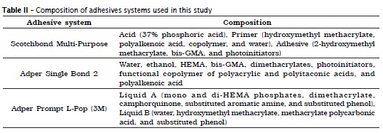
The test specimens were randomly divided into six groups (n = 10) according to adhesive system and dental substrate. The distribution of groups and the adhesive compositions are shown in tables I and II, respectively.
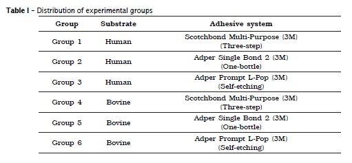
Adhesive tape with a central circular hole measuring 3 mm in diameter was fixed to the dentin surface to delimit the area to be covered with the adhesive. The adhesive systems were applied according to manufacturer's specifications as described below.
Groups 1 and 4: Scotchbond Multi Purpose Adhesive System (3M-ESPE, St Paul, MN, USA).
Dentin etching was performed with 37% phosphoric acid (FGM, São Paulo, SP, Brazil) for 15 s, followed by washing with an air/water spray for 15s and drying with gentle air jet for 5s. The primer was applied and the specimen was again dried with an air jet for 5s. The adhesive was then applied and light-cured for 10s.
Groups 2 and 5: Adper Single Bond 2 Adhesive System (3M-ESPE, St Paul, MN, USA).
Dentin surface was etched with 37% phosphoric acid for 15s followed by washing with water for 20s and removal of water excess with a cotton ball to leave the dentin moist but not wet. Two layers of the adhesive system were applied consecutively for 15s followed by gentle drying for 5s and light-cured for 10s.
Groups 3 and 6: Adper Prompt L-Pop Adhesive System (3M-ESPE, St Paul, MN, USA).
Two applications of the self-etching adhesive system were performed. First, the adhesive was rubbed onto the dentin surface for 15s, followed by gentle drying with an air jet to maintain the surface shiny. In the second application, the adhesive was rubbed onto the surface for 3s, followed by gentle drying with an air jet and light-cured for 10s.
A circular Teflon matrix measuring 20.5 mm in outer diameter and containing a hole (3 mm in diameter and 5 mm in depth) in the center was placed together with the test specimen in a metal device. A composite resin cylinder (Z-100, shade A3; 3M ESPE, St Paul, MN, USA) was fabricated by filling the hole with the composite at three increments. The composite was light-cured on the occlusal surface using a light intensity of 1.000 mW/cm2 (BlueStar, Microdont, São Paulo, SP, Brazil) for 20s per increment.
The specimens were stored for 24h in a closed translucent container with humid gauze at 37°C. The specimens were submitted to the shear strength test using a universal testing machine (Emic, DL2000, São José dos Pinhais, PR, Brazil). A crosshead speed of 0.5 mm/min and a load cell with a maximum capacity of 200 Kgf (ISO/TS 11405) were used. Shear bond strength was calculated using the formula: Rc = F/A, where Rc is the shear strength (MPa), F is the force applied (N), and A is the bonding area (mm2).
The failure mode was examined under an EK3ST binocular stereomicroscope (CQA Nacional, Americana, São Paulo, Brazil) at x35 magnification. Failure patterns were classified as adhesive, cohesive in resin, cohesive in dentin or both, and mixed (adhesive + cohesive in resin or adhesive + cohesive in dentin).
The results were analyzed using SPSS for Windows 10.0 software (1999). Analysis of variance (two-way ANOVA) was used to evaluate shear strength according to adhesive system and substrate, showing an interaction (p<0.05). Therefore, means were compared by Tukey test. For analysis of failure mode, the chi-square test (χ2) for independence was used to evaluate the association of the substrate and adhesive system types with failure mode. The level of significance for rejection of the null hypothesis was set at 5% for all tests.
Results
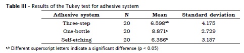
A significant difference in shear bond strength was observed between adhesive systems (p = 0.031) and between human and bovine dentin (p = 0.018). However, the adhesive system/substrate interaction showed no significant difference (p = 0.11).
Analysis by the Tukey test showed a significant difference in shear bond strength between the one-bottle adhesive system and the self-etching adhesive (p<0.05), with the observation of a higher shear strength for the former (table III).
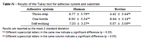
Similar shear strength values on human and bovine substrates were only obtained for the one-bottle adhesive system (p > 0.05). Significant differences in shear bond strength between the two substrates were observed for the three-step and self-etching adhesive systems (table IV).
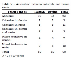
No significant association (p > 0.05) was observed between failure mode and type of substrate (table V).
Discussion
Adhesion to dental substrate is based on the process of hybridization that involves the infiltration of monomers through diffusion and subsequent polymerization of the resinous material in the pores formed by acid etching, with consequent micromechanical imbrications between the dental substrate and resin 8.
One-bottle etch-and-rinse adhesive systems use previous phosphoric acid etching followed by the combined application of primer and adhesive, which provides satisfactory bond strength due to the simultaneous penetration of these components, forming a resistant hybrid layer, showed in some studies 14,24. This present study showed similar results, with significantly higher shear strength for the one-bottle etch-and-rinse adhesive system compared to the self-etching adhesive (p < 0.05). In contrast, promising results of the self-etching adhesive system have been reported, relating to the smaller number of steps required, which reduces the risk of incorrect applications 11,15,18. Similar values of bond strength to dentin for one-bottle etch-and-rinse and self-etching adhesive systems have been also obtained. However, the authors reported lower values for the self-etching systems, probably due to the incomplete infiltration of acid monomers and subsequent partial dissolution of the smear layer, suggesting inconsistent performance in terms of bond quality 7.
Three-step etch-and-rinse adhesive system was similar (p > 0.05) to the one-bottle etch-and-rinse and self-etching adhesive system, in agreement with some studies 6,26. Discrepant results have been reported by some authors 4,14 who observed a significant difference between the three-step Scotchbond Multi-Purpose system and the one-bottle and self-etching adhesives.
Comparison of bovine and human substrates showed a significant difference in shear strength values for the three-step and self-etching adhesive systems (p < 0.05). Significant difference between human and bovine dentin for a three-step adhesive system also was related in literature 2,17,21. Similar values for bovine and human dentin were only obtained with the one-bottle etch-and-rinse system (p > 0.05). This similarity between substrates may be explained by the greater homogeneity of bond strength values for adhesives that contain water, which perform better on drier substrates. This is clinically reflected in the fact that these systems are less susceptible to the lack of surface moisture of dentin 12. However, similar shear bond strength in human and bovine dentin has been reported 3 using a three-step system. These differences between studies might be due to particularities in the methods and materials used.
Several studies have been carried out using teeth from different animals, such as bovines 1,9,10, swine 1,10,13, equines 16 and buffalo 22. Among these, bovine teeth have been most commonly used, due to easy acquisition and to the fact of having several morphological aspects similar to human teeth 5. However, some authors 8,20,21 suggested to reconsider the use of bovine teeth as a substitute of human teeth in bond strength tests since, despite similarities, some differences exist between these substrates. The main difference is that the dentinal tubules of bovine teeth present a larger diameter close to enamel and a smaller diameter close to the pulp unlike to human teeth 8. Some investigations 19,23, found no significant difference between human and bovine dentin. These disagree with the results of this present study, which showed a significant difference (p < 0.05) in shear strength between human and bovine dentin, with the observation of higher shear strength for the former.
In relation to the pattern failure, the three-step and self-etching adhesive systems mainly resulted in adhesive failures. In contrast, a similar frequency of cohesive failure in resin and mixed failure (cohesive in resin) was observed for the one-step etch-and-rinse adhesive system. This finding may be explained by the higher shear bond strength observed in this group. No significant association was observed between the failure patterns observed and substrate type or adhesive system.
Even with the limitations of an in vitro test, the present results enable us to conclude that shear strength differed between human and bovine substrates as well as adhesive system used. The bovine dentin should be used with caution as a substitute of human dentin in shear tests, especially when self-etching adhesive systems are employed.
Further studies are needed to investigate possible substrates that could replace human teeth in shear strength tests to determine the bond strength of different materials. In order to complement the present study, such investigations should not only compare shear bond strength and failure patterns, but also other aspects in the view of the diversity and complexity of the bonding mechanism and the advances in adhesive systems.
References
1. Abuabara A, Santos AJS, Aguiar FHB, Lovadino JR. Evaluation of microleakage in human, bovine and swine enamels. Braz Oral Res. 2004 Oct/Dec;18(4):312-6. [ Links ]
2. Anido-Anido A, Amore R, Lewgoy HR, Anauate-Netto C. Comparative study of bond strength to human and bovine dentine at three different depths. Braz Dent Sci. 2012 Apr/Jun;15(2):56-62.
3. Barkemeier WW, Erickson RL. Shear bond strength of composite to enamel and dentin using Scotchbond Multi-Purpose. Am J Dent. 1994 Jun;7(3):175-9.
4. Bouillaguet S, Gysi P, Wataha JC, Ciucchi B, Cattani M, Godin CH et al. Bond strength of composite to dentin using conventional, one-step, and self-etching adhesive systems. J Dent. 2001 Jan;29(1):55-61.
5. Camargo MA, Marques MM, de Cara AA. Morphological analysis of human and bovine dentine by scanning electron microscope investigation. Arch Oral Biol. 2008 Feb;53(2):105-8.
6 . Cardoso PEC, Braga RR, Carrilho MRO. Evaluation of micro-tensile, shear and tensile tests determining the bond strength of three adhesive systems. 6Dent Mat. 1998 Nov;14(6):394-8.
7. Cavalcante LMA, Erhardt MCG, Bedran-de-Castro AKB, Pimenta LAF, Ambrosano GMB. Influence of different tests used to measure the bond strength to dentin of two adhesive systems. Am J Dent. 2006 Feb;19(1):37-40.
8. Dutra Correa M, Anauante Neto C, Arana Chavez VE. Density and diameter of dentinal tubules in etched and non-etched bovine dentine examined by scanning electron microscopy. Archs Oral Biol. 2007 Sep;52(9):850-5.
9. Fais LMG, Marcelo CC, da Silva RHBT, Guaglianoni DG, Pinelli LAP. Human teeth versus bovine teeth: cutting effectiveness of diamond burs. Braz J Oral Sci. 2010 Jan-Mar;9(1):39-42.
10. Fonseca RB, Haiter Neto F, Fernandes Neto AJ, Barbosa GAS, Soares CJ. Radiodensity of enamel and dentin of human, bovine and swine teeth. Arch Oral Biol. 2004 Nov;49(11):919-22.
11. Issa MH, Silkas N, Watts DC. Strength of a "no-bottle" adhesive systems bonded to enamel and dentine. Dent Mat. 2000 Dec;27(10):484-7.
12. Lopes MB, Sinhoreti MAC, Correr Sobrinho L, Consani S. Comparative study of the dental substrate used in shear bond strength tests. Pesq Odontol Bras. 2003;17(2):171-5.
13. Lopes FM, Markarian RA, Sendyk CL, Duartz CP, Arana-Chavez VE. Swine teeth as potential substitutes for in vitro studies in tooth adhesion: A SEM observation. Arch Oral Biol. 2006 Jul;51(7):548-51.
14. Miranda C, Prates LHM, Vieira R, Calvo MCM. Shear bond strength of different adhesive systems to primary dentin and enamel. J Ped Dent. 2006;31(1):35-40.
15. Mortazavi V, Fathi M, Ataei E, Khodaeian N, Askari N. Shear bond strengths and morphological evaluation of filled and unfilled adhesive interfaces to enamel and dentine. Int J Dent. 2012.
16. Muylle S, Simoens P, Lauwers H. The dentinal structure of equine incisors: a light and scanning electron-microscopic study. Cells Tissues Organs. 2000;167(4):273-84.
17. Nam KY, Kim JB, Jang BC, Kwon TY, Kim KH. Effects of dentin bonding agents on bonding durability of a flowable composite to dentin. Dent Mat J. 2007 Mar;26(2):224-31.
18. Pashley EL, Tao L, Matthews WG, Pashley DH. Bond strengths to superficial, intermediate and deep dentin in vivo with four dentin bonding systems. Dent Mat. 1993 Jan;9(1):19-22.
19. Reis AF, Giannini M, Kavanguchi A, Soares CJ, Line SRP. Comparison of microtensile bond strength to enamel and dentin of human, bovine, and porcine teeth. J Adhes Dent. 2004;6(2):117-21.
20. Retief DH, Mandras RS, Russell CM, Denys FR. Extracted human versus bovine teeth in laboratory studies. Am J Dent. 1990 Dec;3(6):253-8.
21. Rüttermann S, Braun A, Janda R. Shear bond strength and fracture analysis of human vs. bovine teeth. PLoS ONE 2013;8(3):e59181.
22. Santana LNS, Luz MS, Carneiro NCM, Dias AM, Guerra MCS, Lima RR. Ultrastructure of buffalo tooth enamel: a possible replacement for human teeth in laboratory research. Braz J Oral Sci. 2011 Sep;10(3):163-6.
23. Schilke R, Lisson JA, Baub O, Schuckar M, Geurtsen W. Comparison of the number and diameter of dentinal tubules in human and bovine dentine by scanning electron microscopic investigation. Archs Oral Biol. 2000 May;45(5):355-61.
24. Sensi LG, Lopes GC, Monteiro Junior S, Baratieri LN, Vieira LCC. Dentin bond strength of self-etching primers/adhesives. Oper Dent. 2005 Jan-Feb;30(1):63-8.
25. Van Meerbeek B, Munck JD, Yoshida Y, Inoue S, Vargas M, Vijay P et al. Adhesion to enamel and dentin: current status and future challenges. Oper Dent. 2003;28(3):215-35.
26. Wilder Junior AD, Swift Junior EJ, May Junior KN, Waddell SL. Bond strengths of conventional and simplified bonding systems. Am J Dent. 1998 Jun;11(3):114-7.
 Corresponding author:
Clarissa Cabral Trinta
Rua Alto Parnaíba, lote 08, quadra 03, apto. 803, Ed. Varandas de São Marcos – Ponta do Farol
CEP 65075-830 – São Luís – MA – Brasil
E-mail: clarissa.trinta@yahoo.com.br
Received for publication: April 8, 2013
Accepted for publication: June 26, 2013
