


Serviços Personalizados
Artigo
 pdf em Inglês
 Artigo em XML
 Referências do artigo
 Enviar este artigo por email
Links relacionados
Compartilhar

 Permalink
RSBO (Online)
versão On-line ISSN 1984-5685
RSBO (Online) vol.11 no.1 Joinville Jan./Mar. 2014
ORIGINAL RESEARCH ARTICLE
Effect of Er:YAG laser and diamond drill on hybrid layer morphology obtained with self-etch adhesive – analysis by SEM and confocal laser scanning microscopy (CLSM)
Aline de Oliveira GonçalvesI; Cinthia Sawamura KuboI; Osmir Batista Oliveira JúniorI; Edson Alves de CamposI; Marcelo Ferrarezi AndradeI
I Department of Restorative Dentistry, São Paulo State University, School of Dentistry of Araraquara – Araraquara – SP – Brazil
ABSTRACT
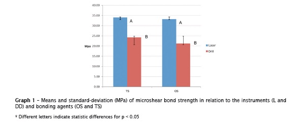
Introduction and Objective: This study aimed to evaluate the effect of Er:YAG (L) and diamond drills (DD) on: 1) the microshear bond strength (MPa); 2) the adhesive interface of two-step (TS) – Adper Scotchbond Multipurpose and one-step (OS) adhesives – Adper EasyOne, both from 3M ESPE. Material and methods: According to the preparation condition and adhesives, the samples were divided into four groups: DD_TS (control); DD_OS; L_TS and L_OS. 60 bovine incisors were randomly divided into experimental and groups: 40 for microshear bond strength (n = 10) and 20 for the adhesive interface morphology [6 to measure the thickness of the hybrid layer (HL) and length of tags (t) by CLSM (n = 3); 12 to the adhesive interface morphology by SEM (n = 3) and 2 to illustrate the effect of the instruments on dentine by SEM (n = 1)]. To conduct the microshear bond strength test, four cylinders (0.7 mm in diameter and 1 mm in height with area of adhesion of 0.38 mm) were constructed with resin composite (Filtek Z350 XT – 3M ESPE) on each dentin surface treated by either L or DD and after adhesives application. Microshear bond strength was performed in universal testing machine (EMIC 2000) with load cell of 500 kgf and a crosshead speed of 0.5 mm / min. Adhesive interface was characterized by thickness of hybrid layer (HL) and length of tags (t) in nm, with the aid of UTHSCSA ImageTool software. Results: Microshear bond strength values were: L_TS 34.10 ± 19.07, DD_TS 24.26 ± 9.35, L_OS 33.18 ± 12.46, DD_OS 21.24 ± 13.96. Two-way ANOVA resulted in statistically significant differences only for instruments (p = 0.047). Mann-Whitney identified the instruments which determined significant differences for HL thickness and tag length (t). Concerning to the adhesive types, these differences were only observed for (t). Conclusion: It can be concluded that 1) laser Er:YAG results in higher microshear bond strength values regardless of the adhesive system (TS and OS); 2) the tags did not significant affect the microshear bond strength; 3) the adhesive interface was affected by both the instruments for cavity preparation and the type of adhesive system used.
Keywords: dental cavity preparation; confocal microscopy; lasers; scanning electron microscopy; smear layer; dentinal adhesives.
Introduction
The technological advancement of the adhesive restorative materials has allowed the execution of more conservative cavity preparations in which the dentist only removes the carious tissue and obtains the convenience form to access and instrument the cavity 13.
The use of diamond drills at high speed to perform cavity preparations has the following main advantages: fast and precise cutting of the tooth structure; obtainment of defined angles and walls; the presence of micro-grooves on the cavity walls, which can contribute to increase the retention of direct restorations 3. However, its disadvantages comprise to cause: pain and physical/emotional discomfort to the patient; pulp damage because of heating the tooth structure due to friction; cross contamination and diseases because of the spray generated during preparation 4. Also, it jeopardizes the visualization of the cavity during instrumentation.
Because Er:YAG laser is silent and does not need the direct contact with the dental structure, there is not pain and discomfort to patient and it enables the execution of minimally invasive cavity preparations, which mostly are impossible to be performed with diamond drills 13,18. Er:YAG laser action is based on the ejection of fragments of tooth structure (ablation), because of the kinetic mechanical action of intense releasing of energy due to micro-explosions of water molecules irradiated by the laser beam 13.
This can affect the performance of adhesive systems since these have been developed to either remove or incorporate the smear layer resulting from the cavity preparations with drills on their surfaces. Considering that the hybrid layer seems to develop a critical role in adhesive Dentistry 7 and that the tooth substrate type can influence on its formation and on the adhesion mechanism 32, it can be stated that the tooth substrate obtained by several instruments may influence on the final result of the adhesive restoration, leading to the formation of different patterns of hybrid layer.
This study aimed to evaluate the effect of Er:YAG laser and drills on the microshear bonding strength and the bonding interface of one- and two-step bonding agents through SEM and confocal laser scanning microscopy (CLSM).
Material and methods
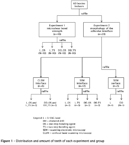
This study was submitted and approved by the Ethical Committee in Research of the São Paulo State University. Sixty bovine incisors without caries were stored into 0.2% thymol solution and kept under refrigeration at temperature of about 7ºC for seven days. The teeth were randomly distributed according to figure 1.
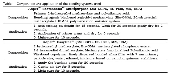
The teeth were prepared with the aid of a diamond drill (DD) no. 3131 (KG Sorensen) coupled to high speed handpiece (Kavo). Er:YAG laser (L) (Twinlight: Fotona, Slovenia) was used. Two-step (TS) bonding system used was Adper Scotchbond SE Multipurpose (3M ESPE, St. Paul, MN, USA), while onestep (OS) bonding system was Adper Easy One (3M ESPE, St. Paul, MN, USA) and both were applied onto the dentin surface of the samples.
Experiment 1 – Microshear bond strength
Sample preparation
The teeth had their roots and distal and mesial surfaces of their crowns removed with the aid of a diamond disc (11-4254, 4"x 0.012" / series 15LC, Diamond Blade, Buehler Ltd., Lake Bluf, IL, USA) and metallographic cutting machine (ISOMET 1000, Buehler, Lake Buff, IL, USA). The labial surface was worn and flattened with the aid of a polishing machine (DP-10 Panambra, Struers, Ballerup, Denmark), to obtain a smooth and flat tooth surface. The fragments were embedded into acrylic resin inside PVC tubes measuring 1.2 cm in height and 2.0 cm in diameter. After the resin setting, the surface was cleaned and again flattened in the polishing machine. Then, the samples were washed with water jet and stored into distilled water for 24 h. After this period, each fragment was submitted to a superficial weariness executed with either (L) or (DD).
Adhesive procedures
The bonding area was delimited onto a double-sided adhesive tape which was guided by four perforation of a tripartite matrix measuring 0.7 mm in diameter. The sequence of application of the bonding systems were performed according to the manufacturers' instructions (table I). All light-curing procedures were carried out with the aid of a LED unit (LED Bluephase, Ivoclar Vivadent, Schann – Liechtenstein), at low intensity (± 800 mW/cm2). Polyethylene tubes were place onto the tripartite matrix with area of 0.38 mm2 (Tygon tubing, R-3603, Saint-Gobain Performance Plastics, Maime Lakes, FL, USA). This set was filled with resin composite (Filtek Z350 XT) and the four cylinders were light-cured for 40 s. The samples were stored into distilled water at 37°C for 24 h. Elapsed that time, the tubes were removed with the aid of a scalpel blade to obtain a resin composite cylinder.
Microshear bond strength testing
Elapsed the storage time period, the samples were individually coupled to a metallic device and fixed to an universal testing machine (EMIC DL 2000, São José dos Pinhais, PR, Brazil). Prior to testing, the device was carefully aligned to allow that the loading was applied as closest as possible to the bonding interface in the base of each cylinder. This loading was applied through a ring made of steel wire (0.2 mm in diameter). The test was performed with a load cell of 500 Kgf and crosshead speed of 0.5 mm/min until the fracture of the specimens. Microshear bond strength was calculated by dividing the maximum force registered during the testing (N) by the bonding area (0.38 mm2) and expressed into MPa. The results obtained were tabulated in an Excel sheet (Excel Microsoft) and analyzed statistically with the aid of SPSS Statistic V 19 software (IBM Corporation, Armonk, NY, USA).
Experiment 2 – Morphology of the bonding interface
Morphology of the bonding interface
Cavity preparations: two bovine incisors had the medium third of the crown prepared measuring 3.0 mm in width, 2.0 mm in height and 2.0 mm in deepness. One tooth was prepared through laser while the other through diamond drills. Next, the teeth were washed and dried with air jet for 1 minute. Aiming to avoid technique artifacts, the surfaces analyzed in SEM were replicated with epoxy resin (Epoxide; Buhler). Impression of the surfaces were taken with the aid of an addition silicone (Express XT Penta – 3MESPM) mixed according to the manufacturer's instructions. After the material setting, the impressions were filled with Epoxide, mixed according to the manufacturer's instructions. The set was stored at environment temperature and controlled humidity for 24 hours.
The samples were longitudinally cut with the aid of metallographic cutting machine (ISOMET 1000, Buehler, Lake Buff, IL, USA) and diamond disc (11-4254, 4"x 0.012"/ series 15LC, Diamond Blade, Buehler Ltd., Lake Bluf, IL, USA), under water cooling.
SEM: the repl icas were removed f rom the impressions, identified and submitted to metallization, in which a layer of 24-carat gold powder with thickness varying from 50 to 100 Angstron was deposited by vaporization onto the samples. The bonding interface of each group was analyzed and characterized with the aid of a scanning electronic microscope (Jeol JSM – 6610 LV, Tokyo, Japan). The dentin morphology was described regarding to its structural characteristics.
CLSM: Six incisors were divided into two groups (n = 3), according to the instrument type employed in cavity preparation (laser or diamond drill). At the medium third, two cavity preparations (3.0 x 2.0 x 2.0 mm) were executed. The incisal cavities (IC) were restored with resin composite (Filtek Z350 – 3M ESPE, St. Paul, MN, USA) and one-step (OS) bonding agent (AdperTM Easy One – 3M ESPE, St. Paul, MN, USA). The cervical cavities (CC) were restored with two-step (TS) bonding agent (AdperTM ScotchbondTM Multipurpose – 3M ESPE, St. Paul, MN, USA) and resin composite (AdperTM Z350 XT – 3M ESPE, St. Paul, MN, USA). Elapsed the 24 h storage in distilled water at 36±1oC, the samples were longitudinally cut to expose the bonding adhesive and characterized through diamond disc (11-4254, 4"x 0.012" / series 15LC, Diamond Blade, Buehler Ltd, Lake Bluf, IL, USA) with the aid of a cutting machine (ISOMET 1000 – Buehler, Lake Buff, IL, USA). The bonding agents for analysis in CLSM were pigmented with rhodamine B – C28H31CIN2O3 – (LABSYNTH Produtos para Laboratórios Ltda., Diadema, SP, Brazil) at 0.001 g/ml ratio.
The right surface of the adhesive interface was polished with 1200-grit silicon carbide sandpaper. The samples were ultrasonically cleaned in water for 3 minutes to remove the debris coming from polishing.
Analysis of confocal laser scanning fluorescence microscopy (CLSFM)
The hybrid layer and the configuration and the medium length of tags were assessed in confocal laser microscopy (Leica TCS SMD – Leica Microsystems – Integrated Research Center of the School of Dentistry of the University of São Paulo, Brazil). The thickness of the hybrid layer was quantitatively measured at different sites of the bonding interface. The obtained results were related to the perpendicular distances between the dentin-composite junction. To perform these measurements, UTHSCSA ImageTool software (http://en.bio-soft.net/draw/ImageTool.html) was used. The thickness of hybrid layer was measured at the incisal, cervical and axial walls of the cavities. Five measurements were executed on each wall, totalizing ten measurements for each sample.
The morphology of the bonding interface by SEM: the labial surfaces of 12 bovine incisors were worn in a polishing machine up to obtain their flattening and dentin exposure. Following, either L or DD weariness were executed according to which is established in figure 1. Next, the bonding sequence was applied and a thin layer of resin composite (Filtek Z350) was inserted and light-cured. To exposure the bonding interface, the samples were identified and longitudinally cut with the aid of a diamond disc (11-4254, 4"x 0.012" series 15LC, Diamond Blade, Buehler Ltd., Lake Bluf, IL, USA) and cutting machine (ISOMET 1000 – Buehler, Lake Buff, IL, USA). Additional cuts left the samples measuring 10.0 mm in width and 2.0 mm in height. The interface was polished with the aid of 1200-grit silicon carbide sandpaper. The samples were ultrasonically cleaned for 3 minutes. Next, the surface was submitted to the action of HCl 4M for 0.5 min, washed and exposed to 5% NaOCl for 10 min. Elapsed this time, the samples were washed and dried with the aid of air jet. Impressions were taken from the specimens with the same technique described above for the obtainment of the images of dentin surface. The morphology of the bonding interface (HL and t) was analyzed through scanning electronic microscope (Jeol JSM – 6610 LV, Tokyo, Japan, of the School of Dentistry of Araraquara – Department of Dental Materials and Prosthesis) at x450 magnification. The bonding interface morphology was comparatively described regarding to its structural features.
Results
Experiment 1 – Effect of Er:YAG laser and diamond drill on microshear bond strength of one- and two-step bonding agents
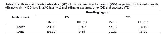
The descriptive statistics of microshear bond strength testing (MPa) is seen in table II.
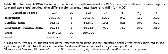
Kolmogorov-Smirnov (p(DD) = 0.20 and p(L) = 0.07) and Levene (p(DD, L) = 0.27 and p(OS and TS) = 0.68) tests confirmed the application of two-way ANOVA. Two-way Anova results are displayed in table III.
These results are presented in graph 1, in which different letters mean statistically significant differences at p < 0.05.
Experiment 2 – Effect of Er:YAG laser and diamond drill on the morphology of the bonding interface of one- and two-step bonding agent – study by CLSFM and SEM
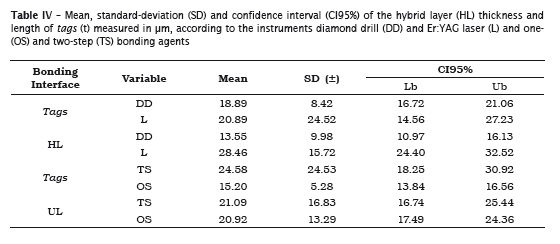
The descriptive statistics for the effect of instruments and bonding agents on the bonding interface (HL) and (t) is seen in table IV.
SEM analysis
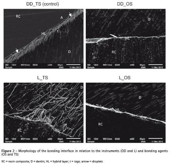
Figure 2 illustrates the effect of (DD) and (L) on the bonding interface (HL) and (t) in the samples.
Different morphologic bonding interfaces were observed. The hybrid layers, although present in all groups/samples, were thin and visually with similar thickness.
The use of one-step bonding agent exhibited a more regular bonding interface, while two-step bonding agent showed thickness variations, deficiencies in the diffusion of the monomers and in the encapsulation of the collagen fibers. The specimens that received the drill action showed the occurrence of many "droplets", which was not observed in the specimens receiving the laser action.
Two-step bonding agent presented a clear formation of tags (t), which could not be observed in the specimens receiving one-step bonding agent.
The group L_TS exhibited longer and irregular tags than those of the other groups.
Analysis of the dentin surface
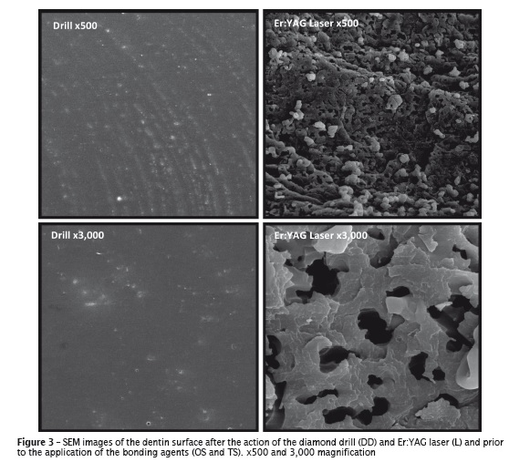
The analysis of the dentin surface by SEM enabled the visualization of an amorphous and uniform smear layer covering the surface of the specimen receiving the action of the diamond drill (figure 3).
The morphology of the cavity preparation executed with Er:YAG laser was very different. It was observed lack of smear layer, dentin with aspect of desquamation, with very irregular margins and superficial waves, and open dentinal tubules in great number. Also, neither the exposure of collagen fibers nor presence of peri- and/or intertubular dentin was observed.
Discussion and Conclusion
It could be observed, similarly to the studies of Antunes et al. 3, Oliveira et al. 30 (2005), Oliveira et al. 29 and Semeraro et al. 34, that the surface treatment with diamond drill (DD) resulted in a regular substrate with some waves and grooves, presence of smear layer over all dentin and the closure of the dentinal tubules (figure 3).
The use of Er:YAG laser (L) resulted in a very regular substrate forming craters, elevations and pores, with rugosities and granulations and with lack of smear layer (figure 3). This same morphological aspect was also described by Delmé et al. 15, Esteves-Oliveira et al. 17, Shirai et al. 36, Nishimoto et al. 27, Shigetani et al. 35, Navarro et al. 26, and Ramos et al. 33.
These differences can be attributed to the mechanism of action of laser, which is characterized by the ablation of the tooth tissues. During its operation, the laser source emits photons inside a water-air jet towards the target tissue. The abrupt heating of the water closer to the surface results in a series of micro-explosions and ejection of hard tissues of the tooth 17,18.20,26,35.
The formation of a morphologically irregular dentin may favor the bonding procedure, since it increases the number of micromechanical retentions. On the other hand, not even the increase of the micromechanical retention results in the increase of the bond strength 5,9,25.
The analysis of the figure 2 enables to observe a clear morphological difference in the hybrid layer formed by the different bonding systems on the surfaces prepared with either the drill or laser. The two-step bonding agent, in both substrates, formed a hybrid layer with similar thickness with the presence of many tags. It was also noted the presence of small lateral rami in these tags, namely micro-tags, indicating that the monomers from the bonding agent could propagate and penetrate deeply in the dentin etched 1. This difference in the performance of one- and two-step bonding agents is explained by the etching of dentin with 37% phosphoric acid, which promoted a greater dentin demineralization, increasing the infiltration of the resin monomers.
The group TS exhibited a hybrid layer less thick and practically without tags. The tendency is this bonding agent form uniform hybrid layers without failures, which is mandatory to prevent nanoleakage. Also, it could be noted in the bonding interface of OS bonding agent the presence of little droplets (figure 2), that is, the separation of a localized zone close to the bonding interface, which resulted from the water absorption coming from the dentin by osmosis due to low molecular weight of HEMA that makes the area fragile and compromises the effectiveness and longevity of the bonding agent 1.
According to Van Meerbeek et al. 39, mild one-step bonding agents dissolve the smear layer without demineralizing the surface in deepness, thus removing the hydroxyapatite from the interface. By preserving the hydroxyapatite, the collagen fibers are protected from external chemical aggression and calcium are available for the chemical bonding to the functional monomer of the bonding agent. The literature demonstrated that two-step agents similar to that used in this present study can produce high bond strength values to dentin 11,36,37. The control group (DD_TS) showed a microshear bond strength mean value of 24.26 MPa, while the group exhibiting the highest mean was L_TS, with mean of 34.10 MPa.
The higher bond strength of group L_TS than that of DD_TS can be explained by the different patterns of smear layer obtained by the instruments and by the greater dentin demineralization promoted by the total etching. However, despite of the favorable mechanical results, the removal of smear layer in the bonding technique of TS bonding agents allowed the exudation of the dentinal fluid 32, which can interfere in the polymerization of the resin monomer and cause dentin hypersensitivity by affecting the hydrodynamic balance of dentin 10. Thus, although group L_TS showed the highest bond strength value (16.10 MPa), its bonding interface presented excessive longer tags that during clinical situation may lead to dentinal hypersensitivity and pulp damage.
Many studies obtained results similar to those of this present study, indicating that generally the bond strength value of one-step bonding agent is statistically similar to that of two-step bonding agents 10,37. Notwithstanding, because of the hydrophilic characteristic of one-step bonding agents, they have a tendency towards the deterioration of the bonding interface since the hydrophilicity of these systems leads to humidity absorption both from the oral environment and the pulp, causing hydrolytic degradation.
This present study suggests the preparation of class V cavity by laser because the results demonstrated that laser produces a substrate with many micro-retentions and lack of smear layer, favoring the penetration of both types of bonding agents and the highest microshear bond strength values were obtained with the use of this instrument. Due to adhesion difficulty, cervical lesions are very employed to evaluate the clinical performance of adhesive systems. Barceleiro et al. 6 and Lizarelli et al. 22 obtained a greater success in laser performance than that of diamond drills through a microleakage study.
In this present study, when two-step bonding agent is used, the preparation with laser favored the bonding process in comparison with those prepared with drills. In the substrate irradiated, the mean hybrid layer was 1.32 μm, while that obtained by drill preparation was 1.03 μm. The same behavior was observed when one-step bonding agent was used, with a greater effect for laser (1.42 μm) than for drill preparation (1.03 μm). It is worth observing that the mean thickness of the hybrid layer obtained through drills was the same for both bonding agents. These data are contrary to those of the studies conducted by Barceleiro et al. 6. These authors by analyzing the surface either irradiated by laser or prepared by drills, obtained a deficient hybrid layer for the former. Barceleiro et al. 7 carried out a study with the same instruments and many adhesive systems and found a hybrid layer less homogenous when the dentin was prepared by drills, which was a result similar to that of this present study.
The literature states that the formation of the hybrid layer on the surface irradiated by Er:YAG laser is more susceptible to the loss of tensile bond strength 17,38, smaller thickness 6,8,16,29,36 and to the increasing of marginal leakage 19,20. The smaller bond strength of the surfaces irradiated by laser comes from the dentin abrasion which promotes the fusion of the collagen fibers, restricting resin diffusion within peritubular dentin 24. Other authors, however, affirmed that laser is a good alternative to conventional drills because it promotes a good marginal sealing 19,26,35 and does not affect the bond strength of adhesive systems 7,10,14.
In the evaluation of the bonding interface by CLSFM, one- and two-step bonding agents showed non-expected behaviors regarding to the hybrid layer formation when submitted to laser, since they displayed similar thickness: 1.32 μm (TS) and 1.42 μm (OS). These results are related to the lack of smear layer and to the dentin morphology after the laser action.
However, this behavior of the hybrid layer and tags of the samples irradiated by laser does not necessarily mean that the bonding to this substrate is more effective than that of the groups treated by drills. The micro-explosions, common features of the mechanism of action of laser could remove and damage the collagen fibers jeopardizing the adhesion. The control group (DD_TS) and group DD_OS showed the same hybrid layer thickness, which was not expected because the mechanism of action of the different bonding agents. Albaladejo et al. 1, Korkmaz et al. 20, Moura et al. 24 and Van Meerbeek et al. 39 already proved that the fact most affecting bonding strength is HL and not the length or number of tags. Additionally, the obtainment of longer tags may indicate the possibility of damaging to both the odontoblastic processes and pulp tissue by the monomers still present within the tags partially or totally unpolymerized.
Because in this present study, groups L_TS and DD_TS exhibited numerous and long tags (means of 28.39 μm and 20.77 μm, respectively) and the laser determined the highest microshear bond strength values, it can be inferred that both mechanically and biologically the use of OS bonding agents is the most recommended 7,29,30. Although the results of this present study showed the higher microshear bond strength of dentin treated by laser than that treated by diamond drills, it was observed a moderate significance (h2 p = 0.10) for clinical practice with a low test power (π = 0.53). These results suggested that the number of specimens for each group (n = 10) could have not be enough to evidence safely the effect of the instruments and bonding systems, despite this sampling number is greater than that of many studies already published: Ramos et al. 33 (n = 4), Mendez et al. 23 (n = 5), Lenzi et al. 21 (n = 5), Carvalho et al. 12 (n = 10), and Ali et al. 2 (n = 10).
References
1. Albaladejo A, Osorio R, Toledano M, Ferrari M. Hybrid layers of etch-and-rinse versus self-etching adhesive systems. Med Oral Patol Oral Cir Bucal. 2010 Jan 1;15(1):e112-8. [ Links ]
2. Ali AA, El Deeb HA, Badran O, Mobarak EH. Bond durability of self-etch adhesive to ethanolbased chlorhexidine pretreated dentin after storage in artificial saliva and under intrapulpal pressure simulation. Oper Dent. 2013 Jul- Aug;38(4):439-46.
3. Antunes LA, Pedro RL, Vieira AS, Maia LC. Effectiveness of high speed instrument and air abrasion on different dental substrates. Braz Oral Res. 2008 Jul-Sep;22(3):235-41.
4. Anusavice KJ, Kincheloe JE. Comparison of pain associated with mechanical and chemomechanical removal of caries. J Dent Res. 1987 Nov;66(11):1680-3.
5. Arrais CA, Giannini M. Morphology and thickness of the diffusion of resin through demineralized or unconditioned dentinal matrix. Pesqui Odontol Bras. 2002 Apr-Jun;16(2):115-20.
6. Barceleiro MO, de Mello JB, de Mello GS, Dias KR, de Miranda MS, Sampaio Filho HR. Hybrid layer thickness and morphology: the influence of cavity preparation with Er:YAG laser. Oper Dent. 2005 May-Jun;30(3):304-10.
7. Barceleiro MO, Dias KR, Sales HX, Silva BC, Barceleiro CG. SEM evaluation of the hybrid layer after cavity preparation with Er:YAG laser. Oper Dent. 2008 May-Jun;33(3):294-304.
8. Bowen RL, Cobb EN, Rapson JE. Adhesive bonding of various materials to hard tooth tissues: improvement in bond strength to dentin. J Dent Res. 1982 Sep;61(9):1070-6.
9. Brudevold F, Buonocore M, Wileman W. A report on a resin composition capable of bonding to human dentin surfaces. J Dent Res. 1956 Dec;35(6):846-51.
10. Cal-Neto JP, de Miranda MS, Dias KR. Comparative SEM evaluation of penetration of adhesive systems in human dentin with a non-rinse conditioner and a self-etching primer. Braz Dent J. 2004;15(1):19-25.
11. Cardoso MV, Coutinho E, Ermis RB, Poitevin A, Van Landuyt K, De Munck et al. Influence of dentin cavity surface finishing on micro-tensile bond strength of adhesives. Dent Mater. 2008 Apr;24(4):492-501.
12. Carvalho RC, de Freitas PM, Otsuki M, de Eduardo CP, Tagami J. Micro-shear bond strength of Er:YAG-laser-treated dentin. Lasers Med Sci. 2008 Apr;23(2):117-24.
13. Cozean C, Arcoria CJ, Pelagalli J, Powell GL. Dentistry for the 21st century? Erbium:YAG laser for teeth. J Am Dent Assoc. 1997 Aug;128(8):1080-7.
14. Dalia MP, Gomes PF, Menezes-Filho RP, Guimarães ALA, Mariz CHV. Ultraconservative dentistry – alternative methods of cavities preparations. RFO. 2009;14(2):168-73.
15. Delmé KI, De Moor RJ. Scanning electron microscopic evaluation of enamel and dentin surfaces after Er:YAG laser preparation and laser conditioning. Photomed Laser Surg. 2007 Oct;25(5):393-401.
16. Ergücü Z, Celik EU, Unlü N, Türkün M, Ozer F. Effect of Er, Cr:YSGG laser on the microtensile bond strength of two different adhesives to the sound and caries-affected dentin. Oper Dent. 2009 Jul-Aug;34(4):460-6.
17. Esteves-Oliveira M, Zezell DM, Apel C, Turbino ML, Aranha AC, Eduardo CP et al. Bond strength of self-etching primer to bur cut, Er, Cr: YSGG and Er:YAG laser dental surfaces. Photomed Laser Surg. 2007 Oct;25(5):373-80.
18. Hibst R, Keller U. Experimental studies of the application of the Er:YAG laser on dental hard substances: I. Measurement of the ablation rate. Lasers Surg Med. 1989;9(4):338-44.
19. Karaarslan ES, Usumez A, Ozturk B, Cebe MA. Effect of cavity preparation techniques and different preheating procedures on microleakage of class V resin restorations. Eur J Dent. 2012 Jan;6(1):87-94.
20. Korkmaz Y, Ozel E, Attar N, Bicer CO, Firatli E. Microleakage and scanning electron microscopy evaluation of all-in-one self-etch adhesives and their respective nanocomposites prepared by erbium:yttrium-aluminum-garnet laser and bur. Lasers Med Sci. 2010 Jul;25(4):493-502.
21. Lenzi TL, Tedesco TK, Soares FZ, Loguercio AD, Rocha RO. Chlorhexidine does not increase immediate bond strength of etch-and-rinse adhesive to caries-affected dentin of primary and permanent teeth. Braz Dent J. 2012;23(4):438-42.
22. Lizarelli RFZ, Silva PCG, Kurachi C, Porto Neto ST, Bagnato VS. Estudo-piloto comparativo da microinfiltração in vitro entre os preparos cavitários classe V, através da ponta diamantada em alta rotação ou laser de Er:Yag seguido ou não de ataque ácido. JBD. 2002;1(1):33-6.
23. Mendez JC, Pabon GE, Hilgenberg SP, Garcia EJ, Arana-Correa B. Effect of water storage on microtensile bond strength of a two-step self-etch adhesive and a two-step etch-and-rinse adhesive Acta Odontol Latinoam. 2012;25(2):176-80.
24. Moura SK, Santos JF, Ballester RY. Morphological characterization of the tooth/adhesive interface. Braz Dent J. 2006;17(3):179-85.
25. Nakabayashi N, Pashley DH. Hibridização dos tecidos dentais duros. São Paulo: Quintessence; 2000.
26. Navarro RS, Gouw-Soares S, Cassoni A, Haypek P, Zezell DM, de Paula Eduardo C. The influence lf erbium:yttrim-aluminum-garnet laser ablation with variable pulse width on morphology and microleakage of composite restorations. Lasers Med Sci. 2010 Nov;25(6):881-9.
27. Nishimoto Y, Otsuki M, Yamauti M, Eguchi T, Sato Y, Foxton RM et al. Effect of pulse duration of Er:YAG laser on dentin ablation. Dent Mater J. 2008 May;27(3):433-9.
28. Oliveira WJ, Pagani C, Rodrigues JR. Comparação da adesividade de dois sistemas adesivos autocondicionantes em esmalte de dentes bovinos. Rev Fac Odontol S J dos Campos. 2001;4(1):43-6.
29. Oliveira SS, Pugach MK, Hilton JF, Watanabe LG, Marshall SJ, Marshall Jr. GW. The influence of the dentin smear layer on adhesion: a self-etching primer vs. a total-etch system. Dent Mater. 2003 Dec;19(8):758-67.
30. Oliveira DC, Manhães LA, Marques MM, Matos AB. Microtensile bond strength analysis of different adhesive systems and dentin prepared with highspeed and Er:YAG laser: a comparative study. Photomed Laser Surg. 2005 Apr;23(2):219-24.
31. Paris S, Bitter K, Renz H, Hopfenmuller W, Meyer-Lueckel H. Validation of two dual fluorescence techniques for confocal microscopic visualization of resin penetration into enamel caries lesions. Microsc Res Tech. 2009 Jul;72(7):489-94.
32. Perdigao J, Swift Jr. EJ, Denehy GE, Wefel JS, Donly KJ. In vitro bond strengths and SEM evaluation of dentin bonding systems to different dentin substrates. J Dental Res. 1994 Jan;73(1):44-55.
33. Ramos TM, Ramos-Oliveira TM, Moretto SG, Freitas PM, Esteves-Oliveira M, de Paula Eduardo C. Microtensile bond strength analysis of adhesive systems to Er:YAG and Er,Cr:YSGG laser-treated dentin. Lasers Med Sci. 2013 Jan 26. [Epub ahead of print].
34. Semeraro S, Mezzanzanica D, Spreafico D, Gagliani M, Re D, Tanaka T et al. Effect of different bur grinding on the bond strength of self-etching adhesives. Oper Dent. 2006 May- Jun;31(3):317-23.
35. Shigetani Y, Tate Y, Okamoto A, Iwaku M, Abu-Bakr N. A study of cavity preparation by Er: YAG laser. Effects on the marginal leakage of composite resin restoration. Dent Mater J. 2002 Sep;21(3):238-49.
36. Shirai K, De Munck J, Yoshida Y, Inoue S, Lambrechts P, Suzuki K et al. Effect of cavity configuration and aging on the bonding effectiveness of six adhesives to dentin. Dent Mater. 2005 Feb;21(2):110-24.
37. Susin AH, Vasconcellos WA, Saad JR, Oliveira Junior OB. Tensile bond strength of self-etching versus total etching adhesive systems under different dentinal substrate conditions. Braz Oral Res. 2007 Jan-Mar;21(1):81-6.
38. Tonami K, Takahashi H, Kato J, Nakano F, Nishimura F, Takagi Y et al. Effects of laser irradiation on tensile strength of bovine dentin, Photomed Laser Surg. 2005 Jun;23(3):278-83.
39. Van Meerbeek B, Inokoshi S, Braem M, Lambrechts P, Vanherle G. Morphological aspects of the resin-dentin interdiffusion zone with different dentin adhesive systems. J Dent Res. 1992 Aug;71(8):1530-40.
 Corresponding author:
Aline de Oliveira Gonçalves Universidade Estadual Paulista (Unesp), Faculdade de Odontologia de Araraquara
Rua Humaitá, n. 1.680, sala 310, 3.º andar
CEP 14801-385 – Araraquara – SP – Brasil
E-mail: alineogoncalves@hotmail.com
Received for publication: June 26, 2013
Accepted for publication:November 11, 2013
