


Serviços Personalizados
Artigo
 pdf em Inglês
 Artigo em XML
 Referências do artigo
 Enviar este artigo por email
Links relacionados
Compartilhar

 Permalink
RSBO (Online)
versão On-line ISSN 1984-5685
RSBO (Online) vol.11 no.1 Joinville Jan./Mar. 2014
ORIGINAL RESEARCH ARTICLE
Adhesion and formation of tags from MTA Fillapex compared with AH Plus® cement
Marina Samara BaechtoldI; Ana Flávia MazaroI; Bruno Monguilott CrozetaI; Denise Piotto LeonardiI; Flávia Sens Fagundes TomazinhoI; Flares Baratto-FilhoI; Gisele Aihara HaragushikuI
I Department of Dentistry, Positivo University – Curitiba – PR – Brazil
ABSTRACT
Introduction and Objective: The aim of this study was two-fold: 1) to evaluate, in vitro, the shear bond strength of two sealers by push-out test and 2) to assess the failures after displacement. Additionally, the formation of tags was observed by SEM. Material and methods: Forty mandibular premolars were selected and the canals were subjected to biomechanical preparation with rotary instruments. These specimens were divided into two groups according to the sealer (n = 20): GI – MTA Fillapex and GII – AH Plus. All roots were filled with sealer only, without gutta-percha. After a period corresponding to three times the setting time of the sealer, the roots were sectioned transversely into slices of 1 mm thickness, to obtain one slice from the cervical third, to be used in the push-out test. Following, two slices of each group were randomly chosen for ultrastructural analysis by scanning electron microscopy (SEM). The data obtained in shear bond strength test were subjected to statistical analysis. Results: AH Plus cement exhibited higher shear bond strength values (1.332±0.75 MPa) than MTA Fillapex (0.071±0.07 MPa), with statistically significant differences. Conclusion: MTA Fillapex has a low bond strength with less formation of tags than AH Plus.
Keywords: endodontic cement; adhesion; scanning electronic microscopy.
Introduction
One of the desirable physical-chemical properties of the endodontic cements is adhesivity to the root canal walls 6. Thus, when meeting this feature, a hermetic filling can be obtained through the sealing of root canal, promoting the apical repair, and avoiding the percolation of fluids to the periapical tissues and consequently preventing endodontic reinfections 3,12.
Currently, the association of the endodontic cement with gutta-percha points is the gold standard in endodontic obturation, mainly because of lack of adhesion of the gutta-percha to the dentinal walls. The flowing property of the endodontic cement should be also taken into consideration, in order to fill the spaces between the gutta-percha and the canal wall, therefore providing a sealing with better quality 4, and enabling the filling of lateral canals and isthmuses 15.
The cements most commonly used today are based on epoxy resin, calcium hydroxide, zinc oxide and eugenol, and glass ionomer. Recently, MTA cement has been also employed and studies have aimed to evaluate the sealing capacity of resin-based cements and the biological repairing of mineral trioxide aggregate which is the new filling material. The following clinical characteristics of MTA-based cement have been reported: higher radiopacity; easy removal in cases of retreatment; excellent f lowing providing the easy filling of depressions and lateral canals; low solubility; releasing of calcium ions, which induces the bone regeneration; high alkalinity, which results in an antibacterial material inducing neoformation of peri-radicular cementum.
Because of its composition, MTA-based cements exhibit an excellent biocompatibility to human tissues, making it an attractive material to both the professionals and researchers. Notwithstanding, little has been known on its adhesivity, which is fundamental for endodontic treatment success.
Most of the endodont ic cement s have demonstrated inadequate biological activity and adhesive capacity 2,7. Consequently, many studies have been constantly conducted to assess their physical, chemical and biological properties, which vary according to the composition of each material.
Therefore, the aim of this study was to evaluate the capacity of adhesion to dentinal walls and the formation of tags of MTA Fillapex compared with AH Plus cement.
Material and methods
This study was submitted and approved by the Ethical Committee in research of the Positivo University under protocol number 088/11.
Forty mandibular human premolars were selected with minimum root length of 11 mm, determined through digital caliper (Starret 799, Athol, USA) and radiographed at buccal-lingual direction. Inclusion criteria comprised: lack of endodontic treatment, bone resorptions and calcifications; and complete formation of root apex. After selection, the teeth were extracted, cleaned with the aid of a periodontal curette and kept into 0.1% thymol solution at temperature of 9ºC. Before the study, the teeth were washed into running water for 24 hours, aiming to eliminate the thymol remnants.
Following, the teeth were cut with the aid of carborundum discs mounted into a straight handpiece at low speed (Kavo do Brasil, Chapecó, Brazil) close to the enamel-cementum junction so that all roots measured 11 mm in length. Then, the specimens were kept into 0.9% saline solution in an incubator at temperature of 9ºC to avoid dehydration.
The working leng th of al l samples was determined at 10 mm. Crown-down technique was used with apical stop of 0.60 mm for all specimens. During all preparation, 2.5% sodium hypochlorite (Asfer Indústria Química Ltda., São Caetano do Sul, Brazil) was used as irrigant solution. Final irrigation was executed with 10 ml of 17% ethylenediaminetetraacetic acid (EDTA) (Farmácia- Escola Universidade Positivo, Curitiba, Brazil), followed by irrigation with 10 ml of distilled water and drying with absorbent paper points (Dentsply- Maillefer, Petrópolis, Brazil).
The specimens were randomly divided into two groups s (n = 20) according with the endodontic cement used: GI – AH Plus (DeTrey Dentsply, Konstanz, Germany), GII – MTA Fillapex (Angelus, Londrina, Brasil). The canals were filled only with endodontic cement, without using gutta-percha points so that gutta-percha/cement interface did not interfere in the shear bond strength test.
The roots were cut at 1 mm slices, with the aid of diamond discs mounted into cutting machine (Isomet 1000 – Buehler, Lake Bluff, USA). A cervical third slice of each specimen was selected to be tested in the universal testing machine (Emic DL2000 – EMIC, São José dos Pinhais, Brazil), at crosshead speed of 0.5 mm/min. A stainless steel device was used to place the samples so that the surface of smaller diameter of the root canal was turned up, aligned with the rod employed to push the cement until the sample displacement. The rods had tips with 1 mm in diameter.
The force (F) required to displace the filling material, in kilonewtons (kN), was transformed into Newton (N), and expressed in megapascal (MPa) by dividing the force value (N) by the adhesion area of the filling material (SL), in mm2. Thus, the formula employed to relate these measures was: s = F / SL.
The calculation of the area (SL) was obtained according to the following formula:
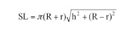
SL = lateral area of the cone trunk; π = 3.14; R = mean radius of the coronal canal, in mm; r = mean radius of the apical canal, in mm; h = height related to the side of the cone trunk, in mm.
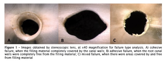
After push-out test, the cuts were assessed with the aid of a stereoscopic lens (ZEISS; Stemi 2000-C, Germany), at x40 magnification, to verify the failure type, which was classified as: 1) adhesive – when the root canals were free of filling material; 2) cohesive – when the filling material completely covered the canal walls; 3) mixed – when there were areas covered by and free from filling material.
Data were submitted to statistical analysis to verify the sample normality and determine the proper statistical test.
Next, two specimens of each group were randomly selected for ultra-structural analysis in scanning electronic microscopy (SEM): one sectioned at the longitudinal direction and other at the cross-sectional direction in order to analyze the tags of cements within the tubules.
The specimens for SEM analysis were kept into 2.5% glutaraldehyde solution, buffered with 0.1 mol/l sodium cacodylate (pH = 7.4) for 12 hours in an incubator at 4ºC. Following, the specimens were submitted to three baths in 0.1 mol/l sodium cacodylate (pH = 7.4) (for 20 min each) and dehydrated in increasing ethanol (Farmácia-Escola Universidade Positivo, Brazil): 25%, 50%, 75%, 95% (for 20 min of immersion into each solution) and 100% for 1 hour.
The specimens were dried in an incubator at 37ºC for 24 hours, placed into a vacuum chamber and covered by gold of about 300 Aº (Bal-Tec SCD 030; Leica Microsystems, Germany). The analysis was performed in scanning electronic microscopy (Jeol JSM-6360LV, JEOL, Milestones, USA).
In the qualitative analysis of the photomicrographies, the formation of cement tags and their aspect were analyzed.
Results
Push-out test
The values obtained by push-out test, in kN were transformed into MPa and submitted to statistical analysis with SPSS software (IBM, Armonk, USA).
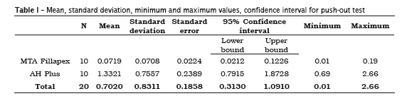
Based on the normality of the samples, one-way ANOVA was chosen (table II).
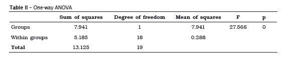
According to one-way ANOVA, there were statistically significant differences (p < 0.05) between AH Plus and MTA Fillapex cements. AH Plus cement exhibited the highest bond strength values (1.332±0.75 MPa) than those of MTA Fillapex cement (0.071±0.07 MPa).
The analysis of the failures observed in stereomicroscopy is seen in graph 1.
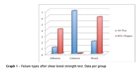
There was the predominance of the cohesive failure for AH Plus and mixed failure for MTA Fillapex. Adhesive failure occurred in both groups, with greater prevalence for MTA Fillapex.
Scanning electronic microscopy (SEM)
SEM analysis showed a greater formation of tags in the teeth filled with AH Plus, while the teeth filled with MTA Fillapex exhibited little or none formation of tags (figure 2).
At higher magnification, it was possible to observe the aspects of each cement: AH Plus was smoother and compact and MTA Fillapex was rougher and sparse.

Discussion
The association of the endodontic cement with gutta-percha points is the gold standard in the filling of root canals. The bonding capacity of the filling material to the dentinal wall is desirable for maintaining the integrity of the cement/dentine interface during displacement forces, as those occurring in the preparation of intraradicular posts, aiming to prevent marginal leakage 8.
In this present study, AH Plus cement exhibited better statistically significant results than those of MTA-based cement. The best adhesion force of epoxy resin-based cements have been studied through the comparison with other endodontic cements 10,11,14.
Prior studies have explained that the highest bond strength values obtained by the epoxy resinbased cements are because the capacity of creating a covalent bonding with an opened epoxy ring to any amine group exposed in collagen, giving longterm dimensional stability and low polymerization tension 5,9,14.
The chemical composition of MTA-based cement could also influence on its bonding capacity 13. A recent study discovered that the rationale behind the low bonding strength of MTA Fillapex is its low bonding capacity to dentinal tubules because of the formation of apatite by MTA, over its own surface, thus creating a similar structure that is different from that of the tag which prevents its leakage 11.
When exposed to scanning electronic microscopy, AH Plus exhibited longer and uniform tags, showing its higher mechanical imbrication and resulting in greater bonding capacity 10, while MTA Fillapex cement displayed little or none formation of tags, confirming the studies of Sagsen et al. 11.
Based on the results of this present studies, it could be observed that the material composition direct ly inf luences on its physical-chemical behavior.
Conclusion
This present study concluded that MTA Fillapex cement has low bond strength and little formation of tags compared with AH Plus cement.
References
1. Assmann E, Scarparo RK, Böttcher DE, Grecca FS. Dentin bond strength of two mineral trioxide aggregate-based and one epoxy resin-based sealers. J Endod. 2012 Feb;38(2):219-21. [ Links ]
2. Bouillaguet S, Shaw L, Barthelemy J, Krejci I, Wataha JC. Long term sealing ability of pulp canal Sealer, AH Plus, GuttaFlow and Epiphany. Int Endod J. 2008 Mar;41(3):219-26.
3. Brosco VH, Bernardineli N, Torres SA, Consolaro A, Bramante CM, Morais IG et al. Bacterial leakage in obturated root canals. Part 2: a comparative histologic and microbiologic analyses. Oral Surg Oral Med Oral Pathol Oral Radiol Endod. 2010 May;109(5):788-94.
4. Cobankara FK, Orucoglu H, Sengun A, Belli S. The quantitative evaluation of apical sealing of four endodontic sealers. J Endod. 2006 Jan;32(1):66-8.
5. Fischer MA, Berzins DW, Bahcall JK. An in vitro comparison of bond strength of various obturation materials to root canal dentine using a push-out test design. J Endod. 2007 Jul;33(7):856-8.
6. Haragushiku GA, Sousa-Neto MD, Silva-Sousa YT, Alfredo E, Silva SC, Silva RG. Adhesion of endodontic sealers to human root dentine submitted to different surface treatments. Photomed Laser Surg. 2010;28(3):405-10.
7. Huang TH, Yang JJ, Li H, Kao CT. The biocompatibility evaluation of epoxy resin-based root canal sealers in vitro. Biomaterials. 2002 Jan;23(1):77-83.
8. Huffman BP, Mai S, Pinna L, Weller RN, Primus CM, Gutmann JL et al. Dislocation resistance of ProRoot Endo Sealer, a calcium silicate-based root canal sealer, from radicular dentine. Int Endod J. 2009 Jan;42(1):34-46.
9. Koh ET, McDonald F, Pitt Ford TR, Torabinejad M. Celular response to mineral trioxide aggregate. J Endod. 1998 Aug;24(8):543-7.
10. Lee KW, Williams MC, Camps JJ, Pashley DH. Adhesion of endodontic sealers to dentin and gutta percha. J Endod. 2002 Oct;28(10):684-8.
11. Sagsen B, Ustün Y, Demirbuga S, Pala K. Pushout bond strength of two new calcium silicate-based endodontic sealers to root canal dentine. Int Endod J. 2011 Dec;44(12):1088-91.
12. Schilder H. Filling root canals in three dimensions. J Endod. 1967 Apr;32(4):281-90.
13. Torabinejad M, Hong CU, McDonald F, Pitt Ford TR. Physical and chemical properties of a new root-end filling material. J Endod. 1995 Jul;21(7):349-53.
14. Vilanova WV, Carvalho-Junior JR, Alfredo E, Sousa-Neto MD, Silva-Sousa YT. Effect of intracanal irrigants on the bond strength of epoxy resin-based and methacrylate resin-based sealers to root canal walls. Int Endod J. 2012 Jan;45(1):42-8.
15. Venturi M, Di Lenarda R, Prati C, Breschi L. An in vitro model to investigate filling of lateral canals. J Endod. 2005 Dec;31(12):877-81.
 Corresponding author:
Gisele Aihara Haragushiku
Rua Professor Pedro Viriato Parigot de Souza, n. 5.300 – Campo Comprido
CEP 81280-330 – Curitiba – PR – Brasil
E-mail: gisele.haragushiku@gmail.com
Received for publication: October 12, 2013
Accepted for publication: November 21, 2013
