


Serviços Personalizados
Artigo
 pdf em Inglês
 Artigo em XML
 Referências do artigo
 Enviar este artigo por email
Links relacionados
Compartilhar

 Permalink
RSBO (Online)
versão On-line ISSN 1984-5685
RSBO (Online) vol.11 no.3 Joinville Jul./Set. 2014
ORIGINAL RESEARCH ARTICLE
Sealing ability of different versions of GuttaFlow2 in comparison to GuttaFlow and AH Plus
Johannes Ebert I; Barbara Holzschuh I; Roland Frankenberger II; Anselm Petschelt I; Matthias Johannes RoggendorfII
I Dental Clinic 1, Operative Dentistry and Periodontology, University of Erlangen-Nuremberg – Erlangen – Germany
II CDepartment of Operative Dentistry and Endodontics, Dental School, Philipps-University of Marburg – Marburg – Germany
ABSTRACT
Introduction and objective: GuttaFlow2 is a further development of the silicone sealer GuttaFlow, exhibiting a stiffer consistency. This is intended to overcome possible problems regarding retention of the apical part of the root canal filling when preparing for a fiber post. GuttaFlow2 is delivered within a capsule, like GuttaFlow, or within an automix syringe. This study compared apical dye leakage of GuttaFlow2 in comparison to GuttaFlow and AH Plus. The null hypothesis tested was that different sealers exhibited similar microleakage. Material and methods: Seventy extracted human lower premolars with fully mature apices were root canal prepared to 45/.04 and divided into seven groups: group 1: AH Plus sealer, group 2: "normal" setting GuttaFlow, group 3: "fast" setting GuttaFlow, group 4: GuttaFlow2 within a capsule, group 5: GuttaFlow2 within an automix syringe, group 6: positive control, group 7: negative control (n = 10 each). Root canals were filled with sealer (except group 7) and a master gutta-percha cone size 40/.04 using the non-compaction technique. A dye penetration test was carried out by centrifugation for 3 min at 30 G within 5 % methylene blue dye. Linear dye penetration was recorded. Statistical evaluation was carried out with IBM SPSS 19.0 (α = 0.05). Results: The positive control was significantly different from all other groups (ANOVA, p < 0.001; Student-Newman-Keuls post-hoc test p < 0.05). When the control groups were disregarded, no significant differences were apparent. Groups 1 to 5 showed low leakage values when compared with results of earlier studies using a similar methodology. Conclusion: All sealers tested exhibited low dye leakage values.
Keywords: AH Plus; dye penetration; GuttaFlow; GuttaFlow2.
Introduction
The objective of root canal filling is to prevent the passage of microorganisms and their byproducts along the root canal 13. Today's state of the art is the combination of a semi-solid material (e.g. gutta-percha) with a root canal sealer 13. The latter has a significant impact on microleakage of root canal fillings 25. The group of silicone sealers exhibited promising results regarding microleakage in different studies besides the wellestablished group of epoxy resins (e.g. AH Plus, DeTrey Dentsply, Konstanz, Germany) 3,4,7,12,23,30,33. This may be due to their slight expansion upon Setting 14.
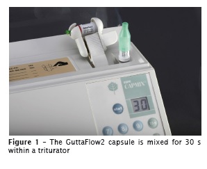
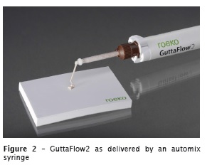
Silicone sealers remain relatively soft after Setting 20, which may cause difficulties when subsequently additional preparation, as for a root canal post, is necessary. This problem may be addressed using a silicone primer and / or special retentive gutta-percha points (Silicone Primer, Roeko Retention Points, both Coltène/Whaledent, Langenau, Germany). Another way to handle this problem is the use of a silicone sealer with an optimized consistency due to variations in inorganic fillers: GuttaFlow2 (Coltène/Whaledent). GuttaFlow2 is delivered in two different ways: a capsule that is to be triturated for 30 s (see figure 1), and an automix syringe (see figure 2) which is well known from other materials like dual-cure composite cements or from the sealer AH Plus Jet (DeTrey Dentsply).The aim of this study is to test microleakage of this newly developed cuttable silicone sealer GuttaFlow2 in comparison to the established sealer materials GuttaFlow, GuttaFlow fast and AH Plus.
The null hypothesis tested was that there is no difference regarding microleakage for different groups.
Material and methods
Seventy straight single-rooted lower premolars with one root canal each and with fully mature apices were selected. Teeth were stored in a 0.5% chloramine-T solution (Merck, Darmstadt, Germany) or water, or were stored in humid conditions (100% humidity) over the whole time of the study. Access cavities were prepared and the lengths of the root canals recorded by passing a size 10 K-file through the apex and subtracting 1 mm. Teeth were randomly divided into five experimental groups and two control groups of ten teeth each.
All root canals were instrumented to size 45/.04 by nickel-titanium instruments (Hyflex, Coltène/ Whaledent, Langenau, Germany). Instrumentation was accompanied by copious irrigation with 3 % NaOCl and 40 % citric acid. A final irrigation with 40 % citric acid followed by 3 % NaOCl and 70 % ethanol was performed (2 mL per root canal for approximately 60 s each) and the root canals were dried with paper points.
For each root canal, a gutta-percha cone size 40/.04 (MTwo Gutta-percha point, VDW, Munich, Germany) was adjusted to fit with tug back at working length. For filling the root canals, a noncompaction technique was applied: the respective sealer was placed with a paper point size 25/.02: group 1: AH Plus sealer, group 2: "normal" setting GuttaFlow, group 3: "fast" setting GuttaFlow, group 4: GuttaFlow2 within a capsule, group 5: GuttaFlow2 within an automix syringe; then the master guttapercha point was placed; additional gutta-percha points size 25/.02 were placed if appropriate, without the use of a spreader; finally, excess gutta-percha was cut off, followed by immediate vertical condensation of the gutta-percha with double-sided hand instruments (HDC 1 and HDC 2; both Deppeler, Rolle, Switzerland). The teeth of the positive control group were only filled with a single gutta-percha cone size 40/.04 without sealer. The teeth of the negative control group were filled similarly to group 5. The floor and the walls of the pulp chamber were cleaned with ethanolmoistened foam pellets until the pulp chamber appeared to be clean as judged by the naked eye. Then a temporary filling with a glass ionomer cement was applied (Fuji IX; GC, Tokyo, Japan) to facilitate the subsequent complete covering of the tooth with nail varnish.
Following the completion of root canal filling and temporary filling, teeth were stored in a wet chamber (37°C / 100% humidity) for one week to allow complete setting of the respective sealer. The roots of the teeth were completely covered with two layers of nail varnish. After drying of the varnish, apices of teeth were cut off (1-2 mm) to expose the root canal fillings of the teeth. Negative control teeth were left completely covered. Then the teeth were placed into test tubes together with 5% methylene blue dye solution (Merck), pipetted to a height of 30 mm. A dye penetration test according apical microleakage was performed using centrifugation for 3 min at 30 G (Varifuge-K, Heraeus Christ, Osterode, Germany; 400 rpm) 25.
Following the dye penetration test, excess of dye was washed off. The teeth were dried and the apical surface gently ground on a fine (250 grit) sand paper to remove superficially adhering dye. Each specimen was then embedded in a resin material and serial sectioned in distances of 1 mm using a Buehler low-speed-saw (Buehler GmbH, Lake Bluff, IL, USA). Transversal cuts were made perpendicular to the long axis under water cooling. Dye penetration was scored using a stereo microscope at x25 magnification. Linear dye penetration was recorded using a simple yes / no decision for presence of dye for each sectioning plane. As the sectioning blade had a thickness of 0.5 mm, and the upper and lower surfaces of each slice could be evaluated, the ingress of dye could be measured near to the next 0.5 mm. The readings were counted until the first sectioning plane without dye. For example, a reading of 3.5 mm of dye penetration results of dye present up to the plane 3 mm from the apex and the first plane without coloration at 3.5 mm from the apex. As the dye that had been adhering on the apical cut-off plane of the root was removed, a reading of 0 mm of dye ingress could be recorded in several specimens.
Data were statistically analyzed with IBM SPSS 19.0 (SPSS, Chicago, IL, USA), using Kolmogorov- Smirnov tests, ANOVA with Student-Newman-Keuls (SNK) post-hoc-tests, Kruskal-Wallis (KW) tests and pairwise Mann-Whitney (MW) tests. The level of significance was set at α = 0.05.
Results
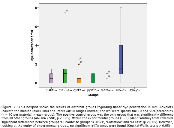
Some of the groups showed no normal distribution (groups 5 and 7: Kolmogorov-Smirnov test, p < 0.05), so additionally to ANOVA with SNK, non-parametric tests were applied as the main statistical tests. According to ANOVA (p = 0.001) and Kruskal-Wallis-test (p < 0.001) significant differences were found regarding the entity of groups, so the test method itself can be regarded as valid. SNK post hoc test indicated that the positive control differed from all other groups (p < 0.05). When positive and negative controls were disregarded, no significant differences could be found (ANOVA: p = 0.150; KW test p = 0.111). However, in some of the pairwise comparisons between groups, significant differences were revealed: group five showed significant lower values than groups 1, 2 and 3 (MW Test p < 0.05) and no significant difference to the negative control (MW Test p = 0.147). Because of the results of the KW test (no significant difference), these results have to be interpreted with care. Results are also shown in figure 3.
Discussion
Arguments towards or against dye penetration within the debate over leakage studies have already discussed in an earlier paper 10. The main reason why we chose apical dye penetration for the present study is that the problem with a large scale of variation within results could not be avoided in any of the published studies, regardless of the applied methodology 1-12,15-19,21,23,25-33. Thus, we chose the method that is the most easy to apply and control and is very cost effective 10,26. Furthermore, the chosen variant of dye penetration test using centrifugation was able to detect significant differences between groups within different earlier studies 9-11,15,25,26. A further point towards apical dye penetration is that it focuses on the apical end of the root canal, rather than looking at the whole root canal filling, similar to which is done in the most of the bacterial leakage 2,6-8,18,19,21,23,29, fluid movement 3-5,27,30-32, or glucose filtration studies 12,16,17. The apical end of the root canal is the region that is most difficult to be cleaned and therefore is crucial regarding a possible treatment failure due to residual bacteria 22,24.
Regarding the entity of experimental groups of the present study no significant differences were found. Thus, the null hypothesis had to be confirmed. However, at least a tendency towards better values for GuttaFlow2 automix compared with GuttaFlow, GuttaFlow Fast and AH plus could be recognized. This can be substantiated by the significant differences found in the comparisons of groups when using pairwise MW tests. This slight improvement maybe derived from an improved handling of GuttaFlow2 automix found during the experiments. Both versions of GuttaFlow2 were not significantly different from each other. However, a trend towards better values for GuttaFlow2 automix was recorded. Favorable results for GuttaFlow2 compared with AH Plus could also be found in a recently published study using a glucose leakage model 12
When looking at the raw data (that partly can be recognized in figure 3), none of the specimens within the GuttaFlow 2 groups exhibited any coloration beyond 1 mm, whereas one specimen of AH Plus reached 1.5 mm, two of the specimens of GuttaFlow Fast ranged 2 mm or more, and one specimen within the "normal" GuttaFlow group exhibited a coloration ranging up to 7.5 mm. These findings – outliers regarding leakage – were also common in earlier studies using a similar methodology 25,26. However, the mean values achieved within the present study are very low for every material tested when compared with the results of earlier studies 25,26. On the other hand, the values recorded within the present study cannot be directly compared with these former studies with a similar methodology, as some slight changes within the evaluation method have been made: lower premolars were used instead of lower incisors; root canals were enlarged to size 45 taper 0.04 instead of size 60 taper 0.02. Furthermore, a finer scale for examination was used: in these former studies, a reading of 0 mm or 0.5 mm would not have been possible, because the first plane for examination was 1 mm from the apex. As our results indicate, this finer scale of evaluation is apparently necessary to examine contemporary very well sealing materials without the need for changing the methodology of the dye penetration test itself.
One effect happened within the posit ive controls: in some specimens of the positive control group dye penetration unexpectedly stopped within the first millimeters of the root canal. This may be attributed to the gutta-percha point used (MTwo gutta-percha). These points seemed to be rather soft and may be prone to swelling due to water uptake 32. This may have been the effect that led to sealing of the apical part of the root canal in spite of using no sealer. However, this effect has to be examined in future experiments.
Conclusion
Within the limits of this study, both forms of GuttaFlow2 showed very good and predictable sealing ability when compared with the former versions of GuttaFlow as well as with the established sealer AH Plus.
References
1. Ahlberg KM, Assavanop P, Tay WM. A comparison of the apical dye penetration patterns shown by methylene blue and India ink in root-filled teeth. Int Endod J. 1995 Jan;28(1):30-4. [ Links ]
2. Barthel CR, Moshonov J, Shuping G, Ørstavik D. Bacterial leakage versus dye leakage in obturated root canals. Int Endod J. 1999 May;32(5):370-3.
3. Bouillaguet S, Shaw L, Barthelemy J, Krejci I, Wataha JC. Long-term sealing ability of Pulp Canal Sealer, AH-Plus, GuttaFlow and Epiphany. Int Endod J. 2008 Mar;41(3):219-26.
4. Brackett MG, Martin R, Sword J, Oxford C, Rueggeberg FA, Tay FR et al. Comparison of seal after obturation techniques using a polydimethylsiloxane-based root canal sealer. J Endod. 2006 Dec;32(12):1188-90.
5. Camps J, Pashley D. Rel iabi l i ty of the dye penet rat ion studies. J Endod. 2003 Sep;29(9):592-4.
6. Chailertvanitkul P, Saunders WP, MacKenzie D. Coronal leakage of obturated root canals after long-term storage using a polymicrobial marker. J Endod. 1997 Oct;23(10):610-3.
7. De-Deus G, Brandão MC, Fidel RA, Fidel SR. The sealing ability of GuttaFlow in oval-shaped canals: an ex vivo study using a polymicrobial leakage model. Int Endod J. 2007 Oct;40(10):794-9.
8. De-Deus G, Leal F, Soares J, Luna AS, Murad C, Fidel S et al. Dye extraction results on bacterial leakproof root fillings. J Endod. 2008 Sep;34(9):1093-5.
9. Ebert J, Frankenberger R, Karl C, Petschelt A, Roggendorf MJ. Adhesive coronal seal of Syntac and Tetric flow following different dentine pretreatment protocols. RSBO. 2010 Oct-Dec;7(4):439-44.
10. Ebert J, Frankenberger R, Petschelt A, Roggendorf MJ. Secondary protective seal of root canal fillings performed under simulated clinical conditions. RSBO. 2011 Jul-Sep;8(3):314-20.
11. Ebert J, Löffler C, Roggendorf MJ, Petschelt A, Frankenberger R. Adhesive sealing of the pulp chamber following endodontic treatment: influence of thermomechanical loading on microleakage. J Adhes Dent. 2009 Aug;11(4):311-7.
12. El Sayed MA, Taleb AA, Balbahaith MS. Sealing ability of three single-cone obturation systems: an in-vitro glucose leakage study. J Conserv Dent. 2013 Nov;16(6):489-93.
13. European Society of Endodontology. Quality guidelines for endodontic treatment: consensus report of the European Society of Endodontology. Int Endod J. 2006 Dec;39(12):921-30.
14. Hammad M, Qualtrough A, Silikas N. Extended setting shrinkage behavior of endodontic sealers. J Endod. 2008 Jan;34(1):90-3.
15. Joseph R, Singh S. Evaluation of apical sealing ability of four different sealers using centrifuging dye penetration method: an in vitro study. J Contemp Dent Pract. 2012 Nov 1;13(6):830-3.
16. Karapinar-Kazandağ M1, Tanalp J, Bayrak OF, Sunay H, Bayirli G. Microleakage of various root filling systems by glucose filtration analysis. Oral Surg Oral Med Oral Pathol Oral Radiol Endod. 2010 Jun;109(6):e96-102.
17. Kececi AD, Kaya BU, Belli S. Corono-apical leakage of various root filling materials using two different penetration models – a 3-month study. J Biomed Mater Res B Appl Biomater. 2010 Jan;92(1):261-7.
18. Khayat A, Lee SJ, Torabinejad M. Human saliva penetration of coronally unsealed obturated root canals. J Endod. 1993 Sep;19(9):458-61.
19. Magura ME, Kafrawy AH, Brown Jr CE, Newton CW. Human saliva coronal microleakage in obturated root canals: an in vitro study. J Endod. 1991 Jul;17(7):324-31.
20. Mokeem-Saleh A1, Hammad M, Silikas N, Qualtrough A, Watts DC. A laboratory evaluation of the physical and mechanical properties of selected root canal sealers. Int Endod J. 2010 Oct;43(10):882-8.
21. Monticelli F, Sadek FT, Schuster GS, Volkmann KR, Looney SW, Ferrari M et al. Efficacy of two contemporary single-cone filling techniques in preventing bacterial leakage. J Endod. 2007 Mar;33(3):310-3.
22. Nair PN, Sjögren U, Krey G, Kahnberg KE, Sundqvist G. Intraradicular bacteria and fungi in root-filled, asymptomatic human teeth with therapy-resistant periapical lesions: a long-term light and electron microscopic follow-up study. J Endod. 1990 Dec;16(12):580-8.
23. Özcan E, Eldeniz AÜ, Aydinbelge HA. Assessment of the sealing abilities of several root canal sealers and filling methods. Acta Odontol Scand. 2013 Nov;71(6):1362-9.
24. Park E, Shen Y, Haapasalo M. Irrigation of the apical root canal. Endod Topics. 2012 Sep;27(1):54-73.
25. Petschelt A, Ebert J, Hickel R. The tightness of root fillings in smear-free root canals. Dtsch Zahnarztl Z. 1988 Aug;43(8):884-6.
26. Roggendorf MJ, Ebert J, Petschel t A, Frankenberger R. Influence of moisture on the apical seal of root canal fillings with five different types of sealer. J Endod. 2007 Jan;33(1):31-3.
27. Sagsen B, Er O, Kahraman Y, Orucoglu H. Evaluation of microleakage of roots filled with different techniques with a computerized f luid f i l trat ion technique. J Endod. 2006 Dec;32(12):1168-70.
28. Souza EM, Pappen FG, Sheme sh H, Bonanato-Estrela C, Bonetti-Filho I. Reliability of assessing dye penetration along root canal fillings using methylene blue. Aust Endod J. 2009 Dec;35(3):158-63.
29. Torabinejad M, Ung B, Kettering JD. In vitro bacterial penetration of coronally unsealed endodontically treated teeth. J Endod. 1990 Dec;16(12):566-9.
30. Vasiliadis L, Kodonas K, Economides N, Gogos C, Stavrianos C. Short- and long-term sealing ability of Gutta-flow and AH-Plus using an ex vivo fluid transport model. Int Endod J. 2010 May;43(5):377-81.
31. Wu MK, De Gee AJ, Wesselink PR, Moorer WR. Fluid transport and bacterial penetration along root canal fillings. Int Endod J. 1993 Jul;26(4):203-8.
32. Wu MK, Fan B, Wesselink PR. Diminished leakage along root canals filled with gutta-percha without sealer over time: a laboratory study. Int Endod J. 2000 Mar;33(2):121-5.
33. Wu MK, van der Sluis LW, Wesselink PR. A 1-year follow-up study on leakage of singlecone fillings with RoekoRSA sealer. Oral Surg Oral Med Oral Pathol Oral Radiol Endod. 2006 May;101(5):662-7.
 Corresponding author:
Dr. Johannes Ebert
Dental Clinic 1 – Operative Dentistry and Periodontology, Glueckstr. 11
91054 Erlangen, Germany
E-mail: ebert@dent.uni-erlangen.de
Received for publication: March 19, 2014
Accepted for publication: April 2, 2014
