


Serviços Personalizados
Artigo
 pdf em Inglês
 Artigo em XML
 Referências do artigo
 Enviar este artigo por email
Links relacionados
Compartilhar

 Permalink
RSBO (Online)
versão On-line ISSN 1984-5685
RSBO (Online) vol.11 no.3 Joinville Jul./Set. 2014
ORIGINAL RESEARCH ARTICLE
Evaluation of adhesive systems in primary dentin by nanoleakage: effects of aging
Carla Miranda I; Ricardo de Sousa Vieira II; Luiz Henrique Maykot Prates II; Marcelo Carvalho Chain II
I School of Dentistry, University of Southern Santa Catarina (Unisul) – Tubarão – SC – Brazil
II School of Dentistry, Federal University of Santa Catarina – Florianópolis – Santa Catarina – Brazil
ABSTRACT
Introduction: Nanoleakage evaluation by silver uptake in permanent teeth provides good spatial resolution of submicron defects at the hybrid layer, which have not been tested in primary teeth. Objective: This study evaluated the nanoleakage at the dentin-adhesive interface in primary teeth by two methods, for three adhesive systems, immediately (IM) and six months (6M) after adhesive procedures. Material and methods: Crowns of primary molars were occlusal flat grounded and divided into three groups according to the adhesive system tested (n = 6-7). Scotchbond Multi-purpose/SMP, Single Bond/SB and Clearfil SE Bond/CSB adhesive systems were applied with a composite resin (Filtek Z-250). Crowns were sectioned so that 0.8 mm² sticks were obtained and subdivided depending on the time of evaluation: IM or 6M. They were immersed into silver nitrate solution and evaluated by SEM-EDS. Data (%) were analyzed by ANOVA (p < 0.05) and the scores by Kruskal-Wallis and U Mann-Whitney tests (p < 0.05). Results: There was no difference between groups regarding to the evaluation time (aging) percentage. In terms of scores, there was a significant difference for the adhesive variable: SMP and SB showed similar results with less leakage, while CSB demonstrated higher leakage. Conclusion: Nanoleakage was not influenced by aging, but noticeable difference was observed between total-etch and self-etching adhesives. Total-etch showed better performance.
Keywords: primary teeth; dentin; aging; adhesive systems; nanoleakage; bonding interface.
Introduction
Adhesion to dent in is primari ly based on the hybridization mechanism, which is a micromechanical bond between adhesive polymers and collagen fibrils from the demineralized dentin 15. Thus, modern adhesive systems may remove total or partially the smear layer as well as the subjacent dentin's mineral, which are replaced by resin monomers 3,27.
Some adhesive systems showed a reduction in the time and number of application steps in order to reduce defects of handling 27, which is attractive for pediatric dentistry 22,23, due to the child's management. However, it is known that this simplification does not necessarily improve bonding effectiveness over time 2-4,32. Adhesion between dentin and resin deteriorates over time, limiting adhesive restorations longevity 28. Studies demonstrated that resin-dentin bond strength decreases over time and that the hydrolysis of collagen fibrils is responsible for such reduction, even in the absence of interfacial gaps. The degradation of the bond could be the result of water movement within the hybrid and adhesive layers. This hydrolysis may extract unconverted monomers from the hybrid layer rendering a weak interface 1,16,19-21,26. Also, not encapsulated collagen fibrils can be hydrolyzed by metalloproteinase enzymes 21,25,29.
Interfacial gaps inside of the hybrid and adhesive layers 21 are explained by nanoleakage, described for the first time by Sano et al. (1995) 19,20. The evaluat ion of nanoleakage by silver uptake provides good spatial resolution of submicron defects in resin infiltration or inadequate polymerizat ion 25,27, which have been evaluated in many studies involving permanent teeth 5,6,10,13,14,16,18,22,14-26,30-32. However, few studies so far, have attempted to show this phenomenon in primary teeth 7,8,11,17.
Therefore, the objective of this study was to evaluate the nanoleakage in primary dentin for three different adhesive systems by means of silver nitrate uptake, immediately and six months after bonding aged specimens.
Material and methods
Teeth selection, storage and preparation
This study was approved by Institutional Review Board (under protocol no. #205/07). Forty extracted caries-free human primary molars were stored into 0.1% thymol solution, 0.9% saline solution, pH = 7 at room temperature. A flat superficial dentin surface of each tooth was exposed after wet grinding the occlusal enamel with a #200 grit silicon carbide paper (SiC). The surface was further wet polished with a #400 and #600 grit SiC paper in four different directions, during 10 seconds each, to standardize the smear layer.
Bonding procedures
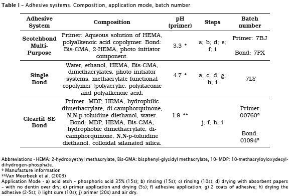
All the bonding procedures were carried out by the same operator, at room temperature. After cleaning with distilled water, specimens were divided into three groups (n= 6-7 teeth) for each adhesive systems: Scotchbond Multi-Purpose (3M ESPE, St. Paul, MN, USA), Single Bond (3M ESPE, St. Paul, MN, USA, and Clearfil SE Bond (Kuraray Medical, Tokyo, Japan)(Table 1) The adhesives were applied according to the manufacturer's instructions and light cured with a LED light unit with a power output of 400 mW/cm² (Radii, SDI, Bayswater, Australia). Resin composite build ups (Filtek Z250 - 3M ESPE, St. Paul, MN, USA) were then constructed on the bonded occlusal surfaces in three increments of 1.5 mm, which were light cured for 20 seconds each with the same light intensity.
Sections of 0.9 mm thickness each were made in a longitudinal direction (perpendicular to the adhesive interface) with a 0.3 mm diamond disc (Buehler, Lake Bluff, IL,USA) in an Isomet 1000 machine (Isomet 1000, Buehler, Lake Bluff, IL, USA), under water refrigeration at 250 rpm. Initially those sections were cut in a mesial-distal direction in order to obtain specimen's slices. A sticky wax was applied in order to keep the slices together. After that, another buccal-lingual sectioning was performed to provide sticks with 0.8 mm² area. The bonded sticks from each tooth were then randomly subdivided into two groups: one assigned to be tested immediately and the other six months after storage in distilled water containing 0.4% sodium azide, at 37°C.
Nanoleakage evaluation
Bonded sticks were coated with two layers of nail varnish applied up to within 1 mm of the bonded interface. They were then rehydrated in distilled water for 10 minutes and immersed in ammoniacal silver nitrate tracer solution for 24h (Cennabras Indústria e Comércio Ltda., Guarulhos, São Paulo, Brazil). The solution was prepared according to the protocol previously described by Tay et al. (2002) 26. The specimens were then rinsed thoroughly in distilled water and immersed in photo developing solution (Caithec Materiais Odontológicos, Rio do Sul, Santa Catarina, Brazil) for 8h, under a fluorescent light to reduce silver ions. All sticks were then placed inside an acrylic ring, which was attached to a double-sided adhesive tape, and embedded in epoxy resin. The specimens were wet polished with #600 SiC paper to remove the nail varnish, and further polished with a #1200 grit SiC paper with a 1; 0.3; and 0.05 μm diamond paste (Buehler, Lake Bluff, IL, USA). They were then ultrasonic cleaned, air dried and gold coated (SCD 005, Bal-tec, Balzers, Liechtenstein) in order to analyse the resin-dentin interfaces by SEM (Philips XL-30, Philips Eletric Corporation, Eindhoven, Netherlands). Two analysis were performed to verify the silver uptake: a) A percentage of silver uptake evaluated 18; b) the silver nitrate uptake expressed by scores 32.
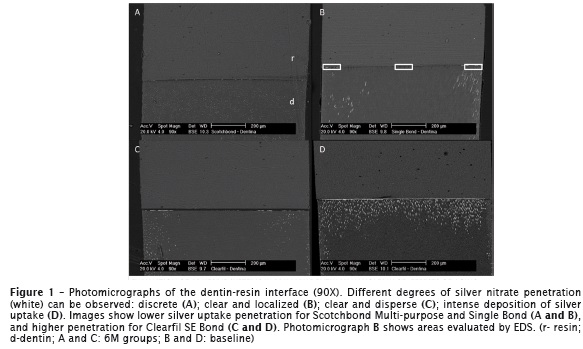
For the first method (a), the analysis was performed in the backscattered electron mode (BSE) and by the use of energy dispersive X-ray spectrometry (EDS) (EDAX, Ametec Inc., USA). Analysis of each stick was performed at three regions (center, right and left) of the bonded stick (adhesive layer, hybrid layer and resin tags) (1000X) (Figure 1). The silver nitrate uptake was expressed as a percentage, according to the mean values observed by EDS for each tooth.
In the second method (b), photomicrographs of the whole stick were taken in BSE mode (90X) and one single operator analysed each one of the photomicrographs in a 14" laptop (Aspire 4520, Acer Inc., China). The evaluation was performed in total area by the use of scores, following an adaptation of ththe method suggested by Yuan et al. (2007) 32:
0 - no leakage;
1 - Mild leakage - less than 25% of the evaluated area;
2 - Clear leakage - between 25 and 50% of the evaluated area;
3 - Large leakage - more than 50% of the evaluated area.
Data treatment
The percentages calculated from the mean values of silver uptake for each tooth were analyzed by ANOVA. Analysis of each stick's score was performed by Kruskal-Wallis and U Mann-Whitnney Test (p < 0.05).
Results
Silver nitrate penetration expressed by percentage
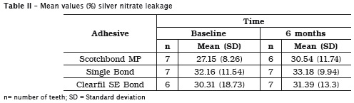
The mean values of silver penetration for each tooth, as well as the results of the statistical analysis are expressed in table 2.
Silver nitrate penetration expressed by scores
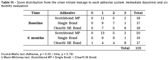
The score distribution according to the silver nitrate penetration for each adhesive system is presented in table 3. Kruskal-Wallis test revealed that there was no significant difference for the variable aging (p = 0.79), but there was a statistical difference among the adhesives (p = 0.00). The U of Mann- Whitney test (p < 0.05) showed that Scotchbond Multi-purpose and Single Bond adhesives had similar performance and allowed less silver nitrate uptake than Clearfil SE Bond adhesive.
Photomicrographs
Figures 1-4 show representative photomicrography observed in SEM.
Discussion
Nanoleakage was first described in 1995 by Sano et al. (1995) 19,20 when a little diffusion of small ions was observed inside of the hybrid layer even with the absence of interfacial gaps. The most commonly technique to analyze these defects uses silver nitrate immersion technique in conjunction with Scanning Electron Microscopy (SEM) and Transmission Electron Microscopy (TEM) analyses 4,6-8,11,16,17,26,31,32.
The results of this study (in percentage) showed that there was no significant difference among the groups studied. Regarding score's evaluation, there was no significant difference for aging, but there was a difference among adhesives.
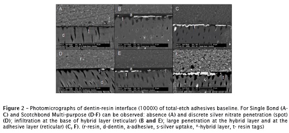
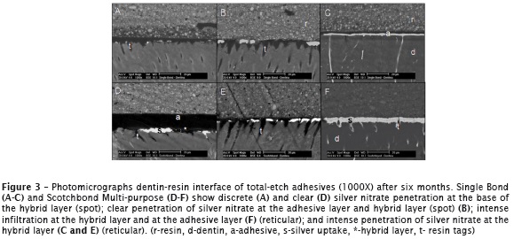
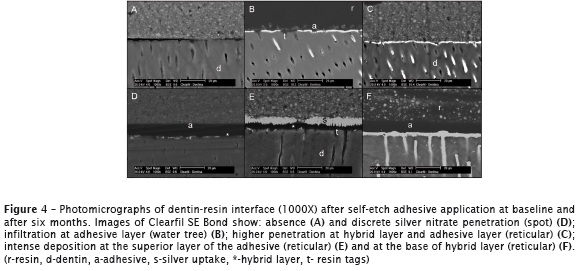
Scotchbond Multi-purpose and Single Bond showed similarity between them with less silver nitrate penetration than Clearfil SE Bond. One can note that the adhesive that demonstrated higher leakage (Clearfil SE Bond) is self-etching adhesive, contrasting with the ones that require total etching (Scotchbond Multi-purpose and Single Bond) which have lower leakage (Figure 1). These findings can be related to higher water content in the self-etching adhesives, once they need it to activate the acidic monomers and produce an efficacious hard tissue demineralization. This necessary water decreases the hydrolytic stability of the adhesive system due to water sorption 7,26.
Some studies have related the influence of aging in nanoleakage 16,18,31, by means of hydrolysis degradation, as well as polymer's plastification, which was not observed in this study. One of the reasons could be the absence of the storage liquid's renewal, since there is an acceleration of adhesion aging when the solution is changed periodically 9.
The short time of evaluation as well as the other factor's interaction over adhesion's aging should be considered, as observed by Ernhardt et al. (2008) 4, who reported a stable adhesion in sound dentin after 6 months, suggesting that other factors may contribute for the physico-chemical degradation of the interface, as pH changes, occlusal load and enzyme's variations 4.
The nanoleakage patterns observed in this study were similar to those reported in the literature (reticular, spot and water tree type") as well as the location of the silver nitrate at the adhesive interface (hybrid layer and adhesive layer) (Figure 2-4). All patterns and location of the nanoleakage were found in all adhesives, except for the water tree type" pattern, observed only for self-etching adhesive, probably due to the higher water content of those systems (Figure 4) 6-7,26. The analyses of resin-dentin interface produced by total-etch adhesives revealed a hybrid layer with high amount and long length of the resin tags, meanwhile the self-etching adhesive showed a hybrid layer with a lower quantity of tags (Figure 2-4) confirming previous studies 12,23. Further studies should be conducted in order to evaluate bonding mechanism in primary dentin.
According to results, the two methods of evaluation showed that nanoleakage at the adhesive interface in primary dentin was not influenced by aging of the three adhesive systems tested. However, the analysis of the scores showed that there was more silver uptake for specimens of the self-etch group than those of the total-etch group.
Acknowledgements
This study was supported by the Dentistry Post Graduation Program at Federal University of Santa Catarina and Coordination for the Improvement of Higher Education Personnel (CAPES).
References
1. Armstrong SR, Vargas MA, Chung I, Pashley DH, Campbell JA, Laffoon JE et al. Resin-dentin interfacial ultrastructure and microtensile dentin bond strength after five-year water storage. Oper Dent. 2004;29:705-12. [ Links ]
2. Breschi L, Mazzoni A, Ruggeri A, Cadenaro M, Di Lenarda R, Dorigo ES. Dental adhesion review: aging and stability of the bonded interface. Dent Mat. 2008;24:90-101.
3. De Munck J, Van Landuyt K, Peumans M, Poitevin A, Lambrechts P, Braem M et al. A critical review of the durability of adhesion to tooth tissue: methods and results. J Dent Res. 2005;84:118-32.
4. Erhardt MCG, Toledano M, Osorio R, Pimenta LA. Histomorphologic characterization and bond strength evaluation of caries-affected dentin/resin interfaces: effects of long-term water exposure. Dent Mat. 2008;24:786-98.
5. Hariri I, Shimada Y, Sadr A, Ichinose S, Tagami J. The effects of aging on shear bond strength and nanoleakage expression of an etch-and-rinse adhesive on human enamel and dentin. J Adhes Dent. 2012;14:235-43.
6. Hashimoto M, De Munck J, Ito S, Sano H, Kaga M, Oguchi H et al. In vitro effect of nanoleakage expression on resin-dentin bond strengths analyzed by microtensile bond test, SEM/EDS and TEM. Biomaterials. 2004;25:5565-74.
7. Hosoya Y, Ando S, Yamaguchi K, Oooka S, Miyazaki M, Tay FR. Quality of the interface of primary tooth dentin bonded with antibacterial fluoride-releasing adhesive. J Dent. 2010;38:423-30.
8. Hosoya Y, Tay FR, Ono T, Miyazaki M. Hardness, elasticity and ultrastructure of primary tooth dentin bonded with a self-reinforcing one-step selfetch adhesive. J Dent. 2010;38:214-21.
9. Kitasako Y, Burrow MF, Nikaido T, Tagami J. The influence of storage solution on dentin bond durability of resin cement. Dent Mat. 2000;16:1-6.
10. Marchesi G, Frassetto A, Visintini E, Diolosa M, Turco G, Salgarello S et al. Influence of ageing on self-etch adhesives: one-step vs. two-step systems. Eur J Oral Sci. 2013;121:43-9.
11. Marquezan M, Skupien JA, da Siveira BL, Ciamponi AL. Nanoleakage related to bond strength in RM-GIC and adhesive restorations. Eur Arch Paediatr Dent. 2011;12:15-21.
12. Miranda C, Prates LHM, Vieira RS, Calvo MCM. Shear bond strength of different adhesives systems to primary dentin and enamel. J Clin Pediat Dent. 2006;31:35-40.
13. Mobarak E, Seyam R. Interfacial nanoleakage and bonding of self-adhesive systems cured with a modified-layering technique to dentin of weakened roots. Oper Dent. 2013;38(5):E154-65.
14. Mobarak EH, Dai fal la LE. Long- term nanoleakage depth and pattern of cervical restorations bonded with different adhesives. Oper Dent. 2012;37:45-53.
15. Nakabayashi N, Kojima K, Masuhara E. The promotion of adhesion by the infiltration of monomers into tooth substrates. J Biomed Mater Res. 1982;16:1240-3.
16. Okuda M, Pereira PRN, Nakajima M, Tagami J, Pashley DH. Long-term durability of resin dentin interface: nanoleakage vs. microtensile bond strength. Oper Dent. 2002;27:289-96.
17. Oznurhan F, Olmez A. Nanoleakage in primary teeth prepared by laser irradiation or bur. Lasers Med Sci. 2013;28(4):1099-105.
18. Reis A, Grande RHM, Oliveira GMS, Lopes GC, Loguercio AD. A 2-year evaluation of moisture on microtensile bond strength and nanoleakage. Dent Mat. 2007;23:862-70.
19. Sano H, Takatsu T, Ciucchi B, Horner JA, Matthews WG, Pashley DH. Nanoleakage: leakage within the hybrid layer. Oper Dent. 1995; 20:18-25.
20. Sano H, Yoshiyama M, Ebisu S, Burrow MF, Takatsu T, Ciucchi B et al. Comparative SEM and TEM observations of nanoleakage with-in the hybrid layer. Oper Dent. 1995;20:160-7.
21. Sano H. Microtensile testing, nanoleakage, and biodegradation of resin-dentin bonds. J Dent Res. 2006;85:11-4.
22. Senawongse P, Harnirattisai C, Shimada Y, Tagami J. Effective bond strength of current adhesive systems on deciduous and permanent dentin. Oper Dent. 2004;29:196-202.
23. Shimada Y, Senawongse P, Harnirattisai C, Burrow MF, Nakaoki Y, Tagami J. Bond strength of two adhesive systems to primary and permanent enamel. Oper Dent. 2002;27:403-9.
24. Taschner M, Nato F, Mazzoni A, Frankenberger R, Falconi M, Petschelt A et al. Influence of preliminary etching on the stability of bonds created by one-step self-etch bonding systems. Eur J Oral Sci. 2012;120:239-48.
25. Tay FR, King NM, Kar-Mun C, Pashley DH. How can nanoleakage occur in self-etching adhesive systems that demineralize and infiltrate simultaneously? J Adhes Dent. 2002;4:255-68.
26. Tay FR, Pashley DH, Yoshiyama M. Two modes of nanoleakage expression in single-step adhesives. J Dent Res. 2002;81:472-6.
27. Van Meerbeek B, De Munck J, Yoshida Y, Inoue S, Vargas M, Vijay P et al. Adhesion to enamel and dentin: current status and future challenges. Oper Dent. 2003;28:215-35.
28. Van Meerbeek B, Perdigão J, Lambrechts P, Vanherle G. The clinical performance of adhesives. J Dent. 1998;26:1-20.
29. Van Meerbeek B. The myth" of nanoleakage. J Adhes Dent. 2007;9:491-2.
30. Vidal CMP, Pavan S, Briso ALF, Bedran-Russo AK. Effects of three restorative techniques in the bond strength and nanoleakage at gingival wall of Class II restorations subjected to simulated aging. Clin Oral Invest. 2013;17:627-33.
31. Yamazaki PCV, Bedran-Russo AKB, Pereira PNR. The effect of load cycling on nanoleakage of deproteinized resin/dentin interfaces as a function of time. Dent Mat. 2008;24:867-73.
32. Yuan Y, Shimada Y, Ichinose S, Tagami J. Qualitative analysis of adhesive interface nanoleakage using FE-SEM/EDS. Dent Mat. 2007;23:561-9.
 Corresponding author:
Carla Miranda
Rua João Pio Duarte e Silva, 94 – ap. 201 – Córrego Grande
CEP 88037-000 – Florianópolis – SC
E-mail: ca_mirand@yahoo.com.br
Received for publication: November 25, 2013
Accepted for publication: December 20, 2013
