


Serviços Personalizados
Artigo
 pdf em Inglês
 Artigo em XML
 Referências do artigo
 Enviar este artigo por email
Links relacionados
Compartilhar

 Permalink
RSBO (Online)
versão On-line ISSN 1984-5685
RSBO (Online) vol.11 no.3 Joinville Jul./Set. 2014
ORIGINAL RESEARCH ARTICLE
Evaluation of three root canal filling techniques through digital radiograph
Caroline Solda I, II; Volmir João Fornari II; Mateus Silveira Hartmann II; Flávia Baldissarelli II; Fabiana Corralo dos Santos III; José Roberto Vanni II
I Department of Dentistry, Lutheran University of Brazil (ULBRA) – Canoas – RS – Brazil
II Department of Endodontics of the School of Dentistry of Meridional School (IMED) – Passo Fundo – RS – Brazil
III School of Dentistry of Meridional School (IMED) – Passo Fundo – RS – Brazil
ABSTRACT
Introduction: Endodontic obturation consists of root canal filling by antiseptic or inert materials that promote a three-dimensional sealing and stimulate the repair process without interfering with it. Different obturation techniques and materials have been proposed to meet this requirement. Objective: To compare the root canal filling promoted by lateral condensation technique, Tagger's hybrid technique and McSpadden technique by assessing the filling quality through digital radiograph. Material and methods: A total of 45 extracted single-rooted human teeth were used and randomly divided into three experimental groups. After instrumentation, the teeth were filled by lateral condensation (n=15), Hybrid Tagger (n=15), and McSpadden techniques (n=15). Then, digital radiographs were taken with projected increased 10 times, at mesial-distal and buccolingual directions. Results: Visually, few empty spaces were detected at the three root thirds of teeth filled by different techniques. However, statistical analysis (Kruskal-Wallis) found no differences among the different groups, neither among the different thirds nor between both incidences evaluated. Conclusion: It was concluded that the three obturation techniques exhibited similar behavior in relation to the sealing of the root canal through digital radiograph.
Keywords: root canal obturation; guttapercha; radiographic image enhancement.
Introduction
According to the American Association of Endodontics (AAE), an adequate filling is defined and characterized by the tridimensional filling of all root canal, as closest as possible to the cementumdentine junction 9.
The filling goal is to seal root canal system (main and accessory canals) within an adequate limit and hermetically, by employing materials and techniques favoring apical and periapical healing process. Many filling techniques and endodontic cements have been proposed to meet these requirements 13.
Over time, different materials have been employed to fill root canals aiming to find the ideal one, that is, to provide both good biological and physicochemical properties 13.
Gutta-percha associated with endodontic cement has been the material of choice of most professionals, employed in different techniques, which can be divided into three groups: lateral and vertical guttapercha compaction, thermomechanical gutta-percha condensation, and injection of thermoplasticized gutta-percha 13.
Currently, root canal filling has been considered as one of the main steps of endodontic therapy. With regard to the techniques (due to either the material used or the conditions of the treatment), all have a common goal: to match quality with practicability 14.
The quality of this obturation is normally evaluated through convent ional radiograph examination, in which the distribution of the filling material is observed all over the canal extension. Notwithstanding, the process of radiographic diagnosis is subjective; thus, complementary resources, such as digitation of the radiographic image and use of digital tools can display architectural changes sometimes not seen during the visual interpretation of the conventional image 4.
The knowledge of the quality and sealing capacity of different filling techniques is of extreme importance and it is necessary an evaluation of Tagger's hybrid and McSpadden's thermomechanical techniques and lateral condensation technique, aiming to the daily clinical application to aid in endodontic treatment success.
Material and methods
This study was submitted and approved by the Institutional Review Board under protocol no. 103.958.
Fifty-four extracted teeth (9 for pilot study and 45 for study analysis) were selected. Inclusion criteria for data collection were: single-rooted human teeth, with one root canal, complete rhizogenesis, without previous endodontic treatment and/or calcifications, and without curvatures. These criteria were verified by conventional radiographic examination, at buccolingual and mesial-distal directions, using com 3x4 cm Ultra-speed Kodak film (Kodak®, Nova York, USA) during sample selection.
Firstly, the teeth were cleaned by toothbrushing and running water and kept into 0.2% thymol solution. At the moment of the use, the teeth were washed in running water and immersed in water for 24 hours to eliminate all thymol traces. Next, tooth crowns were sectioned at cervical level with the aid of no. 3228 bur (KGS, Barueri, SP, Brazil) at high speed, with water spray cooling, followed by size 1 to 3 Gattes Glidden burs (Dentsply Maillefer, Ballaigues, Switzerland), to enlarge the cervical access, and root canal access was complemented with the aid of 20:4 Laxxes bur (Dentsply Maillefer, Ballaigues, Switzerland).
Root canals were copiously irrigated with 2.5% sodium hypochlorite (NaOCl) with the aid of disposable plastic syringe (UltradentProducts Inc., South Jordan, Utah, USA) and NaviTip needle (UltradentProducts Inc., South Jordan, Utah, USA). Prior to odontometry, root canals were negotiated with the aid of size #10 K file (Dentsply Maillefer, Ballaigues, Switzerland) to determine apical patency.
Odontometry was achieved by determining the root length through leveling a size #15 K file with the apical foramen (Dentsply Maillefer, Ballaigues, Switzerland). From this measurement, 1 mm was subtracted resulting in real working length (RWL). Next, apical foramen was standardized by instrumenting root canal at that limit with the aid of size #15 K file (Dentsply Maillefer, Ballaigues, Switzerland).
Root canals were submitted to biomechanical preparation with Hero 642 system in the following instrument order: 20/.02; 25/.02; 25:04; 30/.02; 35/.02; 30:06; 40/.02; 45/02, (memory instrument); driven by rotary motor (X-Smart Dentsply – USA) at 350 rpm speed and 2.8 N torque. All instruments were used at RWL, with irrigation/aspiration at every instrument change.
After instrumentation, a size #15 K file was again introduced up to apical foramen level to confirm the clearing and cleaning. 2.5% sodium hypochlorite was used as instrumentation adjuvant. After root canal shaping, root canals were irrigated with 17% EDTA pH 7.5 (Extratus Farmácia, Passo Fundo, RS, Brazil) followed by the last NaOCl irrigation. Prior to obturation, root canals were dried through aspiration and absorbent paper points (Dentsply Maillefer, Ballaigues, Switzerland) of size matching the memory instrument and according to RWL. Then, main gutta-percha cone (Dentsply Maillefer, Ballaigues, Switzerland) was selected according to the memory instrument and by verifying its lock at RWL. All cones were disinfected with 2.5% NaOCl and dried with the aid of sterilized gauze to verify their adaptation.
Next, the teeth were filled according to the techniques of this present study: G1 – Tagger's hybrid technique; G2 – McSpadden's technique; and G3 lateral condensation.
Group 1 – Tagger's hybrid technique: After selecting the main cone (Dentsply Maillefer, Ballaigues, Switzerland), it was placed into position with the endodontic cement (Endofill, Dentsply Maillefer, Ballaigues, Switzerland), according to the manufacturer's instruction. Then, two accessory points were placed (Dentsply Maillefer, Ballaigues, Switzerland) according to the volume of gutta-percha required to fill the root canal completely, together with the apical third compaction with the aid of an obturator (Dentsply Maillefer, Ballaigues, Switzerland). The guttapercha McSpadden compactor (Dentsply Maillefer, Ballaigues, Switzerland) was selected one or two sizes above that of the main cone. The penetration depth of the compactor inside root canal was 2 mm short of RWL, at 8,000 to 12.000 rpm speed. After the compactor removal, gutta-percha was vertically compacted with the aid of Paiva's condenser (SS White Duflex, Pensilvânia, USA) to obtain a better adaptation to dentinal walls followed by the cleaning of cement remnants with cotton pellet and alcohol (Extratus Pharmacy, Passo Fundo, RS, Brazil).
Group 2 – McSpadden's technique: After selecting the main cone (Dentsply Maillefer, Ballaigues, Switzerland), this was placed into position with endodontic cement (Endofill, Dentsply Maillefer, Ballaigues, Switzerland), according the manufacturer's instruction. The compactor was selected as that of Group 1, placed without pressure with instrument inserted 2 mm short of RWL inside root canals, at 8.000 to 12,000 rpm speed, placed beside the gutta-percha cone. The heat resulting from friction plasticized gutta-percha which concurrently was compacted inside the canal. Vertical condensation was performed according to the group 1 with Paiva's condenser (SS White Duflex, Pensilvânia, USA), and cement remnants was cleaned with the aid of cotton pellet and alcohol (Extratus Farmácia, Passo Fundo, RS, Brazil).
Group 3 – latera l condensat ion: Af ter selecting the main cone (Dentsply Maillefer, Ballaigues, Switzerland), this was placed into position with endodontic cement (Endofill, Dentsply Maillefer, Ballaigues, Switzerland), according the manufacturer's instructions. Next, an accessory point (Dentsply Maillefer, Ballaigues, Switzerland) was placed and lateral condensation at apical third was accomplished with the aid of an obturator (DentsplyMaillefer, Ballaigues, Switzerland), according to the gutta-percha volume required to fill root canal completely verified when the obturator did not enter more than 5 mm. Gutta-percha was vertically condensed with the aid of Paiva's condenser (SS White Duflex, Pensilvânia, USA) to obtain its better adaptation to the dentinal walls and the cement remnants were cleaned with the aid of cotton pellet and alcohol (Extratus Farmácia, Passo Fundo, RS, Brazil).
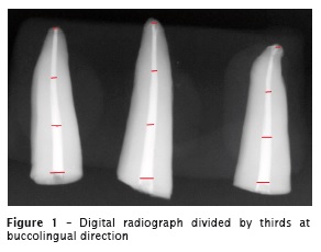
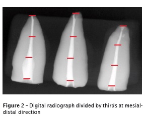
Digital radiograph was executed with Spectro II device (Dabi Atlante, Ribeirão Preto, SP, Brazil), at 70 KW, with exposure time of 0.12 second and focus-film film distance of 5 cm. After the exposure, the sensitized plates were read by the scanner of the digital system and the images were exhibited by the software inside Suarez system (São Paulo, SP, Brazil). Following, digital images were exported to digital media, saved as BMP format, and properly identified as figures 1 and 2.
The previously calibrated examiner visually detected the presence of empty spaces at the three root thirds of the filled canals.
Data were recorded in specific sheets, containing a space to record the items "presence" or "absence" of empty spaces at the different radiographic incidences at the respective root canal thirds.
The following scores were established:
• 0: no space at the analyzed third;
• 1: 1 space at the analyzed third;
• 2: 2 spaces at the analyzed third;
• 3: 3 spaces at the analyzed third, and so on.
Statistical analysis was performed by Kruskal-Wallis test through SPSS version 15.0 software.
Results
Firstly, the amount of empty spaces in relation to the filling technique was verified. Three thirds were evaluated in each one of the 15 specimens from each group, totalizing 45 assessments per surface, thus, obtaining the mean of spaces found at each root third evaluate.
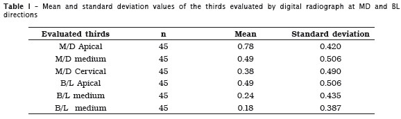
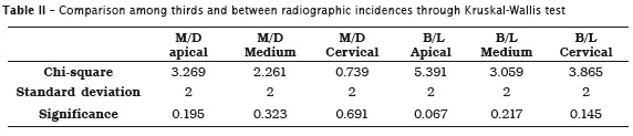
Visually, few empty spaces were detected at the three root thirds filled by different techniques. However, the statistically analysis through Kruskal-Wallis test did not find differences among the studied groups (p > 0.05), neither among the different thirds nor between the radiographic incidences evaluated (table 2).
Although lateral condensation exhibited more empty spaces at medium third, this difference is not statistically relevant.

It is important highlighting that the radiographic images of this study were assessed by a single examiner, but at two times with one-week interval between them.
Discussion
Root canal obturation is the filling of root canal by physical and biological compatible materials, aiming to promote the most hermetically sealing as possible 14.
The final goal of Endodontics is the complete obturation of root canals. The studies revealed that one of the endodontic failures is the incomplete closure of root canal space. It has been determined inappropriate filling of root canal system accounts for a significant percentage of failures 8.
Taking into consideration that the main factor in root canal obturation is filling material adaptation to canal walls, to search the most hermetic sealing as possible and prevent bacterial proliferation, many studies have attempted to determine the most effective obturation technique 5.
Many gutta-percha obturation techniques have been used to close root canal system. Gutta-percha lateral condensation and several thermoplasticized gutta-percha techniques have been the most commonly used. Different studies search to evaluate the behavior of the filling material inside the canal.
Fracassi et al. 7 evaluated the filling techniques employing Thermafil, lateral condensation, and Tagger's hybrid technique, through three radiographic assessments: conventional, digitized with mesial-distal and buccolingual projections. The results showed that in most of the evaluations, Tagger's hybrid technique presented the smaller number of empty spaces, followed by Thermafil and lateral condensation, respectively. It was observed a greater number of endodontic filling failures detected in digital images. By evaluating the detection of the empty spaces, in relation to the radiographic projection, there was no statistically significant difference between buccolingual and mesial-distal projections, which was similar to the results of this present study.
Some authors agree that filling techniques employing heat for thermoplasticization of filling material resulted in better adaptation to dentinal walls and promoted better sealing of all root canal sealing, which did not influence on the results of this present study 1,2,6.
A study conducted by Martins et al. 11 evaluated the endodontic quality of the root canal system by three different techniques: conventional lateral condensation, Tagger's hybrid and Thermafil, through computed microtomography. All techniques showed empty spaces, and Thermafil accounted for the greater volume recorded. Conventional lateral condensation technique presented the best results among the techniques.
Ferraz et al. 4 assessed the quality of root canal filling through measuring the grey level means of the filled canals on the digitized radiographic images. The authors found that there were no statistically significant differences between lateral condensation technique and modified Tagger's hybrid, which corroborates the findings of this present study.
Marciano et al. 10 conducted a study aiming to determine the percentage of empty spaces by four different techniques: lateral condensation, Tagger's hybrid, MicroSeal and GuttaFlow. The images were obtained by digital radiograph. The statistical analysis was verified by Kruskal-Wallis test (α = 0.05). Concerning to the presence of empty spaces, there were no statistically significant differences among the techniques (p > 0.05), which was similar to this present study.
A study conducted by Monteiro et al. 12 suggested that the obturation through Tagger's hybrid technique (thermoplasticized) was more effective than passive condensation technique and none employed techniques prevented apical leakage.
Several studies a l ready compa red the performance of obturation techniques at different aspects, but with different results. Some demonstrated satisfactory results of lateral condensation technique and some did not exhibited differences among the techniques. Thus, little evidence supports one technique over another, considering the advantages and disadvantages of each one 7.
Conclusion
Based on the methodology employed and the results obtained, it can be concluded that:
• through the images obtained by digital radiograph, the three filling techniques exhibited a similar behavior in relation to root canal sealing;
• visually, few empty spaces were detected at the three root thirds by the different techniques;
• the evaluated technique did not demonstrate statistically significant differences in the digital analysis at mesial-distal and buccolingual directions at the different thirds evaluated.
References
1. Baisch SG, Silveira LFM, Martos J. Análise radiográfica da repleção de canais secundários submetidos a duas técnicas de obturação. Revista Pós-Graduação. 2006;13:139-44. [ Links ]
2. Camões GCI, Loretti F, Gomes CC, Freitas FL, Pinto SS. Estudo comparativo entre duas técnicas obturadoras: condensação lateral X híbrida de Tagger. Pesquisa Brasileira de Odontopediatria Clínica Integrada. 2007;7:217-22.
3. Carvalho E, Andrade JJ, Malvar MF, Albergaria S. Avaliação do selamento apical em dentes obturados pela técnica da condensação lateral, híbrida de Tagger e Thermafil. Revista de Ciências Médicas e Biológicas. 2006;5:239-44.
4. Ferraz GE, Fracassi LD, Marques AMC, Albergaria JS, Sarmento VA. Avaliação da qualidade de duas técnicas de obturação do canal radicular por meio de radiografia digitalizada. Revista Faculdade de Odontologia. 2009;14:126-31.
5. Ferreira MC, Gomes AAF, Lessa MJ, Almeida JA, Guimarães LSLN, Vitoriano MM. Análise da espessura da linha de cimento após o uso de diferentes técnicas de obturação de canais radiculares. Revista Faculdade de Odontologia. 2011;16:59-63.
6. Ferreira MC, Gomes AF, Guimarães LSLN, Ximenes AT, Canuto PCSN, Vitoriano MM. Análise da capacidade de preenchimento de canais radiculares com guta-percha promovida por três diferentes técnicas de obturação de canais radiculares. RSBO. 2011;8:19-26.
7. Fracassi LD, Ferraz EG, Albergaria JS, Sarmento VA. Comparação radiográfica do preenchimento do canal radicular de dentes obturados por diferentes técnicas endodônticas. Revista Gaúcha de Odontologia. 2012;58:173-9.
8. Greene HA, Wong M, Ingram IIITA. Comparison of the seal ing abi l i ty of four obturat ion techniques. Journal of Endodontics. 1990; 16:423-8.
9. Leonardo MR, Salgado AAM, Silva LAB, Tanomaru Filho M. Apical and periapical repair of dogs' teeth with periapical lesions after endodontic treatment with different root canal sealers. Pesquisa Odontológica Brasileira. 2003;17:69-74.
10. Marciano MA, Bramante CM, Duarte MA, Delgado RJ, Ordinola-Zapata R, Garcia RB. Evaluation of single root canals filled using the lateral compaction, Tagger's hybrid, Microseal and Guttaflow techniques. Braz Dent J. 2010; 21:411-5.
11. Martins CS, Mello J, Martins CC, Maurício A, Ginjeira A. Comparação da obturação endodôntica pelas técnicas de condensação lateral, híbrida de Tagger e Thermafil: estudo piloto com microtomografia computorizada. Revista Portuguesa de Estomatologia, Medicina Dentária e Cirurgia Bucomaxilofacial. 2011;52:59-69.
12. Monteiro FHL, Vilalba H, Aguiar BHF, Souza SR, Romiti CG, Toledo S. Avaliação da infiltração apical em dentes obturados com a técnica híbrida de Tagger e da condensação lateral passiva. Rev Inst Ciênc Saúde. 2008;26:334-9.
13. Pécora DJ. Descrição e princípio de uso do condensador de guta-percha. [Acesso em: 2012 Jun 5]. Disponível em: http://www.forp.usp.br/ restauradora/mc.htm.
14. Pinheiro BC, Bramante AS, Hussne RP. Influência da penetração de instrumentos, empregados na condensação lateral ativa, na qualidade da obturação de canais radiculares. J Appl Oral Sci. 2003;11:186-91.
 Corresponding author:
Caroline Solda
14 de Julho, 247
CEP 99070-160 – Passo Fundo – RS
E-mail: andre.carol@ibest.com.br
Received for publication: February 1, 2014
Accepted for publication: February 20, 2014
