


Serviços Personalizados
Artigo
 pdf em Inglês
 Artigo em XML
 Referências do artigo
 Enviar este artigo por email
Links relacionados
Compartilhar

 Permalink
RSBO (Online)
versão On-line ISSN 1984-5685
RSBO (Online) vol.12 no.2 Joinville Abr./Jun. 2015
Case Report Article
Dentigerous cyst with atypical mesiodens: an unusual clinical case report
Sulabha A. NarsapurI; Sameer ChoudhariII
I Department of Oral Medicine and Radiology, Al-Ameen Dental College and Hospital, Rajiv Gandhi University – Bijapur – Karnataka – India
II Department of Oral and Maxillofacial Surgery, Al-Ameen Dental College and Hospital, Rajiv Gandhi University – Bijapur – Karnataka – India
ABSTRACT
Introduction: Mesiodens is a common supernumerary tooth occurring in anterior region of maxilla. Its occurrence is more in permanent dentition than the primary dentition. Presence of mesiodens is associated with various complications, cystic changes being less common complication of mesiodens itself. Objective: The aim of the present article is to report an atypical mesiodens with cystic changes causing complication in adjacent structures. Case report: Hereby we report a case of dentigerous cyst associated with atypical mesiodens which was impacted, inverted and had dilaceration in the crown. Surgical removal of cystic lesion along with the mesiodens was carried out. Conclusion: Supernumerary tooth associated with dentigerous cyst may result in considerable bone destruction along with other complications. Dentist should be aware of complications associated with supernumerary structures.
Keywords: inverted; dilacerated; mesiodens.
Introduction
Supernumerary tooth is an extra tooth to normal series of dental formula. Mesiodens is common supernumerary tooth occurring in anterior maxillary region. It can occur as single, multiple, impacted, unilateral or bilateral. Its prevalence varies from 0.9%-2.05% with males being affected twice 6. Presence of mesiodens can be associated with various complications in adjacent areas or in itself. Dentigerous cyst is one such complication affecting the mesiodens itself. Etiology of mesiodens still remains controversial. Phylogenetic reversion, dichotomy, hyperactivity of dental lamina are suggested theories for its development, last being most accepted 9.
Dentigerous cyst is a benign developmental odontogenic cyst associated with impacted tooth. These cysts develop after complication of crown formation 8. These cysts are frequently seen with embedded unerupted teeth, supernumerary teeth and odontomas. However dentigerous cyst associated with supernumerary tooth is uncommon 4. Hereby we report a case of dentigerous cyst in anterior maxilla associated with impacted, inverted atypical mesiodens with dilaceration in crown.
Case report
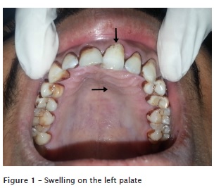
A 40 years-old male patient reported with chief complaint of progressive swelling in palate since 4-5 months. It was jowar size when patient noticed it and was progressive to attain the present size. His past medical and family history were non contributory. Intraoral examination revealed full complement of teeth. Diffuse, solitary swelling was noted in left palate involving the rugae area extending from left central incisor to left first premolar region. (figure 1) The swelling was non-tender, slightly fluctuant. Crowns of left central incisor and left lateral incisor were displaced lingually. Aspiration was positive for the straw coloured fluid.
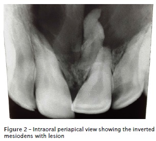
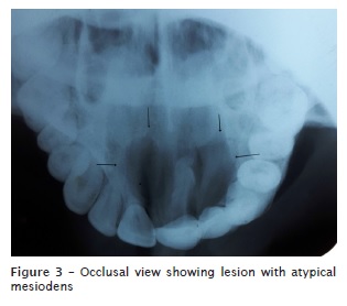
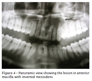
Radiograph supplementation with intra oral periapical radiograph, occlusal, panoramic view (figures 2, 3, 4) revealed well defined unilocular radiolucency in the left maxillary anterior region surrounding the invertedly impacted mesiodens having a beak shaped crown with blunt incomplete root. Radiolucency was measuring approximately 4 x 3 cms and was extending from mesial aspect of right central incisor to mesial aspect of left second premolar anteroposteriorly and superiorly slightly elevating the nasal floor and inferiorly breaking the alveolar crest between the two central incisor. Radiolucency had corticated borders expect at mesiosuperior aspect. Left central incisor and left lateral incisor showed root resorption. Both the left central and lateral incisor responded positively to vitality tests. With these finding a provisional diagnosis of dentigerous cyst with mesiodens was made.
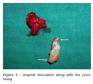
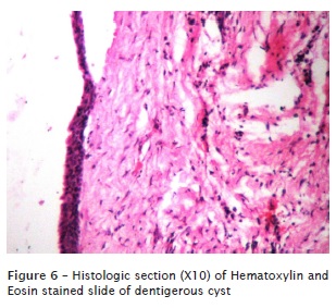
Under local anesthesia palatal crevicular incision was given from right maxillary canine to left maxillary second premolar region. Palatal mucoperiosteal f lap was ref lected, lesion was exposed and invertedly placed di lacerated mesiodens and whole of the cystic lining was removed with surgical curette (figure 5). It was then sutured with 3-0 black silk suture. Post operatively patient was prescribed a course of antibiotics and analgesics for one week. The surgically removed cystic lining was sent for histopathological evaluation. Haematoxylin and Eosin stained section revealed a cystic lumen lined by two to three layers of thin squamous epithelium with underlying wall consisting of loose bundle of collagen and fibroblast confirming the diagnosis of dentigerous cyst (figure 6). Examination of surgically removed mesiodens showed sharp curvature at tip of conical crown giving a beak shaped appearance to it. The root was incomplete, barrel shaped and blunt with open apex. Unfortunately patient never returned for the follow up appointments and was lost to recall.
Discussion
Mesiodens is supernumerary tooth of anterior maxilla. It can be classified as eumorphic and dysmorphic. The eumorphic subclass resembles normal tooth in size and shape where as dysmorphic have different shapes and sizes. These can be further classified as conical, tuberculate, supplemental and odontomas. Most of mesiodens are conical in shape with short complete root. These can be normally aligned or inverted or placed horizontally. Tuberculate mesiodens are barrel shaped with several cusp or tubercles and have incomplete or abnormal root and these rarely erupt in the oral cavity 6, 9. The term dilaceration refers to angulation or sharp bend in root or crown. The condition is thought to be due to acute trauma during the tooth formation 2. In the present case mesiodens showed conical crown with sharp curvature at its tip giving atypical beak shaped mesiodens. Root was barrel shaped, blunt with open apex. Such atypical appearance of mesiodens makes this case report worth reporting.
The presence of mesiodens is often associated with various problems which include prevention of central incisor eruption, ectopic eruption, crowding, root resorption, dilacerations of developing roots, loss of vitality, caries etc. Complication may sometimes involve mesiodens itself such as its eruption into nasal cavity, development of dentigerous cyst etc. 9. In the present case mesiodens was associated dentigerous cyst.
Dentigerous cyst is common developmental cyst formed by fluid accumulation between fully formed tooth crown and reduced enamel epithelium. Dentigerous cyst is more common in males and frequently occurs during second and third decade. According to Mourshed 1.44% of impacted teeth can develop dentigerous cyst 2. It is often associated with impacted teeth and is usually attached to tooth at cementoenamel junction 10. Dentigerous cyst is rarely associated with supernumerary tooth accounting for 5% 1, 3, 4. Literature shows few reports of dentigerous cysts with mesiodens that is placed invertedly 1, 3, 4, 7. In contrast to other reported articles present case is a case of dentigerous cyst associated with atypical mesiodens causing root resorption of adjacent teeth.
Etiology of this cyst is unknown. Three mechanisms for histogensis of dentigerous cyst were proposed by Benn and Altini. Development of dentigerous arises from dental follicle, becomes secondarily inflamed often from non vital tooth. The second type arises when an immature permanent tooth encounters a radicular cyst arising from nonvital deciduous tooth and as permanent tooth moves in radicular cyst it results in extra follicular origin dentigerous cyst. The third type usually results from inflamed exudates from periapical inflammation from nonvital deciduous tooth or other sources 4.
Three types of radiographic presentation are seen with dentigerous cyst. The central variety shows radiolucency surrounding the crown of tooth with its crown projecting into cystic lumen. In lateral variety cyst develops laterally along tooth root and partially surrounds the crown. The circumferential variant exists when cyst surrounds the crown and extends down along the root surface as if entire tooth is located within cyst 7. Resorption of the adjacent roots by mesiodens or its cyst is a rare complication 1. In the present case circumferential variant was noticed with root resorption of adjacent left central and lateral incisors which is very rare. Histopathologically dentigerous cyst is lined by 2-4 cell layer of non keratinized stratified squamous epithelium with thin fibrous capsule containing odontogenic epithelial cell rests 5.
In the present case the possible complications included: esthetic deformity due to the displacement of the incisors by expanding lesion, permanent bone deformation, and nasal cavity perforation. Hence early diagnosis and removal of dentigerous cyst is very important to reduce the morbidity. The treatment modalities range from enucleation to marsupialisation 2. The offending tooth plays a major role in deciding the type of surgical intervention for the dentigerous cyst. Complete enucleation of cyst along with extraction of the tooth forms mainstay of treatment for dentigerous cyst associated with supernumerary tooth. Pathological fractures, ameloblastoma, squamous cell carcinoma, mucoepidemoid carcinoma are frequent other complications associated with dentigerous cyst 4.
To conclude, cystic changes associated with unnoticed impacted supernumerary tooth can cause considerable bone destruction and other associated complications. Dilaceration in crown of tooth may cause difficulty in its removal. Early detection, diagnosis and treatment can reduce the morbidity.
References
1. AD Dinkar, AA Dawasaz, S Shenoy. Dentigerous cyst associated with mult iple mesiodens: a case report. J Indian Soc Pedod Prev Dent. 2007;25(1):56-9. [ Links ]
2. AN Sulabha, C Sameer, G Suchitra. Dentigerous cyst associated with dilacerated and inverted maxillary central incisor: an unusual case report. Arch Oral Res. 2013;9(1):105-9.
3. Gulses A, Karacayli U, Koymen R. Dentigerous cyst associated with inverted and fused supernumerary teeth in a chi ld: a case report. OHDMBSC. 2009;8(1):38-41.
4. Hasan S, Ahmed SA, Reddy LB. Dentigerous cyst in association with impacted inverted mesiodens: report of a rare case with brief review of literature. Int J App Basic Med Res. 2014;4:S61-4.
5. Khambete N, Kumar R, Risbud M, Kale L, Sodhis S. Dentigerous cyst associated with an impacted mesiodens: report of 2 cases. Imaging Sci Dent. 2012;42:255-60.
6. Meighani G, Pakdaman A. Diagnosis and management of supernumerary (Mesiodens): a review of the literature. J Dent Tehran Univ Med Sci. 2010;7(1):41-8.
7. Mohan KR, Natarajan B, Mani S, Sahuthullah YA, Kannan AV, Doraiswamy H. An infected dentigerous cyst associated with an impacted permanent maxillary canine, inverted mesiodens and impacted supernumerary teeth. J Pharm Bioallied Sci. 2013;5(2):S135-8.
8. More CB, Patel H. Dentigerous cyst associated with mesiodens: a rare case report. Int J Dent Clinics. 2011;3(1):77-8.
9. Russel l KA, Folwarczna MA. Mesiodensd i a g no s i s a nd ma n a g ement of c ommon supernumera r y tooth. J Can Dent Assoc. 2003;69(6):362-6.
10. Wang C, Huang P, Wang Y, Shyng YC, Kao W. Dentigerous cyst over maxillary sinus: a case report and literature review. Taiwan J Oral Maxillofac Surg. 2009;20:116-24.
 Correspondence:
Sulabha A. Narsapur
Department of Oral Medicine and Radiology
Al-Ameen Dental College and Hospital, Athani Road
Bijapur 586108 – Karnataka – India
E-mail: sulabha595@gmail.com
Received for publication: January 16, 2015
Accepted: January 27, 2015
