Serviços Personalizados
Artigo
Links relacionados
Compartilhar
RSBO (Online)
versão On-line ISSN 1984-5685
RSBO (Online) vol.13 no.1 Joinville Jan./Mar. 2016
ORIGINAL RESEARCH ARTICLE
Clinical and radiographic assessment of root canal treatments performed by dental students
Andressa Benvenutti I; Micheli Scalvi I; Sinval Adalberto Rodrigues Junior II, III; Carla Battiston II
I Private clinic – Chapecó – SC – Brazil
II School of Dentistry, Universidade Comunitária da Região de Chapecó – Chapecó – SC – Brazil
III Health Sciences Postgraduate Program, Universidade Comunitária da Região de Chapecó – Chapecó – SC – Brazil
ABSTRACT
Introduction: Retrospective assessment of procedures performed in dental school clinics is a valuable tool to critically assess the teaching/ learning process. Objective: This retrospective study assessed the success rate of root canal treatments executed by dental students during a two-year time span. Material and methods: Patients who had undergone root canal treatments at the clinics of the School of Dentistry of Unochapecó during 2011-2013 were recalled for assessment of the quality of the procedure and the associated coronal restoration. The quality of the root canal treatments was assessed clinically and radiographically, while the coronal restorations were assessed clinically by a trained dental student. Data were analyzed by descriptive statistics and chi-square test (α=0.05). Results: Thirtytwo patients returned for evaluation of 38 root canal treatments and coronal restorations. The success of root canal treatments was 89.5%, while 78.5% of the coronal restorations were considered successful. A significant association was observed between the success of the root canal treatments and the quality of the coronal restoration (p<0.005) and the apical length of the root canal filling (p<0.011). The presence of flaws within the filling material was not significantly associated to the success/failure of the root canal treatment (p=0.459). Conclusion: A high success rate of root canal treatment performed by dental students at an average 13-month evaluation period was observed. An adequate coronal restoration and a good apical sealing is required for a good prognosis of teeth undergoing root canal treatment.
Keywords: edental pulp cavity; dental restoration failure; dental clinics.
Introduction
The school clinics is the first practicing space where dentists develop the skills and competences required for an adequate, integral treatment of the patient's needs, including the endodontic therapeutics. For an endodontic treatment to be properly done, the undergraduate student must comprehend and be familiarized with the diagnosis and treatment of pulp and periradicular diseases and must be able to adequately preserve and restore the pulpally-compromised teeth 22.
The main goal of the RCT is cleansing and disinfecting of contaminated root canals followed by the tridimensional filling of the root canal system, avoiding reinfection 25. Since the endodontic procedure is performed in an operatory field with reduced dimensions, with no luminosity and different morphologies and dimensions at each intervention, it is a surgical procedure with highly special characteristics.
The success of the RCT is associated to several technical issues, including the precise diagnosis, the maintenance of the aseptic chain, the knowledge of the anatomy of root canal system, the correct biomechanical preparation, the hermetic filling of the root canals, an adequate coronal restoration, and the periodic treatment follow-up 6,7.
Endodontic therapy has undergone huge technological advances. Nevertheless, the occurrence of adverse clinical situations involving signs and symptoms indicating lack of tissue repair is not uncommon 13. Long-term clinical and radiographic follow-up is the method available to establish the success or failure of the RCT. Lopes and Siqueira Jr. 16 suggest that the outcome of endodontic therapy should be evaluated periodically, every six months. Such control would depict the normal or altered condition of the periapical tissues.
Endodontic and restorative procedures may be more technically challenging and time-demanding for beginner undergraduate students than for experienced operators 5. Besides, the prognostic of the RCT is associated to the quality of the coronal restoration 20. Therefore, long-term prospective and retrospective assessments of these procedures conducted by dental students should be routine for professors and supervisors.
These assessments would funct ion as a pedagogical technolog y that would provide information about the success/failure rate and the causes of failure, allowing the improvement of the teaching/learning process. Also, as an adjunct to the evidence provided by the dental literature, it would help to elect principles, techniques, and materials and to discard others that do not function properly. On that basis, this study aimed at assessing the clinical and radiographic success of endodontic treatments and associated coronal restorations executed by dental students from a Community University in Southern Brazil.
Material and methods
Study design, location and research subjects
The study was designed as a retrospective crosssectional observational analytical study and was developed at the clinics of the School of Dentistry of the Community University of the Region of Chapecó – Unochapecó, after approval by the Institutional Review Board regarding ethical aspects, under the protocol no. #109/13. The research subjects were found by searching the clinical files of the patients of the dental clinics and identifying those who had undergone endodontic treatment from February 2011 to June 2013. Inclusion criteria involved patients whose baseline final radiographs presented good quality, patients who authorized new radiographic exams and patients who were not pregnant. Fiftyeight patients were identified and invited to take part of the study by phone call. Those who attended the dental clinics were clarified about the aims of the study and signed a consent form.
Variables
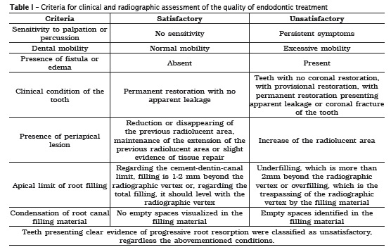
The quality of the root canal treatment was assessed clinically and radiographically and dichotomized as 'satisfactory' or 'unsatisfactory', based on the criteria of the American Association of Endodontics 8 (table 1). Unsatisfactory root canal treatment could have presented more than one reason.
The quality of the coronal restoration was assessed clinically based on the criteria of the FDI published elsewhere 12. The FDI instrument categorizes the criteria into three groups (esthetic, functional, and biological) and f lexibilizes the selection of the criteria according to the investigator's needs 12. The criteria taken into consideration as likely to be associated to failure of root canal treatment were: (i) anatomic form; (ii) fracture of material and retention; (iii) marginal adaptation; (iv) occlusal contour and wear; (v) approximal anatomic form (contact point and contour); (vi) radiographic examination; (vii) recurrence of caries, erosion, abfraction; and (viii) tooth integrity (enamel cracks or tooth fractures).
Originally, each criterion is scored 1 to 5, being 1, 2, and 3 indicative of satisfactory restorations and 4 and 5 indicative of unsatisfactory restorations. In our study, as a means of simplifying assessment criteria, dichotomization was applied using as threshold between "satisfactory" and "unsatisfactory" the difference between the scores 3 and 4.

Training and calibration
Two dental students of the fourth year were trained for clinical and radiographic assessments of root canal treatment and coronal restorations by specialists in endodontics and operative dentistry. Training for endodontic assessment was based on the discussion of radiographs and the calibration was performed comparing the rating of the students with the reference examiner (specialist professors). The inter-examiner diagnostic reproducibility was assessed using Cohen's Kappa coefficient for the success of the root canal treatments and for the reasons of failure (inter-examiner coefficient of 1.00).
As to the training for assessing the quality of coronal restorations, it was conducted through slide exposure meetings. Ten restorations out of five subjects were chosen by the gold-standard examiner and examined as a calibration means. Kappa coefficient for inter-examiner diagnostic reproducibility of the quality of restorations and reasons of failure varied from 0.615 to 1.00.
Clinical exam
Clinical exams were performed at the university dental clinics, during the second semester of 2013, by the student who achieved the highest coefficient of agreement with the gold-standard examiners. The research subjects were examined under artificial light using dental mirror, wooden spatula, sterile gauze and no. 5 explorer. Periapical radiographs were made using radiographic periapical film (Kodak Insight, Rochester, New York) and a radiographic device model Spectro 70x Eletronic (Dabi Atlante, Ribeirão Preto, SP, Brazil). Radiographs were performed using the parallelism technique, with controlled exposure time of 0.5 seconds and were processed in dark environment by the time/temperature method at 20oC with the following times: 3 minutes for revelation, 30s for washing, 3 minutes for fixation and 10 minutes for the final bath. After drying, radiographs were mounted with subject/tooth identification and analyzed with the aid of a hand magnifier under the appropriate illumination.
Data analysis
Data were tabulated and summarized using descriptive statistics. Chi-square test was used to verify whether there were significant associations between variables (α=0.05).
Results
Thirty-two subjects out of the 58 patients identified for recall attended the dental clinics and were examined, yielding 38 root canal treatments for evaluation. Thirteen subjects did not show up to the recall exam and others 13 subjects were excluded due to the following reasons: not found by contact data provided (10 subjects – 17.2%), pregnancy (1 – 1.7%), had the tooth extracted (1 – 1.7%), finished treatment elsewhere (1 – 1.7%).
The age of the subjects ranged from 21 to 56 years, with a mean age of 43.9 (±12.8) years; 47.4% were males and 52.6% were females.
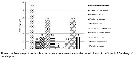
Figure 1 shows the type of tooth that underwent RCT. Maxillary central incisors were the most prevalent tooth type submitted to endodontic treatment, followed by maxillary and mandibular pre-molars.
By searching the subject's files, one observed that 12 teeth (31.6%) presented pulpal vitality, 20 teeth (52.6%) presented pulpal necrosis with periapical lesion and six teeth (15.8%) presented pulpal necrosis without periapical lesion prior to treatment.
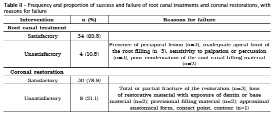
The mean time span from the end of the endodontic treatment was 13.8 (±5.3) months, ranging from 8 to 28 months. Most root canal treatments were classified as satisfactory (table 2).


Our results revealed that 91.6% of the teeth with pulp vitality prior to endodontic treatment and 88.4% of the teeth with pulp necrosis were satisfactory. Chi-square test revealed a significant association between the apical limit of the root filling and the success of the endodontic treatment (χ2=6.513; p=0.011). The association between the condensation of the root filling material and the success of the endodontic treatment, on the other hand, was not significant (χ2=0.549; p=0.459).
Table 2 presents the proportion of success and failure of coronal restorations in the evaluated teeth. Approximately 95% of the teeth were restored with definitive materials. A significant association between the success of the coronal restorations and the success of the endodontic treatment was detected by the chi-square test (χ2=7.828; p=0.005).
Discussion
The results of our study allowed one to check the success of RCTs performed by dental students of a Community University at Southern Brazil. A 55.2% proportion of return of patients was achieved, and denoted the need for improvement of data registration about the patient and the endodontic treatment, since the study sample was reduced due to lack of that information.
Other limitations of the study were the small number of patients, given by the recent character of the dental clinics functioning at the institution, and the short time of assessment. On the other hand, a minimum time of six months has been recommended to suggest the success of the root canal treatment 16. Since the minimum assessment time observed in our study was of eight months, in spite of being low, it can be considered significant for the outcome of the assessed root canal treatments.
The majority of root canal treatment demands were pulpal necrosis. As to the condition of the treatments performed at the school clinics, 89.5% were successful at the evaluation moment. Ng et al. 19 observed a survival rate of 98% after one year in post-graduation endodontic clinics. This difference may be explained by the fact that technique improvement through practice and specialization has been shown to affect the clinical success rate of root canal treatments 1.
Endodontic treatments are unique in their complexity and generate several difficulties to beginner practitioners. Several technical issues are common causes of failure of RCT and may challenge dental students during their first endodontic procedures. The most common are break of aseptic chain during the procedure, incorrect access to the pulp cavity, non-detected canals, deficient chemomechanical preparation, and inadequate apical limit of the root filling and unsatisfactory or absent coronal restorations 2.
Approximately 92% of the teeth with vital pulp prior to RCT presented satisfactory endodontic outcomes after an average 13 months. The results are in agreement with literature, which presented a success rate higher than 90% for teeth in that condition 4. On the other hand, approximately 88% of the teeth with pulp necrosis were successful after RCT, representing a higher rate than those observed in other studies 4. One of the goals of endodontic therapeutics in cases of necrotic pulp tissue is the neutralization, reduction or even the elimination of the infection in the root canal system 6. Teeth under this condition present microorganisms that are not necessarily found in vital teeth, leading to comparatively lower rates of success 7,15.
The success rate has been shown also to decrease as a consequence of the presence of periapical lesion 2,3,24. Our results revealed that 10% of the teeth with periapical lesion prior to treatment presented unsatisfactory outcomes. Periapical radiolucency expresses the extent of periapical destruction by microorganisms derived from the pulp and its size indicates the severity and the extent of the microbiological infestation 3. Resistance to antibacterial therapeutics with either cleansing or medication through mechanisms that involve cooperation between bacteria may partially explain the maintenance of periapical lesions 24.
A significant association was detected between the apical limit of the root canal filling and the success of the endodontic therapeutics. This is in accordance with Heling et al. 11, who observed that root canals filled below the apex presented higher unsuccessful rates. The incomplete filling of the root canal often results of inadequate instrumentation, and allows the maintenance of the necrosis remnants and bacteria close to the periapical region 6. Schaeffer et al. 23 recommended that the root canal filling should achieve the proximities of the apical foramen, approximately 1-2mm short of the apex.
The success rate of root canals filled within the 2-mm limit was of approximately 94%, highlighting the importance of adequately instrumenting the root canal and filling it at the closest level of the apical foramen, assuring adequate disinfection in the highest extent of the root canal 6. Also, the higher prevalence of endodontic failure associated to root canal filling under the 2-mm limit is related to the incapacity of debriding the apical segment or canal, or due to accumulation of contaminated dentin that possess persistent infection agents in the root apexes 18.
Several regions of the root canal system, including ramificat ions, dent in tubules and delta apical are not accessed by endodont ic instrumentation, irrigating chemical agents and medications, allowing the presence of bacterial debris 10. Therefore, the absence of adequate sealing of the obturation would provide nutrients for microorganism's metabolism and growth through percolation of tissue fluid or saliva, thus causing damage to periapical tissues. In spite of that, poor condensation leading to voids within the obturation mass was not associated to failure or the RCTs in this study (p>0.05). In a recent systematic review, Ng et al. 20 found conflicting results of studies that assessed the quality of root filling and its association with RCT failure, with one study having reported a 68% survival rate of teeth with voids in their root fillings at 5 and 10 years.
The analysis of the coronal restorat ions revealed that 78.5% were considered satisfactory at the mean evaluation time of 13 months, which was considered low as compared to studies that assessed retrospectively restorations performed by undergraduate students 17,21. Those studies observed an 85.5% success rate at 3 years 17 and an 87% success rate at 5 years 21.
The reasons of failure identified in our study potentially compromise the success of the RCT and, therefore, are a significant clinical concern. In fact, we observed that the failure of coronal restorations was significantly associated to the failure of the RCT (p<0.05). Both, the partial fracture of the restorative material and the loss of restorative material exposing dentin or base material are situations of potential exposure of the RCT to contaminants, similar to marginal openings, which have been identified as increasing the odds of persistent apical periodontitis and of tooth extraction 14.
As to the other causes of restorative failure, the presence of temporary fillings is a risk factor for contamination, since they undergo degradation more rapidly than the permanent restorative materials 9. Ideally, placing the final coronal restoration immediately after finishing the endodontic procedure would minimize the leakage of oral fluids and bacteria, reducing the odds of a reinfection and improving the prognosis of the endodontic therapeutics 11. Ng et al. 20, studying the factors interfering on the success of endodontic therapy, identified that the execution of the permanent restoration within ninety days after the root canal treatment increased the tooth survival rate. Also, tooth fracture and resulting extraction may result from the absence of approximal contact points in the tooth or of the presence of a defective approximal contact point in the restorative material, since it would lead to unfavorable distribution of occlusal force with an important non-axial stress component 20.
The quality of the coronal restoration has been shown as a paramount factor to ensure the health of periapical tissues. Gillen et al. 9, assessing the influence of the coronal restoration on the quality of endodontic treatments, concluded that the odds of healing increase with both adequate coronal restoration and root canal filling. The authors also observed that whenever one or the other is not adequate, the odds of healing decrease in a similar fashion.
The school clinics is most likely the best place to develop an understanding that every intervention towards the achievement of a health condition for the patient should be periodically assessed as to their quality and longevity, under the risk of generating overtreatment. Also, the criteria used for assessment of the quality of endodontic and restorative procedures should be taught to undergraduate students and explored clinically as a means of revealing what is clinically satisfactory.
Conclusion
Regardless the limitations of the study, a high success rate of RCTs performed by dental students could be observed. Also, one concluded that the success of RCT was conditioned to an adequate apical sealing and to an adequate coronal restoration. The quality of the RCT was not associated with the quality of the condensation of the root canal filling material.
References
1. Alley BS, Kitchens G, Alley LW, Eleazer PD. A comparison of survival of teeth following endodontic treatment performed by general dentists or by specialists. Oral Surg Oral Med Oral Pathol Oral Radiol Endod 2004. Jul;98(1):105-8. [ Links ]
2. Cheung GSP. Endodontic failures – changing the approach. Int Endod J. 1996 Jun;46(3):131-8.
3. Chugal NM, Clive JM, Spangberg LSW. A prognostic model for assessment of the outcome of endodontic treatment: effect of biologic and diagnostic variables. Oral Surg Oral Med Oral Pathol Oral Radiol Endod. 2001 Mar;91(3):342-52.
4. de Quadros I , Gomes BPFA, Zaia AA, Ferraz CCR, Souza-Filho FJ. Evaluation of endodontic treatments performed by students in a Brazilian Dental School. Int Dent Educ. 2005 Oct;69(10):1161-70.
5. Demarco FF, Rosa MS, Tarquínio SBC, Piva E. Influence of the restoration quality on the success of pulpotomy treatment: a preliminary retrospective study. J Appl Oral Sci. 2005 Mar;13(1):72-7.
6. Estrela C, Holland R, Estrela CRA, Alencar AHG, Sousa-Neto MD, Pécora JD. Characterization of successful root canal treatment. Braz Dent J. 2014 Jan-Feb;25(1):3-14.
7. Estrela C, Leles CR, Hollanda AC, Moura MS, Pécora JD. Prevalence of risk factors of apical periodontitis in endodontically treated teeth in a selected population of Brazilian adults. Braz Dent J. 2008;19(1):34-9.
8. Estrela C, De Esponda LCA. Ciência endodôntica. São Paulo: Artes Médicas; 2004. p. 588-617.
9. Gillen BM, Looney SW, Gu LS, Loushine BA, Weller RN, Loushine RJ et al. Impact of the quality of coronal restoration versus the quality of root canal fillings on success of root canal treatment: a systematic review and meta-analysis. J Endod. 2011. Jul;37(7):895-902.
10. Grecca FS, Rosa ARG, Gomes MS, Parolo CF, Bemfica JRD, Frasca LCF et al. Effect of timing and method of post space preparation on sealing ability of remaining root filling material: In vitro microbiological study. J Can Dent Assoc. 2009 Oct;75(8):583.
11. Heling I, Gorfil C, Slutzky H, Kopolovic K, Zalkind M, Slutzky Goldberg I. Endodontic failure caused by inadequate restorative procedures: review and treatment recommendations. J Prosthet Dent. 2002 Jun;87(6):674-8.
12. Hickel R, Peschke A, Tyas M, Mjör I, Bayne S, Peters M et al. FDI World Dental Federation: clinical criteria for the evaluation of direct and indirect restorations – update and clinical examples. Clin Oral Investig. 2010 Aug;14(4):340-66.
13. Imura N, Pinheiro ET, Gomes BP, Zaia AA, Ferraz CC, Souza-Filho FJ. The outcome of endodontic treatment: a retrospective study of 2000 cases performed by a specialist. J Endod. 2007 Nov;33(11):1278-82.
14. Kirkevang LL, Vaeth M, Wenzel A. Ten-year follow-up of root filled teeth: a radiographic study of a Danish population. Int Endod J. 2014 Oct;47(10):980-8.
15. Kojima K, Inamoto K, Nagamatsu K, Hara A, Nakata K, Morita I et al. Success rate of endodontic treatment of teeth with vital and nonvital pulps. A meta-analysis. Oral Surg Oral Med Oral Pathol Oral Radiol Endod. 2004 Jan;97(1):95-9.
16. Lopes HP, Siqueira Junior JF. Endodontia: biologia e técnica. 3. ed. Rio de Janeiro: Guanabara Koogan; 2010. p. 645-91.
17. Moura FRR, Romano AR, Lund RG, Piva E, Rodrigues-Junior SA, Demarco FF. Three-year clinical performance of composite restorations placed by undergraduate dental students. Braz Dent J. 2011;22(2):111-6.
18. Nair PNR. On the causes of persistent apical periodontitis: a review. Int Endod J. 2006 Apr;39(4):249-81.
19. Ng YL, Mann V, Gulabivala K. A prospective study of the factors affecting outcomes of nonsurgical root canal treatment: part 2: tooth survival. Int Endod J. 2011 Jul;44(7):610-25.
20. Ng YL, Mann V, Gulabivala K. Tooth survival following non-surgical root canal treatment: a systematic review of the literature. Int Endod J. 2010 Mar;43(3):171-89.
21. Opdam NJM, Loomans BAC, Roeters FJM, Bronkhorst EM. Five year clinical performance of posterior resin composite restorations placed by dental students. J Dent. 2004 Jul;32(5):379-83.
22. Qualtrough AJE. Undergraduate endodontic education: what are the challenges? Br Dent J. 2014 Mar;216(6):361-4.
23. Schaeffer MA, White RR, Walton RE. Detemining the optimal obturation length: a meta-analysis of literature. J Endod. 2005 Apr;31(4):271-4.
24. Sundqvist G, Figdor D, Persson S, Sjögren U. Microbiologic analysis of teeth with failed endodontic treatment and the outcome of conservative retreatment. Oral Surg Oral Med Oral Pathol Oral Radiol Endod. 1998 Jan;85(1):86-93.
25. Torabinejad M, Kutsenko D, Machnick TK, Ismail A, Newton CW. Levels of evidence for the outcome of nonsurgical endodontic treatment. J Endod. 2005 Sep;31(9):637-46.
 Corresponding author:
Sinval Adalberto Rodrigues Junior
Universidade Comunitária da Região de Chapecó
Área de Ciências da Saúde – Caixa postal 1141
Av. Senador Atílio Fontana, n. 591-E – Efapi
CEP 89809-000 – Chapecó – SC – Brasil
E-mail: rodriguesjunior.sa@unochapeco.edu.br
Received for publication: August 1, 2015
Accepted for publication: March 7, 2016


