


Serviços Personalizados
Artigo
 pdf em Inglês
 Artigo em XML
 Referências do artigo
 Enviar este artigo por email
Links relacionados
Compartilhar

 Permalink
RSBO (Online)
versão On-line ISSN 1984-5685
RSBO (Online) vol.13 no.1 Joinville Jan./Mar. 2016
ORIGINAL RESEARCH ARTICLE
Effect of the use of different periodontal curettes on the topography and roughness of root surface
Matheus André Müller I; Emanuelle Cunha I; Rafaela Scariot I; João Cesar Zielak I; Felipe Rychuv Santos I; Tatiana Miranda Deliberador I; Carmen Lucia Mueller Storrer I
I Program in Dentistry, Positivo University – Curitiba – PR – Brazil
ABSTRACT
Introduction: Periodontal scaling is the treatment approach most used to remove dental calculus, plaque, and altered cementum from root surface. During root decontamination, the instruments used leave the root rougher and more irregular. Objective: To verify the root surface after mechanical scaling with different Gracey curettes steel through SEM and superficial roughness analyses. Material and methods: Twelve teeth were embedded in acrylic resin. The teeth were instrumented with new Gracey curettes Gracey 5/6 from different brands. The groups (n=2) were divided into: control, no instrumentation (GC); carbon steel (CSN); stainless steel Neumar (SSN); stainless steel Millenium (SSM); premium steel Neumar (PSN); Hu-Friedy (HF). An area measuring 3 x 3 mm2 was marked on the distal surface of the root to guide the Reading of the root topography on SEM and rugosimeter. The data were analyzed by a single examiner previously calibrated. SEM analysis was based on scores of the root surface smoothness after scaling. We analyzed the parameters of mean roughness (Ra) and mean roughness deepness (Rz). SEM data were submitted to statistical analysis through Fisher's exact test (p < 0.002) and roughness data by Anova followed by Student t test. Results: The quality of the active surface of the curette demonstrated by SEM and roughness analyses that it can exert difference in the result regarding to the homogeneity produced after the scaling of root surface. Group SSM demonstrated a homogenous root surface (score 0) in SEM and better smoothness in rugosimeter analysis. Conclusion: According to com the methodology used, the group of curettes that provided better smoothness of root surface after scaling was SSM.
Keywords: root scaling; curettes; scanning electronic microscopy; root roughness.
Introduction
Dental plaque, adhered to the tooth surface, accounts for developing and maintaining the periodontal disease 8. The dental plaque distribution on the root surface is non-uniformly, occurring mainly on concave areas or rougher areas of the roots 10,14. The result of the mineralization of the plaque is dental calculus. The treatment for removing these from the root surface is the scaling and polishing of the root surface by hand, ultrasound, piezoelectric or high level laser instruments 1,3,15. Although root scaling is the treatment most used, many studies verified that the complete removal of the dental calculus, plaque, and altered cementum from all root surface is not even reached 5,13, mainly in anatomic areas, as the proximal surfaces of the roots and furcal areas that demanding high skills 12,14. During root decontamination, the instruments used for scaling may leave root surface more irregular and rougher.
The comparisons performed between hand and rotary instruments (ultrasound, piezoelectric, polishing burs, and lasers) do not demonstrate improvements over hand instrumentation 4,7,15. However, after instrumentation, the authors concluded that diamond points promote root roughness equal to that of ultrasound instruments, but higher than that of hand instrumentation 11.
Similar study was also conducted and compared hand, magnetostrictive, and piezoelectric instruments for removing dental calculus on human teeth (n=30) through the reading of root roughness (rugosimeter) and SEM. The Ra values showed that in all cases, the calculus removal was effective, but the piezoelectric instrument left the surface rougher than that of the other instruments 13. In relation to the effects of the instrumentation by curettes, piezoelectric instrument, curettes + piezoelectric instrument, laser, curettes + laser on the alteration of the root morphology and adhesion of the blood components in freshly extracted human teeth demonstrated that the group instrumented by curettes showed the greatest superficial smoothness. Concerning to blood adhesion, no statistically differences among groups occur 15.
The literature demonst rates that hand instrumentation produces less roughness than the sonic, ultrasound, and piezoelectric instruments 13. However, this study lacks information on the type of steel of the instruments used. Many steel types are used without a critical assessment of which would be the most suitable type.
This study aimed to verify the root surface after mechanical scaling with different types of Gracey curettes, through SEM and superficial roughness analyses.
Material and methods
This study was submitted and approved by the Institutional Review Board regarding ethical aspects (CAAE 19471813.4.0000.0093). Twelve maxillary lateral incisors were selected from the tooth bank of the Positivo University. The selected teeth did not have root caries, calculus, debris, and alterations caused due to extraction. These characteristics were examined with the aid of magnifying glasses (Olympus América Inc. model SZX9 Center Valley – Pennsylvania) at x20 magnification. The teeth with any aforementioned characteristic were excluded from the sample.
Preparation of the specimens
The maxillary lateral incisors were embedded in acrylic resin leaving the distal surface of the root exposed. The specimens were numbered from 1 to 12 and randomly divided into groups. The roots of the specimens were instrumented by new Gracey curettes 5/6. The groups (n=2) were divided into: control, without instrumentation (GC); carbon steel Neumar (CSN); stainless steel Neumar (SSN); stainless steel Millenium (SSM); premium steel Neumar (PSN); Hu-Friedy (HF). The root surface of the specimens of each group received tem scaling movements at apex-crown direction. A 3 x 3 mm2 was area marked on the root surface to guide the root topography in SEM and rugosimeter analyses.
Rugosimeter reading
The specimens were submitted to the rugosimeter reading (Mitutoyo SJ-201) before and after scaling. At each reading, the rugosimeter needle crossed a 3 mm long area on the surface with cut-off sampling of 0.25 mm, to maximize the surface undulation filtering. Three readings on different sites of each specimen were performed and the mean of these values were used as the roughness value for each specimen. In this study, two roughness parameters were used: mean roughness (Ra), that is, the arithmetic mean among the recorded peaks and valleys; mean deepness roughness (Rz), that is, the maximum distance between the greatest peak and the greatest valley during the measuring path.
Scanning electronic microscopy (SEM) analysis
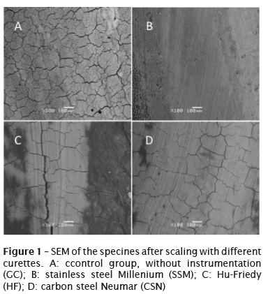
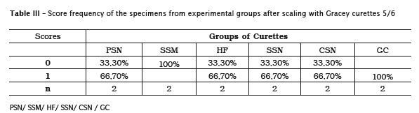
The SEM analysis was based on scores of the smoothness of the root surface after scaling. The SEM images followed a visual pattern per x100 augmentation (figure 1). The area assessed was inside the marked area on the root. Each photomicrograph was analyzed twice by a single calibrated examiner, with one-week interval between the assessments. The data of each assessment was submitted to Spearman's correlation test with level of intra-examiner agreement of 98%.
The photomicrographs were classified according to the scores based on the lack of irregularity and homogeneity of the root surface:
0: homogenous root surface with few grooves produced by the scaling;
1: non homogenous root surface very irregular.
Statistical analysis
SEM data were submitted to statistical analysis through Fisher's exact test and roughness data through Anova and Student t test, with level of significance of 5%.
Results
Roughness reading
The specimens showed a decrease in roughness after instrumentation by curettes in all groups. The result of roughness (Ra) and (Rz) of root surface produced by root instrumentation by different Gracey curettes 5/6 is seen in tables 1 and 2.
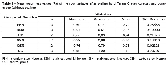
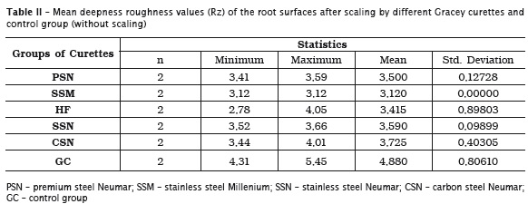
When the results of all curette types were evaluated (Anova), no statistically differences occurred for Ra (p=0.087) and Rz (p=0.128). When the mean roughness values (Ra) were compared two by two (Student t test), we verified that the groups PSN X GC, SSM X SSN, SSM X CSN, SSM X GC demonstrated statistical difference with p values of p=0.008, 0.049, 0.012, ≤0.001, respectively.
When the mean deepness roughness values (Rz) were compared two by two, we verified that the groups SSM X SSN demonstrated statistical significant difference (p=0.021), with group SSM presenting the smallest roughness.
SEM analysis
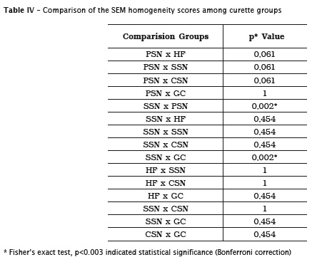
The assessment per score frequency in relation to the root homogeneity after scaling by Gracey curettes and control group is demonstrated in table 3. When the groups were compared two by two, we observed that the score values were statistically different (p<0.002) among the curette groups (SSM X GC and SSM X PSN) (table 4).
Discussion
Root surface roughness is important to avoid new plaque deposits, provide a biocompatible surface to adhesion of periodontal ligament fibroblast, and not favor bacterial adhesion 4,13.
The literature has studies on scaling comparing hand instruments (curettes) with ultrasound, piezoelectric instruments, lasers Er:Cr:YSGG, bicarbonate jets, and curettes associated with diamond points aiming to verify which instrument provided a more homogenous and less rough surface. The results of these studies were unanimous in pointing out that curettes produce a less roughness and more biocompatible surface 9,11,13,15. However, different curette brands and steel types were not compared.
In this study, five curette types were evaluated: CSN, SSN, SSM, PSN and HF and found that SSM was the curette providing the best surface homogeneity in SEM analysis (p<0.002). The root area analyzed both through SEM and rugosimeter was pre-stablished (3 x 3 mm2). Thus, we can affirm that the reading crossed all the marked surface, without possibility of preference for points to be analyzed, corroborating many researches 4,11,15. Although the results regarding to superiority of the curettes versus the ultrasound and piezoelectric devices were also observed in rugosimeter and SEM, the evaluated area was not previously stablished and the analysis occurred on all root surface 13. Still, these aforementioned study teeth were extracted due to periodontal problems and had calculus, so that they underwent scaling prior to the study. In this present study, we used teeth from a tooth bank without calculus or irregularities, all of the same tooth type. It is known that some tooth types have root concavities and others do not. The concavities make scaling difficult and provide a rougher and irregular surface 12,14, and this aspect was not taken into account in other studies 1,4,11,15.
The curettes form group SSM demonstrated significant superiority (p<0.002) compared with GC through SEM, showing that root surface allowed greater root smoothness after instrumentation. This result was not observed in other studies comparing roots underwent scaling with control group 6,15. Such fact could have occurred because of the initial sample homogeneity.
Conclusion
The quality of the active surface of the curette demonstrated in this present study that can exert difference in the result regarding homogeneity produced after scaling. The group SSM demonstrated a homogenous root surface by SEM and better root surface smoothness in rugosimeter analysis.
References
1. Aspriello SD, Piemontese M, Levrini L, Sauro S. Ultramorphology of the root surface subsequent to hand-ultrasonic simultaneous instrumentation during non-surgical periodontal treatments: an in vitro study. J Appl Oral Sci. 2011;19(1):74-81. [ Links ]
2. De Mendonça AC, Maximo MB, Rodrigues JA, Arrais CA, DE Freitas PM, Duarte PM. Er:YAG Laser, ultrasonic system, and curette produce different profiles on dentine root surfaces: an in vitro study. Photomed Laser Surg. 2008;26(2):91-7.
3. Dwivedi S, Verma SJ. Comparison of the effects of periodontal rotary instruments and Gracey curettes on root surface characteristics: an in vivo SEM study. Quintessence Int. 2012;43(10): 135-40.
4. Eick S, Bender P, Flury S, Lussi A, Sculean A. In vitro evaluation of surface roughness, adhesion of periodontal ligament fibroblasts, and Streptococcus gordonii following root instrumentation with Gracey curettes and subsequent polishing with diamond-coated curettes. Clin Oral Investig. 2013;17(2):397-404.
5. Kepic TJ, O'leary TJU, Kafrawy AH. Total calculus removal: an attainable objective? J Periodontol. 1990;61(1):16-20.
6. Khosravi M, Bahrami ZS, Atabaki MS, Shokrgozar MA, Shokri F. Comparative effectiveness of hand and ultrasonic instrumentationsin root surface planing in vitro. J Clin Periodontol. 2004;31(3):160-5.
7. Leal Serra e Silva Filho W, Paullilo LA, Nociti JR FHA, Sallum EA, Casati M Z, Sallum AW. Comparative evaluation of root surface roughness created by curetts, ultrasonic scaler and diamond ultrasonic tips-In vitro study. Periodontia. 2007;17(1):90-4.
8. Löe H, Theilade E, Jensen BS. Experimental gingivitis in man. J Periodontol. 1963;36(3): 177-87.
9. Marda P, Prakash S, Devaraj CG, Vastardis S. A comparison of root surface instrumentation using manual, ultrasonic and rotary instruments: an in vitro study using scanning electron microscopy. Indian J Dent Res. 2012;23(2):164-70.
10. Quirynen M, Bollen CM. The influence of surface roughness and surface-free energy on supra- and subgingival plaque formation in man. A review of the literature. J Clin Periodontol. 1995;22(1):1-14.
11. Ribeiro FV, Casarin RCV, Nociti Jr FH, Sallum EA, Sallum AW, Casati MZ. Comparative in vitro study of root roughness after instrumentation with ultrasonic and diamond tip sonic scaler. J Appl Oral Sci. 2006;14(2):124-9.
12. Rios CM, Pustiglioni FE, Romito G. Biometric study of the width, length and depth of the root trunk groove of human lower second molars. Pesqui Odontol Br. 2002;16:26-36.
13. Singh S, Uppoor A, Nayak DA. Comparative evaluation of the efficacy of manual, magnetostrictive and piezoelectric ultrasonic instruments – an in vitro profilometric and SEM study. J Appl Oral Sci. 2012;20(1):21-6.
14. Storrer CM, Sanchez PL, Romito GA, Pustiglioni FE. Morphometric study of length and grooves of maxillary lateral incisor roots. Arch Oral Biol. 2006;51(8):649-54.
15. Tsurumaki JN, Souto BHM, Oliveira GJPL, Sampaio JEC, Marcantonio Jr E, Marcantonio RAC. Effect of instrumentation using curettes, piezoelectric ultrasonic scaler and Er,Cr:YSGG laser on the morphology and adhesion of blood components on root surfaces – a SEM study. Braz Dent J. 2011;22(3):185-92.
 Corresponding author:
Carmen Lucia Mueller Storrer
Rua Professor Pedro Viriato Parigot de Souza, n. 5.300 – Campo Comprido
CEP: 81280-330 – Curitiba – PR – Brasil
Received for publication: August 12, 2015
Accepted for publication: November 14, 2015
