


Serviços Personalizados
Artigo
 pdf em Inglês
 Artigo em XML
 Referências do artigo
 Enviar este artigo por email
Links relacionados
Compartilhar

 Permalink
RSBO (Online)
versão On-line ISSN 1984-5685
RSBO (Online) vol.13 no.3 Joinville Jul./Set. 2016
ORIGINAL RESEARCH ARTICLE
Fluid-transport evaluation of lateral condensation, GuttaCore™ and continuous wave of obturation techniques
Daniel Silva-HerzogI; Ezequiel MonroyI; Verónica MéndezI; Sandra Maria Alves Sayão MaiaII; Paulo Melo JúniorII; Ricardo OlivaI
I Endodontic Postgraduate Program, School of Stomatology, University of San Luis Potosi – San Luis Potosi – México
II Department of Dentistry, University of Pernambuco – Recife – PE – Brazil
ABSTRACT
Introduction: To achieve success in endodontic treatment, the root canal system should be as sealed as possible with suitable material such as gutta-percha and sealer. Objective: This study compare the apical leakage of roots obturated with GuttaCore™, lateral condensation, and continuous wave obturation through a computerized fluid-transport system. Material and methods: Fifty-two freshly extracted upper molars were used. The disto-buccal roots were cut and standardized to 10 mm long. The root canals were prepared at working length with WaveOne primary reciprocating files. The specimens were randomly divided and filled by one of the following obturation techniques: GuttaCore™, lateral condensation or continuous wave obturation, using SILCO sealer. The positive controls were left unfilled and the negative controls were totally coated with cyanoacrylate and three layers of nail polish. The roots were stored in relative humidity for 72 h at 37°C, allowing the sealer to set. After this period, the roots were connected to a computerized fluid-transport system, and the apical leakage was analyzed. Results: The results were expressed in μL.cmH2O-1.min-1 x10⁻⁴ 1.36 atm. The data were statistically analyzed using one-way ANOVA and Tukey´s tests. The specimens of the positive control group showed extreme amounts of apical leakage. There was no leakage in the negative controls. The statistical analysis indicated that continuous wave obturation and GuttaCore™ showed lower leakage than the lateral condensation technique (p<0.05). No difference was found among the continuous wave obturation and GuttaCore™ (p>0.05). Conclusion: Continuous wave obturation and GuttaCore™ showed lower leakage than the lateral condensation technique. There was no difference between the continuous wave obturation and GuttaCore™. The fluidtransport system used in this study allowed an accurate quantitative measurement of leakage using simpler equipment.
Keywords: apical leakage; continuous wave obturation; fluid filtration; fluid transport; guttacore; lateral condensation.
Introduction
A properly shaped and disinfected root canal system is an environment in which the bacterial communities have been disrupted or eliminated, ensuring that the environment is, unable to promote or sustain periapical disease. The method of preserving this condition over time is by hermetically filling the duct system.
Gutta-percha, derived from resin of Sapotaceae tree family shows most of the characteristics of an ideal sealing material. In its chemically pure state, occurs in two phases: α- found naturally and β- a result of refining, is the predominant phase in dental materials 4. These phases are interchangeable, depending on the temperature of the material.
The lateral condensation of gutta-percha has been widely used for decades and is considered the gold standard in most scientific publications. It is relatively simple, provides good control of the apical sealing level and requires only conventional instruments without the need for an apparatus or complex equipment3.
The continuous wave obturation technique, introduced by Buchanan, is very similar to Schilder's vertical condensation; it uses a heat source or carrier associated with tips of different tapers of 4, 6, 8, 10 and 12%, graduated in 5 mm intervals, which allow rapid heating and cooling and, decreases the contact time between the hot instrument and the canal walls 11.
There are a variety of obturation systems based on a rigid core coated with gutta-percha; Thermafil is one of the most popular systems, although the rigid core of the carrier of these systems made of plastic or metal makes it difficult to create space for posts and to perform a retreatment 7,9.
To overcome the disadvantages of a plastic carrier core and because of advances in polymer chemistry, a core from a thermostable elastomer of crosslinked gutta-percha (GuttaCore™, Dentsply Tulsa Dental Specialties) has been developed. This core is covered by regular gutta-percha that allows it to flow in three dimensions through the root canal when is properly heated 9.
There are numerous leakage studies, using the following as markers: dyes, radioisotopes, and microorganisms and their products 13,21,22. The causes of filtration are as follows: the improper performance of the sealing technique performed, the physical and chemical characteristics of the sealants and the presence or absence of smear layer 12,22.
Fluid transport technique measures the sealing by movement of an air bubble within a capillary tube. It was introduced by Pashley in 1987, and it was modified by Wu in 1993 to apply it to root canals. It consists of connecting a tube filled with water under atmospheric pressure at the coronal portion of the sample and a 20-μm glass capillary tube of 170 mm in length and uniform caliber to the apical portion. Pressure of 0.1 atmospheres is applied at the coronal end, forcing water into the empty spaces between the obturation 24. The results are typically expressed in μl/min 17. One of the major advantages of this system is that the sample is not lost or altered, which allows its evaluation at different intervals of time and, recording of the evolution of the behavior of the samples in the desired timeframe. The results are recorded automatically, eliminating possible errors by the operator.
Orucoglu et al. 15 made a significant contribution to developing a computerized system for measuring the movement of the air bubble. The method considers the refraction of light at the beginning and at the end of the bubble, and movement however minimal, is detected by a laser. This enhancement allows more accurate measurement by a fully electronic and digital system; without the subjectivity of the visual observation of the operator.
The aims of this study were to evaluate through a fluid transport computerized system the sealing quality of the GuttaCore™ system and compare it with two of the most frequently used endodontic techniques regarding apical leakage.
Material and methods
Fifty-two recently extracted caries-free human upper molars were used in this study. The distobuccal roots were sectioned and standardized to 10 mm long. The periodontal ligament was removed with a scalpel and the roots were immersed in a 30 min ultrasonic bath with 1% NaOCl.
After the canal patency was checked and the glide path established with a #10 K file (Dentsply Maillefer, Ballaigues, Switzerland), the roots were instrumented with WaveOne™ Primary reciprocating files (Dentsply Maillefer, Ballaigues, Switzerland) using a pecking motion and irrigated with 2 ml of 1% NaOCl between files delivered with a 27G needle. The dedicated reciprocating motor (Dentsply Maillefer) of the WaveOne™ file was used with the manufacturer configuration setup. The final irrigation protocol was accomplished with 2 ml of 18% EDTA and 2 ml of 1% NaOCl. The canals were dried with sterile paper points. The roots that needed an apical instrumentation larger than 25/.08 were discarded. All of the specimens were prepared by the identical operator using new instruments for each specimen.
After instrumentation, one coat of cyanoacrylate and three of nail polish were applied to the external root surfaces within 3 mm from the apex to avoid sealing the foramen. The fifty-two roots were randomly assigned to three experimental groups (n=14) and two control groups (n=5) and filled.
In the first group, the roots were filled with GuttaCore™ (Dentsply, Tulsa Dental Specialties, Tulsa, OK, USA) as specified by the manufacturer. The size and taper was checked with the #25 size. Sterile paper points were used to coat the walls of the coronal half of the canal with a thin layer of a zinc oxide-eugenol based sealer (Silco, Productos Endodónticos Especializados, SLP, Mexico). After disinfection and adjustment of the proper length, a #25 obturator was heated with the GuttaCore™ oven using the #1 setup. Once heated, the obturator was placed in a slow, non-twisting movement until the working length was reached and held firmly with one finger. The handle was cut off with a sharp spoon excavator.
In the second group, the canals were filled using a lateral condensation technique. A #25 master cone (Hygienic Corp, Akron, Ohio, USA) was fitted to the working length. The inner walls of the canal were coated with a thin layer of SILCO sealer (Productos Endodónticos Especializados, San Luis Potosí, México) using a #20 hand file (Dentsply Maillefer, Ballaigues, Switzerland) in a counterclockwise motion. The master cone was coated with sealer and seated into place. A #7 spreader (Hu-Friedy, Chicago, IL, USA) was inserted 1 mm shorter than the working length and kept in the canal until a fine gutta-percha accessory cone (Hygienic Corp, Akron, Ohio, USA) was placed. The subsequent condensation was accomplished with an MA-57 spreader (Hu-Friedy, Chicago, IL, USA) until it could no longer penetrate beyond the coronal one-third of the canal. The coronal guttapercha was removed with a hot instrument and apical pressure was applied with a cold #1 Glick (Hu-Friedy, Chicago, IL, USA).
The roots in the third group were filled with the continuous wave obturation technique, conducted in two steps. First, a non-standardized fine-medium gutta-percha cone was adjusted with a scalpel 1 mm short of working length until tugback was achieved. The B&L alpha (B&L Biotech, PA, USA) heat source was set to 200°C with a 40/.08 plugger, which was bound within 3 mm from the working length. After coating the canal walls with SILCO sealer using a sterile paper point, the apical part of the master cone was coated with a thin film of sealer and placed into the canal. The heated plugger was inserted slowly 5 mm short of the working length. The plugger was then deactivated; applying apical pressure for 10 sec., the plugger was reactivated for 1 sec. and drawn back slowly. Finally, the apical pressure was held with a #60 Ni-Ti plugger (S-Kondenser, Obtura Spartan, Earth City, MO, USA) calibrated 4 mm short of the working length. After the down-packing procedure was completed, the backfill was performed with the B&L beta (B&L Biotech, PA, USA) obturation gun set at 200°C. The injection was made in a single increment and condensed with a #100 stainless steel plugger (S-Kondenser, Obtura Spartan, Earth City, MO, USA).
The roots of the positive control group were left unfilled, and the negative controls were totally coated with one layer of cyanoacrylate and three of nail polish, including the apical foramina.
Following the obturation, the roots were stored in relative humidity for 72 h at 37°C, allowing the sealer to set.
For the leakage study, the computerized fluid transport system described by Orucoglu et al. 15 was modified. The pressure was provided by an O2 tank and maintained at 20 psi (1.36 atm) throughout the experiment with a pressurized buffer reservoir. A plastic tube was connected from the reservoir to the left side of a 1 mm diameter glass capillary tube (Chase Scientific Glass Inc., Rockwood, TN, USA) mounted horizontally on a circuit board. The left end of the capillary tube was adapted to a bifurcated connector; the first end of the bifurcation was connected to a microsyringe and the other to the apical portion of the specimen to be evaluated. All of the connections were sealed with cyanoacrylate and inspected to avoid leakage. The system was filled with distilled water. An air bubble was inserted in the microsyringe and placed in the capillary tube before each measurement.
For the apical leakage quantification two lightsensitive photodiodes were arranged beside the capillar, with a separation of 26 mm between them. The measurements of the fluid movement were determined by the time needed for the air bubble to travel the 26 mm between the two photodiodes.
All of the operations were controlled with PCLabVIEW software (National Instruments, Austin, TX, USA). This software calculated the speed of the air bubble expressed in mm/s. The results were converted to filtration units and expressed as μL.cmH2O-1.min-1 x10⁻⁴ 1.36 atm and the means were determined.
The data from the experiments were statistically analyzed by one-way ANOVA followed by Tukey´s test. The confidence level used was 95% (p<0.05).
Results
The means and standard errors of the control and experimental groups are presented in table I , expressed as μL.cmH2O-1.min-1 x10⁻⁴ 1.36 atm.
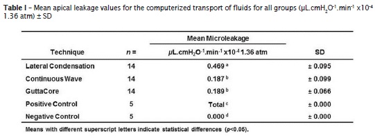
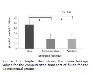
Continuous wave obturation showed the least amount of leakage (0.187 ± 0.099 μL.cmH2O-1.min-1 x10⁻⁴ 1.36 atm), followed by GuttaCore™ (0.189 ± 0.066 μL.cmH2O-1.min-1 x10⁻⁴ 1.36 atm). Lateral condensation exhibited the greatest amount of leakage (0.469 ± 0.095 μL.cmH2O-1.min-1 x10⁻⁴ 1.36 atm) among the tested techniques. Although the continuous wave obturation group showed the least apical leakage, no significant difference was found compared with that of the GuttaCore™ group (p>0.05). There was a significant difference between the lateral condensation and the other two techniques (p<0.05). The results are presented in figure 1.
The negative controls demonstrated no leakage as any movement of the air bubble was detected after 30 min. The positive controls showed extreme amounts of leakage and the photodiode sensors were unable to detect the passage of the air bubble, expressing the results as total.
Discussion
Multiple studies are in agreement that a poor seal has a negative influence on the prognosis of endodontic treatment 1,5,8,19. More than half of the failures are caused by problems associated with obturation because a poor seal is susceptible to leakage. GuttaCore™ is an obturation system through a gutta-percha carrier, characterized by a resilient core from a crosslinked, thermostable elastomer covered with regular gutta-percha, which can make a difference among other systems that use plastic in the carrier construction. Although filtration studies allow evaluation of apical or coronal filtration and comparison between obturation techniques available, the methodology that better fulfills the goals of evaluation should be selected 13,21,22. Pommel et al. 18 assessed the sealing techniques by dye penetration, fluid transport, and an electrochemical method without finding correlation of the results between the assessment methods. He attributed the differences to physical and chemical laws that govern each technique. Camps and Pashley 2 compared the penetration of dyes, the ink removal, and the transport of fluids without finding a correlation between these three assessment techniques, suggesting that the multiple limitations of dye penetration were the reason that their results are not comparable with other techniques. Computerized fluid transport represents an objective, sensitive, reproducible method that offers quantifiable results without the alteration or destruction of samples. The pressure exerted on the fluid used in the technique overcomes the limitations of the methods of the diffusion of dyes, radioisotopes and bacterial toxins because of air trapped in the cracks that does not allow those elements to leak at the interface between the sealing material and the dentin 13,17,20,24. The results are expressed in μL.cmH2O-1.min-1, allowing highly comparable hard data to be obtained. Other important elements in this type of evaluation are the pressurization and the time it is exercised, and the pressure measurement for this study was 20 psi (1.36 atm). A short measurement time could affect the results; because the fluid pressure could expand the hoses containing it. The liquid moved by such expansion could cause movement of the bubble without this movement representing the actual filtration 13,17. In this study, a pressure measurement for 5 min was taken. Based on the results obtained in this study, it can be inferred that the plasticizing of gutta-percha inherent to the GuttaCore™ system and continuous wave obturation allows better sealing compared with lateral condensation technique. These results agree with those found by Gencoglu et al. 7, who compared Thermafil, Js Quick-Fill, Soft Core, Microseal, System B and lateral condensation including 60 single-rooted teeth that were divided into six groups and evaluated at two years. This study showed that lateral condensation has greater filtration than Thermafil and Soft Core; both carrier based guttapercha systems. Thermafil showed the lowest rate of filtration, with no significant difference between the Thermafil and Soft Core groups. Pommel and Camps 16 analyzed the sealing quality of the single cone technique, lateral condensation, vertical condensation, Thermafil and System B using fluid filtration. After 24 h, the single cone obturation showed the highest filtration; Thermafil, System B and vertical condensation had the lowest. Gencoglu 6 compared six obturation techniques, and found no significant differences, noting that the carrier based techniques showed the highest amount of gutta-percha, with minimal amounts of sealant. In a similar study, Kececi et al. 11 compared the amount of gutta-percha in the lateral technique compared with continuous wave obturation and found no statistically significant differences. De-Deus et al. 4 compared three techniques, sectioning at 2 and 4 mm from the apex and taking photomicrographs; the study showed that at the two levels studied, the carrier based technique showed greater density of gutta-percha than the lateral technique and the continuous wave. According to our results, the techniques that require less sealant are less susceptible to leakage. Chu et al.3 evaluated the prognosis of 64 teeth treated with Thermafil and lateral condensation using radiographic control at 3 years. The lateral condensation group included 34 teeth of which 7 (21%) were considered failures. The Thermafil group consisted of 37 teeth of which 7 (9%) failed. Hale et al. 10 conducted a study in 71 teeth, 35 filled by gutta-percha carriers and 36 by lateral condensation, with a range of 18 to 37 months of observation; 6 teeth filled by carrier, (17 %) and 7 by lateral condensation, (19%) failed. The results from this study suggest that the differences in in vitro filtration studies do not correlate with the prognosis of techniques in the clinical environment and highlight the need to develop methodologies for applying in vivo filtration analysis.
By the computerized fluid transport technique, GuttaCore™ and continuous wave obturation showed leakage values statistically lower than those of the lateral condensation technique. No significant differences were found between GuttaCore™ and the continuous wave obturation technique.
References
1. Bergenholtz G, Spangberg L. Controversies in endodontics. Crit Rev Oral Biol Med. 2004;15(4)99-114. [ Links ]
2. Camps J, Pashley D. Reliability of the dye penetration studies. J Endod. 2003 Sep;29(9):592-4.
3. Chu CH, Lo ECM, Cheung GSP. Outcome of root canal treatment using Thermafil and lateral condensation filling techniques. Int Endod J. 2005 Mar;38(3):179-85.
4. De-Deus G, Gurgel-Filho ED, Magalhães M, Coutinho-Filho. A laboratory analysis of guttapercha- filled area obtained using Thermafil, System B and lateral condensation. Int Endod J. 2006;29:378-83.
5. Friedman S, Abitbol S, Lawrence H. Treatment outcome in endodontics: the Toronto study. Phase I: initial treatment. J Endod. 2003 Dec;29(2):787- 93.
6. Gencoglu N. Comparison of 6 different guttapercha techniques (part II): Thermafil, Js Quick- Fill, Soft Core, Microseal, System B and lateral condensation. Oral Surg Oral Med Oral Path Oral Radiol and Endod. 2003;96:91-5.
7. Gencoglu N, Orucoglu H, Helvacioglu D. Apical leakage of different gutta-percha techniques: Thermafil, Js Quick-Fill, Soft Core, Microseal, System B and lateral condensation with a computerized fluid filtration meter. Eur J Dent. 2007;1:97-103.
8. Grossman LI, Shepard LI, Pearson LA. Roentgenological and clinical evaluation of endodontically treated teeth. Oral Surg Oral Med Oral Path Oral Radiol and Endod. 1964;17:368-74.
9. Gutmann JL. The future of root canal obturation. Dent Today. 2011;30:130-1.
10. Hale R, Gatti R, Glickman GN, Opperman LA. Comparative analysis of carrier-based obturation and lateral compaction: a retrospective clinical outcomes study. Int J Dent. 2012;8. Available from: http://dx.doi.org/10.1155/2012/954675.
11. Kececi AD, Celik-Unal G, Sen BH. Comparison of cold lateral compaction and continuous wave of obturation techniques following manual or rotary instrumentation. International J Dent. 2005;38:381-8.
12. Madison S, Wilcox LR. An evaluation of coronal microleakage in endodontically treated teeth. Part 3. In vivo study. J Endod. 1988 Sep;14(9):455-8.
13. Moreira-Veríssimo D, Sampaio do Vale M. Methodologies for assessment of apical and coronal leakage of endodontic filling materials: a critical review. J Oral Sci. 2006;48:93-8.
14. Orstavik D. Materials used for root canal obturation: technical, biological and clinical testing. Endod Topics. 2005 Nov;12(1):25-38.
15. Orucoglu H, Sengun A, Yilmaz N. Apical leakage of resin based root canal sealers with a new computerized fluid filtration meter. J Endod. 2005 Dec;31(12):886-90.
16. Pommel L, Camps J. In vitro apical leakage of System B compared with other filling techniques. J Endod. 2001 Jul;27(7):449-51.
17. Pommel L, Camps J. Effects of pressure and measurement time on the fluid filtration method in endodontics. J Endod. 2001 Apr;27(4):256-8.
18. Pommel L, Jacquot B, Camps J. Lack of correlation among three methods for evaluation of apical leakage. J Endod. 2001 May;27(5):347-50.
19. Sjögren U, Hägglund B, Sundqvist G, Wing K. Factors affecting the long-term results of endodontic treatment. J Endod. 1990 Oct;16(10):498-504.
20. Spangberg LSW, Acierno TG, Yongbum Cha B. Influence of entrapped air on the accuracy of leakage studies using dye penetration methods. J Endod. 1989;15:548-51.
21. Swanson K, Madison S. An evaluation of coronal microleakage in endodontically treated teeth. Part 1. Time periods. J Endod. 1987 Feb;13(2):56-9.
22. Torabinejad M, Ung B, Kettering JD. In vitro bacterial penetration of coronally unsealed endodontically treated teeth. J Endod. 1990 Dec;16(2):566-9.
23. Whitworth J. Methods of filling root canals: principles and practices. Endod Topics. 2005 Nov;12(1):2-24.
24. Wu MK, De Gee AJ, Wesselink PR. Fluid transport and dye penetration along root canal fillings. Int Endod J. 1994 Sep;27(5):233-8.
 Corresponding author:
Sandra Maria Alves Sayão Maia
Av. Rua Santo Elias, n. 67
CEP 52020-090 – Recife – PE – Brasil
E-mail: sandrinhasayao@hotmail.com
Received for publication: February 12, 2016
Accepted for publication: June 20, 2016
