


Serviços Personalizados
Artigo
 pdf em Inglês
 Artigo em XML
 Referências do artigo
 Enviar este artigo por email
Links relacionados
Compartilhar

 Permalink
RSBO (Online)
versão On-line ISSN 1984-5685
RSBO (Online) vol.13 no.3 Joinville Jul./Set. 2016
ORIGINAL RESEARCH ARTICLE
Anatomical analysis of the pulp chamber of artificial teeth
Maiara GiongoI; Poliana GaonaI; Fausto Rodrigo VictorinoI
I Department of Dentistry, University Center of Maringá – Maringá – PR – Brazil
ABSTRACT
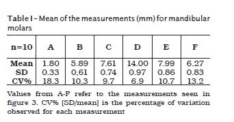
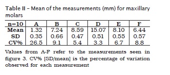
Introduction: The anatomy of root canals is very variable, with the presence of ramifications, side canals, accessory canals, and interconnections. Therefore, the knowledge of the internal tooth morphology has a fundamental importance for the localization and treatment of root canals, thus achieving success in endodontic treatment. In Endodontics, the preclinical teaching requires human teeth, but because of the current difficulty in obtaining them, the students have used artificial dental elements. Objective: This study aimed to evaluate the anatomy of the pulp chamber of artificial teeth, used in practical activities in Endodontics. Material and methods: Therefore, the artificial teeth were X-rayed through the digital system and the internal anatomy of the pulp chamber was measured through the Image Tool software. Results: The height of the mandibular pulp chamber: mandibular molar = 1.8 mm and maxillary molar = 1.32 mm; distance from the furcation to the pulp chamber floor: mandibular molar = 5.89 mm and maxillary molar = 7.24 mm; distance from the furcation to the pulp chamber's roof: mandibular molar = 7.61 mm and maxillary molar = 8.59 mm; distance from the tip of the buccal cusps to the furcation: mandibular molar = 14 mm and maxillary molar = 15 mm; distance from the tip of the buccal cusps to the pulp chamber's floor: mandibular molar = 7.9 mm and maxillary molar = 8.1 mm; distance from the tip of the buccal cusps to the pulp chamber's roof: mandibular molar = 6.2 mm and maxillary molar = 6.4 mm. The measures were homogeneous with maximum percentage variation of 26.5%. Also, the artificial teeth were similar to natural teeth, except for the relationship between the pulp chamber's roof and cementum-enamel junction. Thus, it is recommended the use artificial teeth with caution in preclinical teaching of Endodontics.
Keywords: Endodontics; anatomy; artificial tooth.
Introduction
The aim of the endodontic treatment is to achieve the adequate cleaning and antisepsis of the root canal system and the proper mechanical preparation, obtaining the taper shape towards the apex 1. The morphology of the root canals is very variable, with side canals, accessory canals, and interconnections. Thus, the knowledge of this morphology is of fundamental importance to locate and treat of root canals 17. Accordingly, the detailed view the pulp cavity is indispensable the study and practical learning of Endodontics practically 19, consequently leading to treatment success.
In Endodontics, the clinical practice requires previous laboratorial training in manikins with extracted human teeth, aiming at mimicking the real conditions in which the undergraduate will face during clinical practice. For that purpose, extracted human teeth are asked for the undergraduates to provide along with the list of materials and instruments before the laboratorial activities. However, currently, by adopting a minimally invasive dental practice and due to the absence of legal human tooth banks in most dental schools, to obtain human teeth very difficulty. Moreover, since 1997, the Brazilian law on organ donation advocates that teeth are organs and the donators have to sign a donors form 11. Thus, nowadays, the use of human teeth in dental education comes up against ethical questions about its illegal trade, such as buying teeth in cemeteries and in private clinics. Furthermore, other relevant issue is to know which decontamination and storage procedures are being employed by the students, because some microorganisms can survive for a long period on extracted teeth and they may cause several infections 14. To avoid or reduce the crosscontamination in using extracted human teeth in laboratorial activities, the dental schools developed guidelines on biosecurity and specific protocols for storage to avoid alterations 5,3.
Therefore, artificial teeth made of opaque and colorless resin have been employed in the preclinical teaching of Endodontics 18. For that purpose, these artificial teeth should reliably mimic the possible complexity exhibited by natural root canals. This study aimed to evaluate the pulp chamber of the maxillary and mandibular artificial first molar used in practical activities in Endodontics.
Material and methods
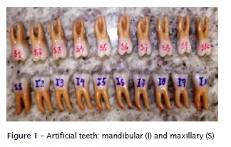
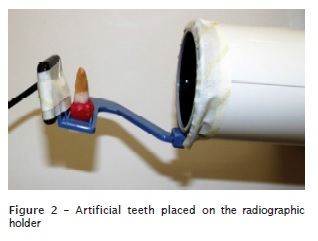
Ten mandibular and ten maxillary artificial first molars (IM do Brasil, São Paulo, SP, Brazil) were used. Each tooth received a number and the letter regarding a dental arch, that is, maxillary or mandibular (figure 1). The artificial teeth were adapted on a radiographic holder, in which a digital sensor (Kodak) was positioned. The x-ray device (Kodak 220) was matched to the radiographic holder to maintain a standardized position (figure 2). The exposure time was 0.107 s, and standard contrast control panel.
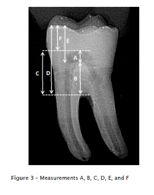
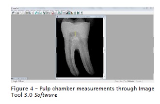
The following measurements were performed on the digital radiographs: height of the mandibular pulp chamber (measurement A); distance from the furcation to the pulp chamber floor (B); distance from the furcation to the pulp chambes roof (C); distance from the tip of the buccal cusps to the furcation (D); distance from the tip of the buccal cusps to the pulp chambers floor (measurement E); distance from the tip of the buccal cusps to the pulp chambers roof (measurement F) (figure 3). The measurements were obtained through Image Tool 3.0 software (figure 4).
Descriptive statistical analysis was performed using the mean of the obtained results.
Results
The mean values, standard deviation, and coefficient of variation of all measurements analyzed of the mandibular and maxillary molars are available in tables I and II . The measurements were homogenous with little variation. The height of the pulp chamber (measurement A) exhibited the greater percentage range (26.5%). The following tables (tables I and II) show the mean values in millimeters of the different analyzed distances of the artificial mandibular and maxillary molars.
Discussion
Teaching in the discipline of Endodontics requires knowledge of the internal and external tooth anatomy and the development of ability and sensitivity of the student to perform a satisfactory treatment. Due to the difficulty in obtaining natural teeth and with the purpose of facilitating the teaching / learning process in the endodontic preparation in preclinical activities, some authors raised alternatives such as: the replacement of one of the walls of the pulp chamber and root canal by colorless resin 8, the use of a colorless plastic tube with 30 mm of height and 0.8 mm of diameter 8, and the creation of root canals in colorless polyester 21 and epoxy resins 2,20. In the latter, the student learns by visualizing the intra-channel procedures and uses a smaller number of extracted natural teeth. In addition to being used in the teaching of endodontic techniques, simulated channels have also been useful in scientific research 10,15. Other author attempted to reproduce models simulating natural teeth, mimicking the bone density of the mandibular and maxillary arches 7.
In Brazil, Nassri et al. 12 published a study in which they presented artificial teeth for the teaching of the discipline of Endodontia, made of opaque resin that mimics natural human teeth.. Nassri et al. 13 evaluated the morphological, physical, and radiographic features of these artificial teeth and observed their potential in replacing the natural teeth in preclinical training.
Concerning to the pulp chamber morphology of natural teeth, Deutsch and Musikant 4 reported the measurement of the pulp chamber of 100 maxillary and 100 mandibular molars and provided information that may be used as a guideline for crown opening. Generally, the measurement from the cusp to the pulp chambers roof was approximately 6 mm, the measurement from the pulp chambers floor to the furcation was approximately 3 mm and the measurement of the pulp chamber height was from 1.5 to 2 mm. Moreover, the pulp chambers roof was at the same level of cementum-enamel junction in 97 to 98% for the maxillary and mandibular molars. The knowledgement of the pulp chamber morphology should be integrated with the information provided by the initial radiographic for the correct planning of the crown opening and access to the root canals. Based on this study of Deutsch and Musikant 4, we opted to similarly verify, the radiographic morphology of the pulp chamber of the artificial teeth available in the Brazilian dental market and compare the results.
Therefore, in the present study, we observed that the pulp chamber height of the maxillary was similar to the mandibular molars, with mean of 1.5 mm for both, compared to the natural teeth of the study of Deutsch and Musikant 4, who found 1.5 mm for the maxillary and 2 mm for the mandibular molar. However, the measurements from the furcation and the pulp chambers floor were different because the artificial tooth mean was of 5.5 mm for the mandibular molars and 7 mm for the maxillary molars; the natural teeth exhibited a mean of approximately 3 mm for both mandibular and maxillary molars. In the artificial teeth, in 100% of the times, the pulp chambers floor matched the cementum-enamel junction, differently from what was found in natural teeth that the pulp chambers floor was below the cementum-enamel junction 4,16. The fact that the pulp chambers floor is at the level of the cementum-enamel junction may confuse the undergraduate during the crown opening in artificial teeth.
What was most noticeable in the anatomy of the artificial teeth was the measurement B, which was very high, making the floor of the pulp chamber very high, at the cement-enamel junction level, which differs from the natural teeth. It was also observed, in some teeth, the presence of radiolucent images with the appearance of bubbles, resulting from the process of manufacturing the teeth.
It is worth noting that in the present study, only the radiographic aspect of the pulp chamber of the artificial teeth was evaluated, since it is the first contact that the professional has of the pulp chamber. However, future assessments of vestibulolingual and mesio-distal dimensions are required. It should also be emphasized the difficulty in comparing the results found due to the scarcity of studies in the literature
Conclusion
In general, artificial teeth are very similar to natural teeth, radiographically and externally. However, the artificial teeth should be used with caution in the preclinical teaching of Endodontics bue to the presence of bubbles, reduced pulp chamber, and large area between the floor of the pulp chamber and furcation region, which may with the teaching of Endodontic procedures. It is also understood that further studies such as the crosssectional and longitudinal section of the artificial elements should be carried out to measure the diameter of the root canal, the diameter of the apex and the location of the root canal.
References
1. Aucélio RN. Comparação de duas técnicas de instrumentação mecanizada ut i l izando limas Quantec e o aparelho Endo-Pró (Driller). Monografia [Especialização em Endodontia] – Associação Brasileira de Odontologia, Distrito Federal; 2000.
2. Carvalho MGP, Duarte GCP, Amaral MM, Milano, NF. Poder de absorção das pontas de papel. Revista Gaúcha de Odontologia. 1995:171-4.
3. Costa AMDD, Costa JRV, Costa MD, Costa RD, Botrel TEA. Contribuição do perfil do aluno de graduação em odontologia para a redefinição dos recursos usados pelo professor no processo ensino-aprendizagem. Revista da Faculdade de Odontologia Lins. 2002;14(1):30-4.
4. Deutsch AS, Musikant BL. Morphological measurement of anatomic landmarks in human maxillary and mandibular molar pulp chambers. Journal of Endodontics. 2004;3:388-90.
5. Diegoli NM, Bottan ER, Stuker H, Imianowski S. Estratégia da pesquisa como princípio educativo e o processo de integração curricular e de ações de extensão. Associação Brasileira de Ensino Odontológico. 2005:162-3.
6. Jackson AP, Tidmarsh G. Simulation models for teaching endodontic surgical procedures. Int Endod J. 1993:198-200.
7. Kahn HA. Preclinical dentec for teaching endodontic procedures. Journal of Endodontics. 1983;9(11):506-9.
8. La Turno SAL, Corcoran JF, Ellison RL. An evaluation of a teaching aid in endodontics. Journal of Endodontics. 1984;10:507-11.
9. Leonardo MR, Leal JM. Endodontia: tratamento de canais radiculares. São Paulo: Medicina Panamericana; 1998.
10. Miranzi BAS, Miranzi MAS, Miranzi AJS, Oliveira WJ, Borges GA, Araújo LCR. Avaliação in vitro das distorções promovidas em canais radiculares artificiais curvos comparando o preparo cervical com limas de níquel-titânio acionadas a motor e brocas de Gates-Glidden. Revista Odonto Ciência. 2005;20(49):245-50.
11. Nassif ACS, Ramos DLP, Tieri F, Matsumoto IT, Franchim GH, Marin G et al. Banco de dentes humanos. Curitiba: Maio; 2003.
12. Nassri MRG, Carlik J, Souza NJA, Montezel JL, Maekawa LE, Oliveira S. Modelo de dentes artificiais com canais simulados para treinamento da técnica endodôntica por alunos de graduação. Braz Oral Res. 2005;19:19-21.
13. Nassri MRG, Carlik J, Silva CRN, Okagawa RE, Lins S. Critical analysis of artificial teeth for endodontic teaching. J Appl Oral Sci. 2008;16(1):43-9.
14. Pantera EA, Schuster GS. Sterilization of extracted human teeth. Dent Mater. 1990:321-3.
15. Pasqualine D, Scotti N, Tamagnone L, Ellena F, Berutti E. Hand-operated and rotary ProTaper instruments: a comparison of working time and number of rotations in simulated root canals. Journal of Endodontics. 2008;34.
16. Pereira ER, Carnevalli B, Franco de Carvalho EMO. Anatomy of the pulp-champer floor of maxillary molars: part I. Rev Odontol UNESP. 2011;40(2):73-7.
17. Silveira LFM, Danesi VC, Baisch GS. Estudo das relações anatômicas entre os canais mesiais de molares inferiores. Revista de Endodontia Pesquisa e Ensino online. 2005 Jul-Dec;1.
18. Siqueira RMP, Si lva Júnior AC, Si lva JRS, D'Assunção FLC, Ferreira JRA. Dentes artificiais no ensino da prática endodôntica – uma experiência preliminar. 2007. Available from: URL:http://www.prac.ufpb.br/anais/IXENEX/ iniciacao/documentos/catalogresumo/6.SAUDE/ 6CCSDORMTAS.pdf.
19. Soares IJ, Goldberg F. Endodontia técnica e fundamentos. Porto Alegre: Artmed; 2001.
20. Soo WKM, Thong YL. Construct ion of standardized simulated root canals in resin blocks for pre-clinical teaching. Annals Dent Univ Malaya. 2002;9:7-10.
21. Schulz-Bongert U, Weine FS. Method for constructing standardized simulated root canals. Journal of Dental Education. 1990:328-30.
 Corresponding author:
Maiara Giongo
Rua Formosa, 489 - Centro
CEP: 86990-000
Marialva – PR – Brasil
E-mail: maiara_giongo@hotmail.com
Received for publication: September 6, 2016
Accepted for publication: June 3, 2016
