<?xml version="1.0" encoding="ISO-8859-1"?><article xmlns:mml="http://www.w3.org/1998/Math/MathML" xmlns:xlink="http://www.w3.org/1999/xlink" xmlns:xsi="http://www.w3.org/2001/XMLSchema-instance">
<front>
<journal-meta>
<journal-id>1519-4442</journal-id>
<journal-title><![CDATA[Stomatos]]></journal-title>
<abbrev-journal-title><![CDATA[Stomatos]]></abbrev-journal-title>
<issn>1519-4442</issn>
<publisher>
<publisher-name><![CDATA[ULBRA]]></publisher-name>
</publisher>
</journal-meta>
<article-meta>
<article-id>S1519-44422011000100009</article-id>
<title-group>
<article-title xml:lang="en"><![CDATA[Occlusal adjustment in the treatment of primary traumatic injury]]></article-title>
<article-title xml:lang="pt"><![CDATA[Equilíbrio oclusal no tratamento de injúria traumática primária]]></article-title>
</title-group>
<contrib-group>
<contrib contrib-type="author">
<name>
<surname><![CDATA[Borges]]></surname>
<given-names><![CDATA[Raulino Naves]]></given-names>
</name>
<xref ref-type="aff" rid="A01"/>
</contrib>
<contrib contrib-type="author">
<name>
<surname><![CDATA[Arantes]]></surname>
<given-names><![CDATA[Bárbara Morais]]></given-names>
</name>
<xref ref-type="aff" rid="A02"/>
</contrib>
<contrib contrib-type="author">
<name>
<surname><![CDATA[Vieira]]></surname>
<given-names><![CDATA[Denise Ferreira]]></given-names>
</name>
<xref ref-type="aff" rid="A02"/>
</contrib>
<contrib contrib-type="author">
<name>
<surname><![CDATA[Guedes]]></surname>
<given-names><![CDATA[Orlando Aguirre]]></given-names>
</name>
<xref ref-type="aff" rid="A03"/>
</contrib>
<contrib contrib-type="author">
<name>
<surname><![CDATA[Estrela]]></surname>
<given-names><![CDATA[Carlos]]></given-names>
</name>
<xref ref-type="aff" rid="A04"/>
</contrib>
</contrib-group>
<aff id="A01">
<institution><![CDATA[,UFGO Dental Occlusion ]]></institution>
<addr-line><![CDATA[ ]]></addr-line>
</aff>
<aff id="A02">
<institution><![CDATA[,UFGO School of Dentistry ]]></institution>
<addr-line><![CDATA[ ]]></addr-line>
</aff>
<aff id="A03">
<institution><![CDATA[,UFGO  ]]></institution>
<addr-line><![CDATA[ ]]></addr-line>
</aff>
<aff id="A04">
<institution><![CDATA[,UFGO Endodontics ]]></institution>
<addr-line><![CDATA[ ]]></addr-line>
</aff>
<pub-date pub-type="pub">
<day>00</day>
<month>12</month>
<year>2011</year>
</pub-date>
<pub-date pub-type="epub">
<day>00</day>
<month>12</month>
<year>2011</year>
</pub-date>
<volume>17</volume>
<numero>33</numero>
<fpage>71</fpage>
<lpage>77</lpage>
<copyright-statement/>
<copyright-year/>
<self-uri xlink:href="http://revodonto.bvsalud.org/scielo.php?script=sci_arttext&amp;pid=S1519-44422011000100009&amp;lng=en&amp;nrm=iso"></self-uri><self-uri xlink:href="http://revodonto.bvsalud.org/scielo.php?script=sci_abstract&amp;pid=S1519-44422011000100009&amp;lng=en&amp;nrm=iso"></self-uri><self-uri xlink:href="http://revodonto.bvsalud.org/scielo.php?script=sci_pdf&amp;pid=S1519-44422011000100009&amp;lng=en&amp;nrm=iso"></self-uri><abstract abstract-type="short" xml:lang="en"><p><![CDATA[A major concern in dentistry is the correct distribution of occlusal forces to promote balance among the elements of the stomatognathic system. Occlusal trauma may develop in situations where the magnitude of the load exerted by occlusion exceeds the ability of the periodontium surrounding the involved teeth to resist and distribute the resulting forces without moving. A 41-year-old female patient was referred to the Occlusion and Orofacial Pain Research Center at the School of Dentistry, Universidade Federal de Goiás, Brazil, with headache, temporomandibular joint pain, toothache, and bone resorption on the distal aspect of tooth #33. During clinical examination, a 2-mm difference between centric relation and habitual occlusion was detected, with interference between teeth #28 and #38, causing anterior projection of the mandible to the right. Probing depth of the lower canine was within normal limits, with a positive pulp vitality test. We concluded that pain and (grade II) mobility in tooth #33 were caused by interference of third molars, which exerted a distal pressure on the lower canine, characterizing primary occlusal trauma. Occlusal adjustment by selective grinding was then indicated to eliminate premature contact. Ten sessions were required to obtain optimal occlusion. Three months after treatment, follow-up radiograph showed newly formed bone tissue between teeth #33 and #34, with absence of mobility and symptoms. The case reported here indicates that occlusal adjustment is recommended for the treatment of periodontal injuries caused by traumatic occlusion. The treatment allows the achievement of an optimal occlusion by directing occlusal forces to the long axis of the teeth.]]></p></abstract>
<abstract abstract-type="short" xml:lang="pt"><p><![CDATA[Uma das principais preocupações na odontologia é a distribuição correta das forças oclusais para que haja equilíbrio entre os elementos do complexo estomatognático. O trauma oclusal pode ocorrer quando a magnitude da carga exercida pela oclusão excede a capacidade do tecido periodontal ao redor dos elementos dentários envolvidos de resistir e distribuir as forças resultantes sem se mover. Uma paciente de 41 anos foi encaminhada ao Centro de Pesquisa em Oclusão e Dor Orofacial da Faculdade de Odontologia, Universidade Federal de Goiás, Brasil, com quadro clínico de cefaleia, dor nas articulações temporomandibulares, dor de dente e reabsorção óssea na face distal do dente 33. Ao exame clínico, detectou-se 2 mm de diferença entre a relação central e a oclusão habitual, com interferência entre os dentes 28 e 38, causando projeção mandibular anterior e para a direita. A profundidade de sondagem do canino inferior encontrava-se dentro dos limites normais, respondendo positivamente ao teste de vitalidade pulpar. Concluiu-se que a dor e a mobilidade (grau II) no dente 33 foram causadas por interferência existente entre os terceiros molares, fazendo com que o canino inferior fosse pressionado distalmente, caracterizando trauma oclusal primário. Para eliminar o contato prematuro, o tratamento indicado foi o ajuste oclusal por desgaste seletivo. Foram necessárias dez sessões para a obtenção de uma oclusão equilibrada. Após três meses de acompanhamento verificou-se ao exame radiográfico neoformação óssea entre os dentes 33 e 34, com desaparecimento da mobilidade e dos sintomas. O caso aqui relatado indica que o ajuste oclusal é recomendado para o tratamento de injúria periodontal causada por trauma oclusal. O tratamento permite atingir ótima oclusão ao direcionar as forças oclusais para o eixo longo dos elementos dentários.]]></p></abstract>
<kwd-group>
<kwd lng="en"><![CDATA[Occlusal adjustment]]></kwd>
<kwd lng="en"><![CDATA[traumatic dental occlusion]]></kwd>
<kwd lng="en"><![CDATA[maxillomandibular relation record]]></kwd>
<kwd lng="en"><![CDATA[occlusion]]></kwd>
<kwd lng="pt"><![CDATA[Ajuste oclusal]]></kwd>
<kwd lng="pt"><![CDATA[oclusão dentária traumática]]></kwd>
<kwd lng="pt"><![CDATA[registro da relação maxilomandibular]]></kwd>
<kwd lng="pt"><![CDATA[oclusão]]></kwd>
</kwd-group>
</article-meta>
</front><body><![CDATA[ <p align="right"><font size="2" face="Verdana, Arial, Helvetica, sans-serif"><b>SCIENTIFIC ARTICLE</b></font></p>     <p>&nbsp;</p>     <p><font size="4" face="Verdana, Arial, Helvetica, sans-serif"><a name="top"></a><B>Occlusal adjustment in the treatment of primary traumatic injury</B></font></p>     <p>&nbsp;</p>     <p><font size="3" face="Verdana, Arial, Helvetica, sans-serif"><b>Equil&iacute;brio oclusal no tratamento de inj&uacute;ria traum&aacute;tica prim&aacute;ria</b></font></p>     <p>&nbsp;</p>     <p>&nbsp;</p>     <p><font size="2" face="Verdana, Arial, Helvetica, sans-serif"><b>Raulino Naves Borges<sup>1</sup>; B&aacute;rbara Morais Arantes<sup>2</sup>;  Denise Ferreira Vieira<sup>2</sup>; Orlando Aguirre Guedes<sup>3</sup>; Carlos Estrela<sup>4</sup></b></font></p>     <p><font size="2" face="Verdana, Arial, Helvetica, sans-serif"><sup>1</sup>Professor of Dental Occlusion &ndash; UFGO, Goi&acirc;nia, Brazil.    <br> <sup>2</sup>Dental student, School of Dentistry &ndash; UFGO, Goi&acirc;nia, Brazil.    ]]></body>
<body><![CDATA[<br> <sup>3</sup>Post-graduate student &ndash; UFGO, Goi&acirc;nia, Brazil.    <br> <sup>4</sup>Chairman and Professor of Endodontics &ndash; UFGO, Goi&acirc;nia, Brazil.</font></p> </font><font size="2" face="Verdana, Arial, Helvetica, sans-serif"><a href="#back">Corresponding Author:</a></font> </p> </p>     <p>&nbsp;</p>     <p>&nbsp;</p> <hr size="1" noshade>     <p><font size="2" face="Verdana, Arial, Helvetica, sans-serif"><b>ABSTRACT</b> </font></p>     <p><font size="2" face="Verdana, Arial, Helvetica, sans-serif">A major concern in dentistry is the correct distribution of occlusal forces to promote   balance among the elements of the stomatognathic system. Occlusal trauma may develop   in situations where the magnitude of the load exerted by occlusion exceeds the ability of   the periodontium surrounding the involved teeth to resist and distribute the resulting forces   without moving. A 41-year-old female patient was referred to the Occlusion and Orofacial   Pain Research Center at the School of Dentistry, Universidade Federal de Goi&aacute;s, Brazil, with   headache, temporomandibular joint pain, toothache, and bone resorption on the distal aspect   of tooth #33. During clinical examination, a 2-mm difference between centric relation and   habitual occlusion was detected, with interference between teeth #28 and #38, causing anterior   projection of the mandible to the right. Probing depth of the lower canine was within normal   limits, with a positive pulp vitality test. We concluded that pain and (grade II) mobility in tooth   #33 were caused by interference of third molars, which exerted a distal pressure on the lower   canine, characterizing primary occlusal trauma. Occlusal adjustment by selective grinding was   then indicated to eliminate premature contact. Ten sessions were required to obtain optimal   occlusion. Three months after treatment, follow-up radiograph showed newly formed bone   tissue between teeth #33 and #34, with absence of mobility and symptoms. The case reported   here indicates that occlusal adjustment is recommended for the treatment of periodontal injuries   caused by traumatic occlusion. The treatment allows the achievement of an optimal occlusion by directing occlusal forces to the long axis of the teeth.</font></p>     <p><font size="2" face="Verdana, Arial, Helvetica, sans-serif"><B>Keywords: </B>Occlusal adjustment, traumatic dental occlusion, maxillomandibular relation record, occlusion.</font></p> <hr size="1" noshade>     <p><font size="2" face="Verdana, Arial, Helvetica, sans-serif"><B>RESUMO</B> </font></p>     <p><font size="2" face="Verdana, Arial, Helvetica, sans-serif">Uma das principais preocupa&ccedil;&otilde;es na odontologia &eacute; a distribui&ccedil;&atilde;o correta das for&ccedil;as   oclusais para que haja equil&iacute;brio entre os elementos do complexo estomatogn&aacute;tico. O trauma   oclusal pode ocorrer quando a magnitude da carga exercida pela oclus&atilde;o excede a capacidade   do tecido periodontal ao redor dos elementos dent&aacute;rios envolvidos de resistir e distribuir as   for&ccedil;as resultantes sem se mover. Uma paciente de 41 anos foi encaminhada ao Centro de   Pesquisa em Oclus&atilde;o e Dor Orofacial da Faculdade de Odontologia, Universidade Federal de   Goi&aacute;s, Brasil, com quadro cl&iacute;nico de cefaleia, dor nas articula&ccedil;&otilde;es temporomandibulares, dor   de dente e reabsor&ccedil;&atilde;o &oacute;ssea na face distal do dente 33. Ao exame cl&iacute;nico, detectou-se 2 mm   de diferen&ccedil;a entre a rela&ccedil;&atilde;o central e a oclus&atilde;o habitual, com interfer&ecirc;ncia entre os dentes 28   e 38, causando proje&ccedil;&atilde;o mandibular anterior e para a direita. A profundidade de sondagem   do canino inferior encontrava-se dentro dos limites normais, respondendo positivamente ao   teste de vitalidade pulpar. Concluiu-se que a dor e a mobilidade (grau II) no dente 33 foram   causadas por interfer&ecirc;ncia existente entre os terceiros molares, fazendo com que o canino   inferior fosse pressionado distalmente, caracterizando trauma oclusal prim&aacute;rio. Para eliminar   o contato prematuro, o tratamento indicado foi o ajuste oclusal por desgaste seletivo. Foram   necess&aacute;rias dez sess&otilde;es para a obten&ccedil;&atilde;o de uma oclus&atilde;o equilibrada. Ap&oacute;s tr&ecirc;s meses de   acompanhamento verificou-se ao exame radiogr&aacute;fico neoforma&ccedil;&atilde;o &oacute;ssea entre os dentes 33   e 34, com desaparecimento da mobilidade e dos sintomas. O caso aqui relatado indica que   o ajuste oclusal &eacute; recomendado para o tratamento de inj&uacute;ria periodontal causada por trauma   oclusal. O tratamento permite atingir &oacute;tima oclus&atilde;o ao direcionar as for&ccedil;as oclusais para o eixo longo dos elementos dent&aacute;rios.</font></p>     <p><font size="2" face="Verdana, Arial, Helvetica, sans-serif"><B>Palavras-chave: </B>Ajuste oclusal, oclus&atilde;o dent&aacute;ria traum&aacute;tica, registro da rela&ccedil;&atilde;o maxilomandibular, oclus&atilde;o.</font></p> <hr noshade size="1">     ]]></body>
<body><![CDATA[<p>&nbsp;</p>     <p>&nbsp;</p>     <p><font size="3" face="Verdana, Arial, Helvetica, sans-serif"><B>INTRODUCTION</B></font></p>     <p><font size="2" face="Verdana, Arial, Helvetica, sans-serif">The health of the stomatognathic system depends mostly on the correct   distribution of occlusal forces. Functional balance among all elements contributes   to patient satisfaction with function and aesthetics. Changes in the direction   of occlusal forces, i.e., the mismatch between the physiological and geometric   axes of the tooth, create resultant vectors outside the long axis of the tooth. The   more frequently an intermittent force is applied, the greater the damage to the   periodontium. When occlusal forces exceed the adaptive capacity of the periodontal   tissue, injury occurs, and this is called occlusal trauma<sup>1-4</sup>. The effect of these   forces on the periodontium is influenced by their intensity, direction, duration, and frequency<sup>2</sup>.</font></p>     <p><font size="2" face="Verdana, Arial, Helvetica, sans-serif">Tissue damage associated with occlusal trauma can be divided into two types:   primary and secondary. Primary trauma occurs in teeth with a normal periodontal   structure, while secondary trauma is related to (normal or excessive) occlusal forces   causing damage to an already compromised periodontium<sup>2-4</sup>.</font></p>     <p><font size="2" face="Verdana, Arial, Helvetica, sans-serif">In addition to periodontal injury, excessive occlusal forces may also affect the   masticatory muscles, leading to muscle spasms and damage to the temporomandibular joint (TMJ) and dental tissues<sup>2,5,6</sup>.</font></p>     <p><font size="2" face="Verdana, Arial, Helvetica, sans-serif">This study aimed to discuss the strategy adopted for the treatment of a patient   with a clinical diagnosis of primary occlusal trauma, associated with occlusal interferences, TMJ sensitivity and headache.</font></p>     <p>&nbsp;</p>     <p><font size="3" face="Verdana, Arial, Helvetica, sans-serif"><B>CASE REPORT</B></font></p>     <p><font size="2" face="Verdana, Arial, Helvetica, sans-serif">A 41-year-old female patient was referred to the Occlusion and Orofacial Pain   Research Center at the School of Dentistry, Universidade Federal de Goi&aacute;s, Goi&acirc;nia, Brazil, with complaints of pain in tooth #33 and severe headache.</font></p>     ]]></body>
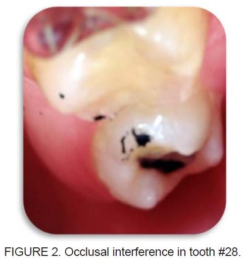
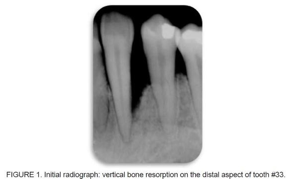
<body><![CDATA[<p><font size="2" face="Verdana, Arial, Helvetica, sans-serif">At the first visit, medical and dental history taking and clinical and radiographic   examination were performed. TMJ and the following muscles were sensitive to   palpation: inferior lateral pterygoid, temporal, left masseter, left medial pterygoid,   and left sphenomandibular. A 2-mm difference between the centric relation (CR)   and habitual occlusion (HO) was observed. Clinical examination of the lower left   canine (tooth #33) revealed grade II mobility, probing depth within normal limits,   and a positive pulp vitality test. Radiographic examination detected extensive vertical   bone resorption on the distal aspect of tooth #33, thus confirming mobility (<a href="#fig01">Figure   1</a>). The absence of tooth #36 resulted in the mesial movement of teeth #37 and #38,   causing the extrusion of tooth #28 and leading to an increased vertical dimension   of occlusion (VDO) in CR. Evaluation of mandibular dynamics revealed occlusal   interference between teeth #28 and #38 (<a href="#fig02">Figure 2</a>), causing anterior projection of the   mandible to the right. As a result, tooth #33 in HO was pushed distally due to forced   contact with the upper tooth, causing occlusal trauma and consequent vertical bone   resorption on the distal aspect of tooth #33 (<a href="#fig01">Figure   1</a>).</font></p>       <p>&nbsp;</p>     <p><a name="fig01"></a></p>     <p>&nbsp;</p>     <p align="center"><img src="/img/revistas/sto/v17n33/a09fig01.jpg">     <p>&nbsp;</p>     <p><a name="fig02"></a></p>     <p>&nbsp;</p>     <p align="center"><img src="/img/revistas/sto/v17n33/a09fig02.jpg">     <p>&nbsp;</p>     ]]></body>
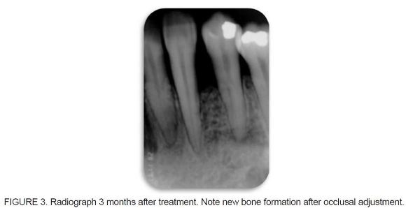
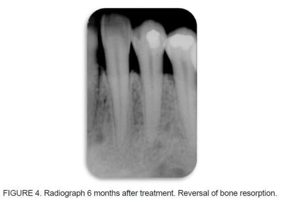
<body><![CDATA[<p><font size="2" face="Verdana, Arial, Helvetica, sans-serif">A two-phase molding process was carried out using condensation silicone impression   materials and a stock tray in the upper and lower arches. Casts were poured with type IV   improved dental stone and mounted on a semi-adjustable articulator (SAA) in the CR   position. Thus, it was possible to confirm occlusal interference between teeth #28 and #38, increased VDO, and occlusal overload in the lower left canine.</font></p>     <p><font size="2" face="Verdana, Arial, Helvetica, sans-serif">Treatment was initiated with removal of occlusal interference through occlusal   adjustment by selective grinding in CR. Occlusal contacts were marked using a thin carbon   articulating paper (Accufi lm II) held by Miller clamping tweezers, noting the presence   of interceptive or deflective contacts. High-speed carbide drills with a spherical-shaped   tip were selected for carrying out the grinding. Ten sessions were required to achieve a   balanced occlusion. There was remission of muscle and TMJ symptoms and headache.   Tooth #33 showed an improvement in mobility within 3 months of treatment (<a href="#fig03">Figure   3</a>). After 6 months, all symptoms, including pain and mobility, were no longer present.   During this 6-month period, there was radiographic evidence of bone healing on the distal   aspect of tooth #33 (<a href="#fig04">Figure   4</a>). This study is part of a research protocol that was approved   by the Local Ethics Committee (protocol no. 140/2003).</font></p>       <p>&nbsp;</p>     <p><a name="fig03"></a></p>     <p>&nbsp;</p>     <p align="center"><img src="/img/revistas/sto/v17n33/a09fig03.jpg">     <p>&nbsp;</p>     <p><a name="fig04"></a></p>     <p>&nbsp;</p>     <p align="center"><img src="/img/revistas/sto/v17n33/a09fig04.jpg">     ]]></body>
<body><![CDATA[<p>&nbsp;</p>     <p><font size="3" face="Verdana, Arial, Helvetica, sans-serif"><B>DISCUSSION</B></font></p>     <p><font size="2" face="Verdana, Arial, Helvetica, sans-serif">The influence of occlusal factors on injuries to the various elements of the   stomatognathic system, such as muscles, TMJ, teeth, and periodontal structures, should   be thoroughly studied. Under nonpathological conditions, the periodontal structures   would absorb incident forces in the axial direction. However, a change in the direction   of these forces would be detrimental, because horizontal loads are not well supported by the periodontium.</font></p>     <p><font size="2" face="Verdana, Arial, Helvetica, sans-serif">Occlusal trauma occurs due to interference, as interference leads to an increased   VDO. This type of injury is the result of occlusal forces applied outside the long axis of   the tooth and is called traumatic occlusion<sup>2-6</sup>. Bone resorption is often observed as a   result of trauma.</font></p>     <p><font size="2" face="Verdana, Arial, Helvetica, sans-serif">Tissue injury associated with occlusal trauma is divided into primary and secondary   trauma. The primary form occurs when tissue reactions affect teeth with a normal   periodontium, while the secondary form is related to damage to an already compromised   periodontium with reduced height<sup>1-4,7-9</sup>. Any type of occlusal interference may cause   abnormal muscle activity. Muscle hyperactivity can characterize damage to muscles, as   their normal function is disrupted, causing pain and tenderness<sup>1,10,11</sup>, as in the case   reported here.</font></p>     <p><font size="2" face="Verdana, Arial, Helvetica, sans-serif">During the functional course of mandibular closure, mechanical interference   may cause a deviation in the movement of the mandible, leading one or more teeth to   have abnormal occlusal contacts, causing detrimental effects on periodontal structures.   In some cases, incorrect tooth positioning causes the teeth to receive all occlusal   forces, thus being predisposed to changes in their supporting structures<sup>5,8,10,11</sup>. Abnormal muscle activity can cause traumatic/painful injury to joint structures. Other   damage caused by traumatic occlusion occurs in the dental pulp. Several studies   have reported the occurrence of pulp calcification and pulp necrosis when occlusal forces continue to act in a degenerative manner<sup>2,6,12,13</sup>. The periodontal tissues   respond to this increase in occlusal force by adaptive or degenerative changes, which   are reversible when the trauma is removed. The effect of occlusal forces on the   periodontium is influenced by their direction, duration, and frequency.</font></p>     <p><font size="2" face="Verdana, Arial, Helvetica, sans-serif">Repair occurs when occlusal forces are reduced or the tooth is moved away   from them. If these forces continue to act in a chronic fashion, tissues remodel to   better absorb the impact. This fact results in changes in the periodontal ligament,   alveolar bone, cementum and dental pulp, periapical inflammation, and root resorption<sup>1-9,13-16</sup>. The periodontium may also become more resistant to withstand the   conditions created. The consequences of this phenomenon can be seen at the tooth   level, leading to the formation of wear facets, severe attrition of the occlusal surfaces,   and even tooth fractures<sup>1</sup>.</font></p>     <p><font size="2" face="Verdana, Arial, Helvetica, sans-serif">Occlusal adjustment by selective grinding was indicated, because this   technique can promote an equal incidence of all forces on the teeth, which causes   the physiological and geometric axes to match, thus establishing a harmonious   relationship among the elements of the stomatognathic system through the elimination   of occlusal interferences<sup>17-24</sup>.</font></p>     <p><font size="2" face="Verdana, Arial, Helvetica, sans-serif">Removal of interference and the establishment of a balanced occlusion   resulted in reversal of periodontal injury (traumatic occlusion) and remission of all   symptoms.</font></p>     <p>&nbsp;</p>     ]]></body>
<body><![CDATA[<p><font size="3" face="Verdana, Arial, Helvetica, sans-serif"><B>FINAL CONSIDERATIONS</B></font></p>     <p><font size="2" face="Verdana, Arial, Helvetica, sans-serif">The correct diagnosis of primary occlusal trauma allows treatment with occlusal adjustment.</font></p>     <p>&nbsp;</p>     <p><font size="3" face="Verdana, Arial, Helvetica, sans-serif"><B>REFERENCES</B></font></p>     <!-- ref --><p><font size="2" face="Verdana, Arial, Helvetica, sans-serif">1. Lascala NT, Moussalli NH. Compendio Terap&ecirc;utico Periodontal 2a ed. S&atilde;o Paulo: Artes M&eacute;dicas; 1995.    &nbsp;&nbsp;&nbsp;&nbsp;&nbsp;&nbsp;&nbsp;&nbsp;[&#160;<a href="javascript:void(0);" onclick="javascript: window.open('/scielo.php?script=sci_nlinks&ref=485655&pid=S1519-4442201100010000900001&lng=','','width=640,height=500,resizable=yes,scrollbars=1,menubar=yes,');">Links</a>&#160;]<!-- end-ref --></font></p>     <!-- ref --><p><font size="2" face="Verdana, Arial, Helvetica, sans-serif">2. Carranza FA. Periodontia Cl&iacute;nica. 9a ed. Rio de Janeiro: Guanabara Koogan; 2004.    &nbsp;&nbsp;&nbsp;&nbsp;&nbsp;&nbsp;&nbsp;&nbsp;[&#160;<a href="javascript:void(0);" onclick="javascript: window.open('/scielo.php?script=sci_nlinks&ref=485657&pid=S1519-4442201100010000900002&lng=','','width=640,height=500,resizable=yes,scrollbars=1,menubar=yes,');">Links</a>&#160;]<!-- end-ref --></font></p>     <!-- ref --><p><font size="2" face="Verdana, Arial, Helvetica, sans-serif">3. Hallmon WH, Harrel SK. Occlusal analysis, diagnosis and management in the practice   of periodontics. Periodontology. 2000; 34: 151-164.    &nbsp;&nbsp;&nbsp;&nbsp;&nbsp;&nbsp;&nbsp;&nbsp;[&#160;<a href="javascript:void(0);" onclick="javascript: window.open('/scielo.php?script=sci_nlinks&ref=485659&pid=S1519-4442201100010000900003&lng=','','width=640,height=500,resizable=yes,scrollbars=1,menubar=yes,');">Links</a>&#160;]<!-- end-ref --></font></p>     ]]></body>
<body><![CDATA[<!-- ref --><p><font size="2" face="Verdana, Arial, Helvetica, sans-serif">4. Lindhe J. Tratado de Periodontia Cl&iacute;nica e Implantodontia Oral 3&ordf; ed. Rio de Janeiro:   Guanabara Koogan; 2004.    &nbsp;&nbsp;&nbsp;&nbsp;&nbsp;&nbsp;&nbsp;&nbsp;[&#160;<a href="javascript:void(0);" onclick="javascript: window.open('/scielo.php?script=sci_nlinks&ref=485661&pid=S1519-4442201100010000900004&lng=','','width=640,height=500,resizable=yes,scrollbars=1,menubar=yes,');">Links</a>&#160;]<!-- end-ref --></font></p>     <!-- ref --><p><font size="2" face="Verdana, Arial, Helvetica, sans-serif">5. Paiva HJ. Oclus&atilde;o; No&ccedil;&otilde;es e Conceitos B&aacute;sicos 1&ordf; ed: Santos, 1997.    &nbsp;&nbsp;&nbsp;&nbsp;&nbsp;&nbsp;&nbsp;&nbsp;[&#160;<a href="javascript:void(0);" onclick="javascript: window.open('/scielo.php?script=sci_nlinks&ref=485663&pid=S1519-4442201100010000900005&lng=','','width=640,height=500,resizable=yes,scrollbars=1,menubar=yes,');">Links</a>&#160;]<!-- end-ref --></font></p>     <!-- ref --><p><font size="2" face="Verdana, Arial, Helvetica, sans-serif">6. Harn WM et al. Effect of occlusal trauma on healing of periapical pathoses: report of   two cases. International Endodontic Journal. 2001; 34: 554-561.    &nbsp;&nbsp;&nbsp;&nbsp;&nbsp;&nbsp;&nbsp;&nbsp;[&#160;<a href="javascript:void(0);" onclick="javascript: window.open('/scielo.php?script=sci_nlinks&ref=485665&pid=S1519-4442201100010000900006&lng=','','width=640,height=500,resizable=yes,scrollbars=1,menubar=yes,');">Links</a>&#160;]<!-- end-ref --></font></p>     <!-- ref --><p><font size="2" face="Verdana, Arial, Helvetica, sans-serif">7. Gher ME. Changing concepts. The effects of occlusion on periodontitis. Dent Clin   North Am. 1998; 42(2): 285-299.    &nbsp;&nbsp;&nbsp;&nbsp;&nbsp;&nbsp;&nbsp;&nbsp;[&#160;<a href="javascript:void(0);" onclick="javascript: window.open('/scielo.php?script=sci_nlinks&ref=485667&pid=S1519-4442201100010000900007&lng=','','width=640,height=500,resizable=yes,scrollbars=1,menubar=yes,');">Links</a>&#160;]<!-- end-ref --></font></p>     <!-- ref --><p><font size="2" face="Verdana, Arial, Helvetica, sans-serif">8. Ishigaki S, Kurozumi T, Morishige E, Yatani H. Occlusal interference during mastication   can cause pathological tooth mobility. J Periodont Res. 2006; 41: 189-192.    &nbsp;&nbsp;&nbsp;&nbsp;&nbsp;&nbsp;&nbsp;&nbsp;[&#160;<a href="javascript:void(0);" onclick="javascript: window.open('/scielo.php?script=sci_nlinks&ref=485669&pid=S1519-4442201100010000900008&lng=','','width=640,height=500,resizable=yes,scrollbars=1,menubar=yes,');">Links</a>&#160;]<!-- end-ref --></font></p>     ]]></body>
<body><![CDATA[<p><font size="2" face="Verdana, Arial, Helvetica, sans-serif">9. Segura&ndash;Egea JJ, Jim&eacute;nez&ndash;Rubio A, Velasco&ndash;Ortega e, R&iacute;os&ndash;Santos JV. Talon Cusp   causing occlusal trauma and acute apical periodontitis: report of a case. Dental   Traumatol. 2003; 19: 55-59.</font></p>     <!-- ref --><p><font size="2" face="Verdana, Arial, Helvetica, sans-serif">10. Ash, Ramfjord, Schmidseder. Oclus&atilde;o 2a ed: Santos; 2007.    &nbsp;&nbsp;&nbsp;&nbsp;&nbsp;&nbsp;&nbsp;&nbsp;[&#160;<a href="javascript:void(0);" onclick="javascript: window.open('/scielo.php?script=sci_nlinks&ref=485672&pid=S1519-4442201100010000900010&lng=','','width=640,height=500,resizable=yes,scrollbars=1,menubar=yes,');">Links</a>&#160;]<!-- end-ref --></font></p>     <!-- ref --><p><font size="2" face="Verdana, Arial, Helvetica, sans-serif">11. Garcia AR. Fundamentos Te&oacute;ricos e Pr&aacute;ticos da Oclus&atilde;o S&atilde;o Paulo: Cid Editora,   2003.    &nbsp;&nbsp;&nbsp;&nbsp;&nbsp;&nbsp;&nbsp;&nbsp;[&#160;<a href="javascript:void(0);" onclick="javascript: window.open('/scielo.php?script=sci_nlinks&ref=485674&pid=S1519-4442201100010000900011&lng=','','width=640,height=500,resizable=yes,scrollbars=1,menubar=yes,');">Links</a>&#160;]<!-- end-ref --></font></p>     <!-- ref --><p><font size="2" face="Verdana, Arial, Helvetica, sans-serif">12. Harrel SK. Occlusal forces as a risk factor for periodontal disease. Periodontology   2000. 2006; 32: 111-117.    &nbsp;&nbsp;&nbsp;&nbsp;&nbsp;&nbsp;&nbsp;&nbsp;[&#160;<a href="javascript:void(0);" onclick="javascript: window.open('/scielo.php?script=sci_nlinks&ref=485676&pid=S1519-4442201100010000900012&lng=','','width=640,height=500,resizable=yes,scrollbars=1,menubar=yes,');">Links</a>&#160;]<!-- end-ref --></font></p>     <!-- ref --><p><font size="2" face="Verdana, Arial, Helvetica, sans-serif">13. Kumazawa, M., Kohsaka T, Yamasaki M, Nakamura H, Kameyama Y. Effect of   traumatic occlusion on periapical lesions in rats. Journal of Endodontics. 1995;</font> <font size="2" face="Verdana, Arial, Helvetica, sans-serif">21(7): 372-375.    &nbsp;&nbsp;&nbsp;&nbsp;&nbsp;&nbsp;&nbsp;&nbsp;[&#160;<a href="javascript:void(0);" onclick="javascript: window.open('/scielo.php?script=sci_nlinks&ref=485678&pid=S1519-4442201100010000900013&lng=','','width=640,height=500,resizable=yes,scrollbars=1,menubar=yes,');">Links</a>&#160;]<!-- end-ref --></font></p>     <p><font size="2" face="Verdana, Arial, Helvetica, sans-serif">14. Silva Filho CE, Silva EMM. Periodontite &ndash; Traumatismo oclusal. Rev Bras Odontol.   1986; 43(5): 8-12.</font></p>     ]]></body>
<body><![CDATA[<p><font size="2" face="Verdana, Arial, Helvetica, sans-serif">15. Scott WR. Application of &ldquo;cusp writer&rdquo; findings to practical and theoretical occlusal   problems. Part I. J Prosthet Dentistry. 1976; 35(2): 211-221.</font></p>     <!-- ref --><p><font size="2" face="Verdana, Arial, Helvetica, sans-serif">16. Pompei VC, Pacca CAA. Participa&ccedil;&atilde;o do Trauma Oclusal na Etiologia da Doen&ccedil;a   Periodontal. Rev Bras Odontol. 1988; 45(1): 48-50.    &nbsp;&nbsp;&nbsp;&nbsp;&nbsp;&nbsp;&nbsp;&nbsp;[&#160;<a href="javascript:void(0);" onclick="javascript: window.open('/scielo.php?script=sci_nlinks&ref=485682&pid=S1519-4442201100010000900016&lng=','','width=640,height=500,resizable=yes,scrollbars=1,menubar=yes,');">Links</a>&#160;]<!-- end-ref --></font></p>     <!-- ref --><p><font size="2" face="Verdana, Arial, Helvetica, sans-serif">17. Gray HS. Occlusal adjustment: principles and practice. New Zealand Dental Journal.   1994; 90:13-19.    &nbsp;&nbsp;&nbsp;&nbsp;&nbsp;&nbsp;&nbsp;&nbsp;[&#160;<a href="javascript:void(0);" onclick="javascript: window.open('/scielo.php?script=sci_nlinks&ref=485684&pid=S1519-4442201100010000900017&lng=','','width=640,height=500,resizable=yes,scrollbars=1,menubar=yes,');">Links</a>&#160;]<!-- end-ref --></font></p>     <!-- ref --><p><font size="2" face="Verdana, Arial, Helvetica, sans-serif">18. Bailey JO. Occlusal Adjustment. Dent Clin North Am.1995; 39(2): 441-458.    &nbsp;&nbsp;&nbsp;&nbsp;&nbsp;&nbsp;&nbsp;&nbsp;[&#160;<a href="javascript:void(0);" onclick="javascript: window.open('/scielo.php?script=sci_nlinks&ref=485686&pid=S1519-4442201100010000900018&lng=','','width=640,height=500,resizable=yes,scrollbars=1,menubar=yes,');">Links</a>&#160;]<!-- end-ref --></font></p>     <!-- ref --><p><font size="2" face="Verdana, Arial, Helvetica, sans-serif">19. Selaimen CMP, et al. Occlusal Risk Factors for Temporomandibular Disorders. Angle   Orthodontist. 2007; 77(3): 471-477.    &nbsp;&nbsp;&nbsp;&nbsp;&nbsp;&nbsp;&nbsp;&nbsp;[&#160;<a href="javascript:void(0);" onclick="javascript: window.open('/scielo.php?script=sci_nlinks&ref=485688&pid=S1519-4442201100010000900019&lng=','','width=640,height=500,resizable=yes,scrollbars=1,menubar=yes,');">Links</a>&#160;]<!-- end-ref --></font></p>     <!-- ref --><p><font size="2" face="Verdana, Arial, Helvetica, sans-serif">20. Au AR, Klineberg IJ. A new Approach for Accurate pre-planned occlusal adjustment.   Aust Dental Journal. 1994; 39(1): 11-14.    &nbsp;&nbsp;&nbsp;&nbsp;&nbsp;&nbsp;&nbsp;&nbsp;[&#160;<a href="javascript:void(0);" onclick="javascript: window.open('/scielo.php?script=sci_nlinks&ref=485690&pid=S1519-4442201100010000900020&lng=','','width=640,height=500,resizable=yes,scrollbars=1,menubar=yes,');">Links</a>&#160;]<!-- end-ref --></font></p>     <!-- ref --><p><font size="2" face="Verdana, Arial, Helvetica, sans-serif">21. Ash Jr M, Ward ML. Occlusal Adjustment: Quo Vadi? The Journal of Craniomandibular   Practice. 2003; 21(1): 1-4.    &nbsp;&nbsp;&nbsp;&nbsp;&nbsp;&nbsp;&nbsp;&nbsp;[&#160;<a href="javascript:void(0);" onclick="javascript: window.open('/scielo.php?script=sci_nlinks&ref=485692&pid=S1519-4442201100010000900021&lng=','','width=640,height=500,resizable=yes,scrollbars=1,menubar=yes,');">Links</a>&#160;]<!-- end-ref --></font></p>     <!-- ref --><p><font size="2" face="Verdana, Arial, Helvetica, sans-serif">22. Milosevic A. Occlusion 2: Occlusal splints, Analysis and Adjustment. Dental Update.   2003; 30; 416-422.    &nbsp;&nbsp;&nbsp;&nbsp;&nbsp;&nbsp;&nbsp;&nbsp;[&#160;<a href="javascript:void(0);" onclick="javascript: window.open('/scielo.php?script=sci_nlinks&ref=485694&pid=S1519-4442201100010000900022&lng=','','width=640,height=500,resizable=yes,scrollbars=1,menubar=yes,');">Links</a>&#160;]<!-- end-ref --></font></p>     <!-- ref --><p><font size="2" face="Verdana, Arial, Helvetica, sans-serif">23. Kerstein RB. Is patient Confirmation an Adequate Indicator of Occlusal Adjustment   Completion? Dentistry Today. 1997; 16(10): 72-75.    &nbsp;&nbsp;&nbsp;&nbsp;&nbsp;&nbsp;&nbsp;&nbsp;[&#160;<a href="javascript:void(0);" onclick="javascript: window.open('/scielo.php?script=sci_nlinks&ref=485696&pid=S1519-4442201100010000900023&lng=','','width=640,height=500,resizable=yes,scrollbars=1,menubar=yes,');">Links</a>&#160;]<!-- end-ref --></font></p>     <!-- ref --><p><font size="2" face="Verdana, Arial, Helvetica, sans-serif">24. McCullock AJ. Making Occlusion Work: 2. Practical Considerations. Dental Update.   2003; 30: 211-219.    &nbsp;&nbsp;&nbsp;&nbsp;&nbsp;&nbsp;&nbsp;&nbsp;[&#160;<a href="javascript:void(0);" onclick="javascript: window.open('/scielo.php?script=sci_nlinks&ref=485698&pid=S1519-4442201100010000900024&lng=','','width=640,height=500,resizable=yes,scrollbars=1,menubar=yes,');">Links</a>&#160;]<!-- end-ref --></font></p>     <p>&nbsp;</p>     ]]></body>
<body><![CDATA[<p>&nbsp;</p>     <p><font size="2" face="Verdana, Arial, Helvetica, sans-serif"><a name="back"/></a><a href="#top"><img src="/img/revistas/sto/v17n33/seta.jpg" border="0" align="absmiddle"/></a><b>Corresponding Author:</b></a>    <br> Professor Raulino Naves Borges    <br>  Faculdade de Odontologia &ndash; UFG    <br>  Av.  Universit&aacute;ria Esquina com 1&ordf; Avenida, s/n&ordm;, Setor Universit&aacute;rio, 3&ordm; andar    <br> CEP 74605&ndash;220 &ndash; Goi&acirc;nia &ndash; Goi&aacute;s &ndash; Brasil     <br> Tel./fax: +55 (54) 3313.3925    <br> E-mail:<a href="mailto:raulinonaves@hotmail.com">raulinonaves@hotmail.com</a></font>    <p>      ]]></body>
<back>
<ref-list>
<ref id="B1">
<nlm-citation citation-type="book">
<person-group person-group-type="author">
<name>
<surname><![CDATA[Lascala]]></surname>
<given-names><![CDATA[NT]]></given-names>
</name>
<name>
<surname><![CDATA[Moussalli]]></surname>
<given-names><![CDATA[NH]]></given-names>
</name>
</person-group>
<source><![CDATA[Compendio Terapêutico Periodontal]]></source>
<year>1995</year>
<edition>2</edition>
<publisher-loc><![CDATA[São Paulo ]]></publisher-loc>
<publisher-name><![CDATA[Artes Médicas]]></publisher-name>
</nlm-citation>
</ref>
<ref id="B2">
<nlm-citation citation-type="book">
<person-group person-group-type="author">
<name>
<surname><![CDATA[Carranza]]></surname>
<given-names><![CDATA[FA]]></given-names>
</name>
</person-group>
<source><![CDATA[Periodontia Clínica]]></source>
<year>2004</year>
<edition>9</edition>
<publisher-loc><![CDATA[Rio de Janeiro ]]></publisher-loc>
<publisher-name><![CDATA[Guanabara Koogan]]></publisher-name>
</nlm-citation>
</ref>
<ref id="B3">
<nlm-citation citation-type="journal">
<person-group person-group-type="author">
<name>
<surname><![CDATA[Hallmon]]></surname>
<given-names><![CDATA[WH]]></given-names>
</name>
<name>
<surname><![CDATA[Harrel]]></surname>
<given-names><![CDATA[SK]]></given-names>
</name>
</person-group>
<article-title xml:lang="en"><![CDATA[Occlusal analysis, diagnosis and management in the practice of periodontics]]></article-title>
<source><![CDATA[Periodontology]]></source>
<year>2000</year>
<volume>34</volume>
<page-range>151-164</page-range></nlm-citation>
</ref>
<ref id="B4">
<nlm-citation citation-type="book">
<person-group person-group-type="author">
<name>
<surname><![CDATA[Lindhe]]></surname>
<given-names><![CDATA[J]]></given-names>
</name>
</person-group>
<source><![CDATA[Tratado de Periodontia Clínica e Implantodontia Oral]]></source>
<year>2004</year>
<edition>3</edition>
<publisher-loc><![CDATA[Rio de Janeiro ]]></publisher-loc>
<publisher-name><![CDATA[Guanabara Koogan]]></publisher-name>
</nlm-citation>
</ref>
<ref id="B5">
<nlm-citation citation-type="book">
<person-group person-group-type="author">
<name>
<surname><![CDATA[Paiva]]></surname>
<given-names><![CDATA[HJ]]></given-names>
</name>
</person-group>
<source><![CDATA[Oclusão: Noções e Conceitos Básicos]]></source>
<year>1997</year>
<edition>1</edition>
<publisher-name><![CDATA[Santos]]></publisher-name>
</nlm-citation>
</ref>
<ref id="B6">
<nlm-citation citation-type="journal">
<person-group person-group-type="author">
<name>
<surname><![CDATA[Harn]]></surname>
<given-names><![CDATA[WM]]></given-names>
</name>
</person-group>
<article-title xml:lang="en"><![CDATA[Effect of occlusal trauma on healing of periapical pathoses: report of two cases]]></article-title>
<source><![CDATA[International Endodontic Journal]]></source>
<year>2001</year>
<volume>34</volume>
<page-range>554-561</page-range></nlm-citation>
</ref>
<ref id="B7">
<nlm-citation citation-type="journal">
<person-group person-group-type="author">
<name>
<surname><![CDATA[Gher]]></surname>
<given-names><![CDATA[ME]]></given-names>
</name>
</person-group>
<article-title xml:lang="en"><![CDATA[Changing concepts: The effects of occlusion on periodontitis]]></article-title>
<source><![CDATA[Dent Clin North Am]]></source>
<year>1998</year>
<volume>42</volume>
<numero>2</numero>
<issue>2</issue>
<page-range>285-299</page-range></nlm-citation>
</ref>
<ref id="B8">
<nlm-citation citation-type="journal">
<person-group person-group-type="author">
<name>
<surname><![CDATA[Ishigaki]]></surname>
<given-names><![CDATA[S]]></given-names>
</name>
<name>
<surname><![CDATA[Kurozumi]]></surname>
<given-names><![CDATA[T]]></given-names>
</name>
<name>
<surname><![CDATA[Morishige]]></surname>
<given-names><![CDATA[E]]></given-names>
</name>
<name>
<surname><![CDATA[Yatani]]></surname>
<given-names><![CDATA[H]]></given-names>
</name>
</person-group>
<article-title xml:lang="en"><![CDATA[Occlusal interference during mastication can cause pathological tooth mobility]]></article-title>
<source><![CDATA[J Periodont Res]]></source>
<year>2006</year>
<volume>41</volume>
<page-range>189-192</page-range></nlm-citation>
</ref>
<ref id="B9">
<nlm-citation citation-type="journal">
<person-group person-group-type="author">
<name>
<surname><![CDATA[Segura-Egea]]></surname>
<given-names><![CDATA[JJ]]></given-names>
</name>
<name>
<surname><![CDATA[Jiménez-Rubio]]></surname>
<given-names><![CDATA[A]]></given-names>
</name>
<name>
<surname><![CDATA[Velasco]]></surname>
<given-names><![CDATA[Ortega e]]></given-names>
</name>
<name>
<surname><![CDATA[Ríos-Santos]]></surname>
<given-names><![CDATA[JV]]></given-names>
</name>
</person-group>
<article-title xml:lang="en"><![CDATA[Talon Cusp causing occlusal trauma and acute apical periodontitis: report of a case]]></article-title>
<source><![CDATA[Dental Traumatol]]></source>
<year>2003</year>
<volume>19</volume>
<page-range>55-59</page-range></nlm-citation>
</ref>
<ref id="B10">
<nlm-citation citation-type="book">
<person-group person-group-type="author">
<name>
<surname><![CDATA[Ash]]></surname>
</name>
<name>
<surname><![CDATA[Ramfjord]]></surname>
</name>
<name>
<surname><![CDATA[Schmidseder]]></surname>
</name>
</person-group>
<source><![CDATA[Oclusão]]></source>
<year>2007</year>
<edition>2</edition>
<publisher-name><![CDATA[Santos]]></publisher-name>
</nlm-citation>
</ref>
<ref id="B11">
<nlm-citation citation-type="book">
<person-group person-group-type="author">
<name>
<surname><![CDATA[Garcia]]></surname>
<given-names><![CDATA[AR]]></given-names>
</name>
</person-group>
<source><![CDATA[Fundamentos Teóricos e Práticos da Oclusão]]></source>
<year>2003</year>
<publisher-loc><![CDATA[São Paulo ]]></publisher-loc>
<publisher-name><![CDATA[Cid Editora]]></publisher-name>
</nlm-citation>
</ref>
<ref id="B12">
<nlm-citation citation-type="journal">
<person-group person-group-type="author">
<name>
<surname><![CDATA[Harrel]]></surname>
<given-names><![CDATA[SK]]></given-names>
</name>
</person-group>
<article-title xml:lang="en"><![CDATA[Occlusal forces as a risk factor for periodontal disease]]></article-title>
<source><![CDATA[Periodontology 2000]]></source>
<year>2006</year>
<volume>32</volume>
<page-range>111-117</page-range></nlm-citation>
</ref>
<ref id="B13">
<nlm-citation citation-type="journal">
<person-group person-group-type="author">
<name>
<surname><![CDATA[Kumazawa]]></surname>
<given-names><![CDATA[M]]></given-names>
</name>
<name>
<surname><![CDATA[Kohsaka]]></surname>
<given-names><![CDATA[T]]></given-names>
</name>
<name>
<surname><![CDATA[Yamasaki]]></surname>
<given-names><![CDATA[M]]></given-names>
</name>
<name>
<surname><![CDATA[Nakamura]]></surname>
<given-names><![CDATA[H]]></given-names>
</name>
<name>
<surname><![CDATA[Kameyama]]></surname>
<given-names><![CDATA[Y]]></given-names>
</name>
</person-group>
<article-title xml:lang="en"><![CDATA[Effect of traumatic occlusion on periapical lesions in rats]]></article-title>
<source><![CDATA[Journal of Endodontics]]></source>
<year>1995</year>
<volume>21</volume>
<numero>7</numero>
<issue>7</issue>
<page-range>372-375</page-range></nlm-citation>
</ref>
<ref id="B14">
<nlm-citation citation-type="journal">
<person-group person-group-type="author">
<name>
<surname><![CDATA[Silva Filho]]></surname>
<given-names><![CDATA[CE]]></given-names>
</name>
<name>
<surname><![CDATA[Silva]]></surname>
<given-names><![CDATA[EMM]]></given-names>
</name>
</person-group>
<article-title xml:lang="pt"><![CDATA[Periodontite: Traumatismo oclusal]]></article-title>
<source><![CDATA[Rev Bras Odontol]]></source>
<year>1986</year>
<volume>43</volume>
<numero>5</numero>
<issue>5</issue>
<page-range>8-12</page-range></nlm-citation>
</ref>
<ref id="B15">
<nlm-citation citation-type="journal">
<person-group person-group-type="author">
<name>
<surname><![CDATA[Scott]]></surname>
<given-names><![CDATA[WR]]></given-names>
</name>
</person-group>
<article-title xml:lang="en"><![CDATA[Application of &#8220;cusp writer&#8221; findings to practical and theoretical occlusal problems: Part I]]></article-title>
<source><![CDATA[J Prosthet Dentistry]]></source>
<year>1976</year>
<volume>35</volume>
<numero>2</numero>
<issue>2</issue>
<page-range>211-221</page-range></nlm-citation>
</ref>
<ref id="B16">
<nlm-citation citation-type="journal">
<person-group person-group-type="author">
<name>
<surname><![CDATA[Pompei]]></surname>
<given-names><![CDATA[VC]]></given-names>
</name>
<name>
<surname><![CDATA[Pacca]]></surname>
<given-names><![CDATA[CAA]]></given-names>
</name>
</person-group>
<article-title xml:lang="pt"><![CDATA[Participação do Trauma Oclusal na Etiologia da Doença Periodontal]]></article-title>
<source><![CDATA[Rev Bras Odontol]]></source>
<year>1988</year>
<volume>45</volume>
<numero>1</numero>
<issue>1</issue>
<page-range>48-50</page-range></nlm-citation>
</ref>
<ref id="B17">
<nlm-citation citation-type="journal">
<person-group person-group-type="author">
<name>
<surname><![CDATA[Gray]]></surname>
<given-names><![CDATA[HS]]></given-names>
</name>
</person-group>
<article-title xml:lang="en"><![CDATA[Occlusal adjustment: principles and practice]]></article-title>
<source><![CDATA[New Zealand Dental Journal]]></source>
<year>1994</year>
<volume>90</volume>
<page-range>13-19</page-range></nlm-citation>
</ref>
<ref id="B18">
<nlm-citation citation-type="journal">
<person-group person-group-type="author">
<name>
<surname><![CDATA[Bailey]]></surname>
<given-names><![CDATA[JO]]></given-names>
</name>
</person-group>
<article-title xml:lang="en"><![CDATA[Occlusal Adjustment]]></article-title>
<source><![CDATA[Dent Clin North Am]]></source>
<year>1995</year>
<volume>39</volume>
<numero>2</numero>
<issue>2</issue>
<page-range>441-458</page-range></nlm-citation>
</ref>
<ref id="B19">
<nlm-citation citation-type="journal">
<person-group person-group-type="author">
<name>
<surname><![CDATA[Selaimen]]></surname>
<given-names><![CDATA[CMP]]></given-names>
</name>
</person-group>
<article-title xml:lang="en"><![CDATA[Occlusal Risk Factors for Temporomandibular Disorders]]></article-title>
<source><![CDATA[Angle Orthodontist]]></source>
<year>2007</year>
<volume>77</volume>
<numero>3</numero>
<issue>3</issue>
<page-range>471-477</page-range></nlm-citation>
</ref>
<ref id="B20">
<nlm-citation citation-type="journal">
<person-group person-group-type="author">
<name>
<surname><![CDATA[Au]]></surname>
<given-names><![CDATA[AR]]></given-names>
</name>
<name>
<surname><![CDATA[Klineberg]]></surname>
<given-names><![CDATA[IJ]]></given-names>
</name>
</person-group>
<article-title xml:lang="en"><![CDATA[A new Approach for Accurate pre-planned occlusal adjustment]]></article-title>
<source><![CDATA[Aust Dental Journal]]></source>
<year>1994</year>
<volume>39</volume>
<numero>1</numero>
<issue>1</issue>
<page-range>11-14</page-range></nlm-citation>
</ref>
<ref id="B21">
<nlm-citation citation-type="journal">
<person-group person-group-type="author">
<name>
<surname><![CDATA[Ash Jr]]></surname>
<given-names><![CDATA[M]]></given-names>
</name>
<name>
<surname><![CDATA[Ward]]></surname>
<given-names><![CDATA[ML]]></given-names>
</name>
</person-group>
<article-title xml:lang="en"><![CDATA[Occlusal Adjustment: Quo Vadi?]]></article-title>
<source><![CDATA[The Journal of Craniomandibular Practice]]></source>
<year>2003</year>
<volume>21</volume>
<numero>1</numero>
<issue>1</issue>
<page-range>1-4</page-range></nlm-citation>
</ref>
<ref id="B22">
<nlm-citation citation-type="journal">
<person-group person-group-type="author">
<name>
<surname><![CDATA[Milosevic]]></surname>
<given-names><![CDATA[A]]></given-names>
</name>
</person-group>
<article-title xml:lang="en"><![CDATA[Occlusion 2: Occlusal splints, Analysis and Adjustment]]></article-title>
<source><![CDATA[Dental Update]]></source>
<year>2003</year>
<volume>30</volume>
<page-range>416-422</page-range></nlm-citation>
</ref>
<ref id="B23">
<nlm-citation citation-type="journal">
<person-group person-group-type="author">
<name>
<surname><![CDATA[Kerstein]]></surname>
<given-names><![CDATA[RB]]></given-names>
</name>
</person-group>
<article-title xml:lang="en"><![CDATA[Is patient Confirmation an Adequate Indicator of Occlusal Adjustment Completion?]]></article-title>
<source><![CDATA[Dentistry Today]]></source>
<year>1997</year>
<volume>16</volume>
<numero>10</numero>
<issue>10</issue>
<page-range>72-75</page-range></nlm-citation>
</ref>
<ref id="B24">
<nlm-citation citation-type="journal">
<person-group person-group-type="author">
<name>
<surname><![CDATA[McCullock]]></surname>
<given-names><![CDATA[AJ]]></given-names>
</name>
</person-group>
<article-title xml:lang="en"><![CDATA[Making Occlusion Work: 2. Practical Considerations]]></article-title>
<source><![CDATA[Dental Update]]></source>
<year>2003</year>
<volume>30</volume>
<page-range>211-219</page-range></nlm-citation>
</ref>
</ref-list>
</back>
</article>