Toxic effects of carbamide peroxide on the dentin-pulp complex
Efeitos tóxicos produzidos pelo peróxido de carbamida sobre o complexo dentino-pulpar
Alessandra Dutra da Silva I; Márcia Kijner II; André Wiltgen III; Paulo Oliva de Borba IV
I Doctor in Oral Pathology, Professor of Dentistry at the Lutheran University of Brazil, Canoas Campus, RS, Brazil
II Master's in Oral Pathology, Professor of Dentistry at the Lutheran University of Brazil, Canoas Campus, RS, Brazil ]]>
III Master's in Genetic and Molecular Diagnosis, Professor in the Dentistry Program at the Lutheran University of Brazil, Canoas Campus, RS, Brazil
IV Doctor in Physiology, Pharmacology and Toxicology, Professor in the Dentistry Program at the Lutheran University of Brazil, Canoas Campus, RS, Brazil
The authors have no conflicts of interest to declare concerning the publication of this manuscript.
]]> ABSTRACT
The objective of the present study was to evaluate the toxicity of carbamide peroxide on the dentin-pulp complex in the teeth of young, male rats using this medication in different doses that are considered clinical and subclinical. The methodology used was a comparative histological analysis between the control group and the test groups. The test groups were characterized by the ingestion of carbamide peroxide, in different concentrations, in the water supplied to them for a period of 40 days. The concentrations were: 1.9 mg/ml (test 1), 0.95 mg/ml (test 2), 0.71 mg/ml (test 3), 0.47 mg/ml (test 4) and 0.24 mg/ml (test 5). The results in the test groups showed a decrease in the odontoblast layer as well as changes in its morphology, and thickening of the pre-dentin layer. This demonstrates that different concentrations of carbamide peroxide may cause significant changes in the dentin-pulp complex.
Keywords: Carbamide peroxide; Pulp; Toxicity.
RESUMO
O objetivo deste estudo foi avaliar a toxicidade do peróxido de carbamida sobre o complexo dentino-pulpar de dentes de ratos machos jovens, com o uso desta medicação em diferentes doses consideradas clínicas e subclínicas. A metodologia utilizada foi uma análise histológica comparativa entre o grupo controle e grupos testes, que se caracterizavam pela ingestão de peróxido de carbamida na água de abastecimento em diferentes concentrações: 1,9 mg/ml (teste1), 0,95 mg/ml (teste 2), 0,71 mg/ml (teste 3), 0,47 mg/ml (teste 4) e 0,24 mg/ml (teste 5) durante um período de 40 dias. Os resultados mostraram que os grupos testes apresentavam uma diminuição na camada de odontoblastos, assim como alterações na sua morfologia e espessamento da camada de pré-dentina, demonstrando que o peróxido de carbamida em diferentes concentrações pode provocar alterações significativas no complexo dentino-pulpar.
Palavras-chave: Peróxido de Carbamida; Polpa; Toxicidade.
INTRODUCTION
]]> Several methods of dental whitening have been used since the end of the 17th century and carbamide peroxide, in the most different concentrations, is the medication most used for this purpose. When metabolized in our body, carbamide peroxide breaks down into hydrogen peroxide (H2O2) and ureia, the latter being easily degraded, therefore innocuous. However, H2O2 has, as its final product, the radical (OH*), the second most powerful free radical, which causes premature aging processes and systemic disorders1-4.Hydrogen peroxide is neutralized in the saliva by the peroxidase enzymes and salivary catalase, producing a protective effect against the aggression that this substance may cause in the oral mucosa5-8.
Dental whiteners that have carbamide peroxide as a base are extremely effective. However, they are not always used in a safe way and, often, are used without proper guidance from the dentist. Hydrogen peroxide can cause the destruction of the patient's protective mechanisms such as the epithelial and salivary barrier and tissue protective enzymes, in addition to significant changes in the pulp-dentin complex. This produces toxic effects in fibroblasts and odontoblasts, such as reversible inflammatory reactions or even pulp tissue necrosis7,9,10.
Therefore, this study aims to evaluate the toxic effect of carbamide peroxide in different concentrations on the dentin-pulp complex.
MATERIALS AND METHODS
Forty-eight albino Wistar rats (Rattus norvegicus), weighing between 290 and 300g, were selected from the vivarium at the Lutheran University of Brazil. They were from the same lineage and had minimal weight changes. Later, they were randomly divided into six groups: a control group, which received only water and ration, and five test groups subjected to carbamide peroxide ingestion in the supply water in the following concentrations, during a 40-day period: 1.9 mg/ml (test 1), 0.95 mg/ml (test 2), 0.71 mg/ ml (test 3), 0.47 mg/ml (test 4) and 0.24 mg/ml (test 5).
Euthanasia was performed on the animals with subsequent removal of the mandible and teeth, which were placed in a solution of 10% formaldehyde and then in a solution for decalcification (5% nitric acid). After 5 days, they were removed, washed with water and placed in 70% alcohol. Thereafter, the process of neutralization with NaCO3H was initiated and, subsequently, the pieces were processed. Microtomy and staining with hematoxylin and eosin were conducted, and histological slides were obtained for pulp tissue analysis. The histological slides were prepared in the Physiology, Mutagenesis and Vivarium laboratories of the Lutheran University of Brazil, Canoas/POA.
Evaluation of the histological slides was performed using an Axiolable photomicroscope, model MC 80- (Zeiss-Germany) and analyses of the effect of carbamide peroxide on the dentin-pulp complex were performed on the histological slides. A random selection of the slide was done in each group by a blind and calibrated examiner.
]]> RESULTS
Effects of carbamide peroxide on the dentin-pulp complex
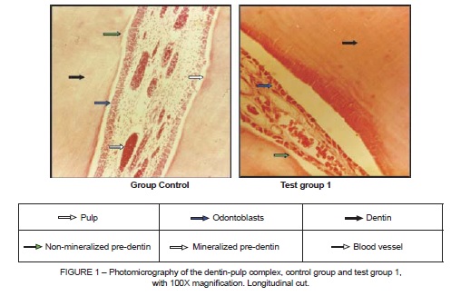
Figure 1 shows the population reduction of the odontoblast layer in test group 1, in addition to irregular thickness of the pre-dentin and odontoblasts which were cuboid in shape. This characterizes a reduction of the secretory phase of mineralized and collagenous tissue; that is, metabolic change of the dentin-pulp complex which determines a qualitative and quantitative change in the dental collagen and mineral secretion of this tissue.
Figure 2 also shows thickening of the non-mineralized pre-dentin layer, which determines an increase in the mineralization time of the dental tissue and odontoblast layer. This shows an indefinite and irregular aspect, with a cuboid shape, that is nonsecretory.
]]>
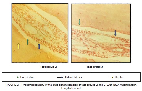
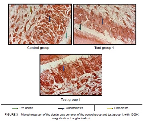
Figure 3 shows the dentin-pulp complex in the coronal extension, showing a reduction in the odontoblast layer in the central peripheral part of the pulp in test group 1. This practically became a row with only one cell connected to another cell, in addition to these cells presenting an inactive format; that is, cuboid, without formative conditions and much less reactive, besides an increase in the fibroblasts in the region near the odontoblasts. The lower part of the figure shows the irregular shape of the pre-dentin, which may be understood as a change in its mineralization while using carbamide peroxide.
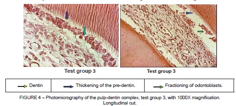
]]> Figure 4 shows a reduced number of odontoblasts, with migration to the pre-dentin layer, odontoblasts with cuboid shape. In the upper right part of the figure, the pre-dentin layer and dentinal tubules with an intense permeability aspect can be seen. The figure on the right shows morphological changes in the pre-dentin and odontoblast layer (migration of odontoblasts to the pre-dentin layer, and some showing a cuboid shape), a fractured odontoblast layer, a reduced odontoblast count and pre-dentin thickening.
DISCUSSION
The objective of the present study was to evaluate the effects of carbamide peroxide on the dentin-pulp complex. This was an innovative study, since there are no studies in the literature which performed a morphological analysis of the dental tissues related to the action of carbamide peroxide using an in vivo model. Currently, studies have evaluated the effect of carbamide peroxide in different concentrations, and its association with substances such as sodium fl uoride, in order to minimize the toxic effects and dentin sensitivity11-13.
Results of the present study showed changes caused by carbamide peroxide on the odontoblasts, such as modifications of the morphology and in the number of cells. This may promote mineralizing, secretory inactivity, which is shown by the changes in dimension of the dentinal tubules seen in the test groups, when compared to the control group. OSBORNI and TENCATE14 report that if there were damage to the dentin-pulp complex, however without death of the odontoblasts, dentinal changes like the deposition of scleral dentin and of reparative or tertiary dentin may occur. However, if there were death of the odontoblasts, such as found in the present study, then reparation of this tissue would not occur because the odontoblast is a terminal cell.
In the pre-dentin region, a thickening of this layer was found in the test groups. This indicates an increase in the mineralization time, indicating reactive difficulties. In addition, the loss of odontoblasts reflects a reduction in the production of pre-dentin, thus permitting the pulpar macrophages to become odontoclasts performing dentinal reabsorption. This is aggravated with the prolonged use of carbamide peroxide, causing increased dental sensitivity15.
]]>CONCLUSION
The toxicity of carbamide peroxide on the dentin-pulp complex was confirmed through the changes observed in the odontoblast and pre-dentin layers of the test groups. Thus, new studies are necessary in order to evaluate the potential for local and systemic pathogenicity of carbamide peroxide and its impact on public health.
REFERENCES
1. Timiras PS. Bases Fisiológicas del envejecimiento y geriatría. Barcelona: Editora Masson; 1997. 75p. [ Links ]
2. Woolverton CJ, Haywood VB, Heymann HO. Toxicity of two carbamide peroxide products used in night guard vital bleaching. Am J Dent. 1993;6: 310-314.
3. Hummert TW, Osborne JW, Norling BK, Cardenas HL. Mercury in solution following exposure of various amalgams to carbamide peroxides. Am J Dent. 1996: 305-309.
4. Berry, JH. What about whiteness safety concerns explored. JADA 1990; 121:223-225.
]]> 5. Salome P, Bueno RP, Nascimento PC, Pozzobon RT. Residual oxygen releasing time from dental structure after carbamide peroxide exposure: study of the effects of a neutralizer gel. Gen Dent. 2012;60(2):147-50.6. Pligina KL, Rodina IA, Shevchenko TV, Bekchanova ES, Tikhonov VP, Sirota NP. DNA damaging effects of dental bleaching agents. Bull Exp Biol Med. 2012;153(1):57-60.
7. Tipton DA, Braxton SD, Dabbous MK. Role of saliva and salivary components as modulators of bleaching agent toxicity to human gingival fibroblasts in vitro. J Periodontol. 1995; 66(9):766-74.
8. Leonard RH, Haywood VB, Phillips C. Risk factors for developing tooth sensitivity and gingival irritation associated with night guard vital bleaching. Quint Int. 1997;28: 527-534.
9. Almeida LC, Soares DG, Azevedo FA, Gallinari MO, Costa CA, Santos PH, Briso AL. At-Home Bleaching: Color Alteration, Hydrogen Peroxide Diffusion and Cytotoxicity. Braz Dent J. 2015; 26(4): 378-383.
10. Bowles WH, Burns H. Catalase/peroxidase activity in dental pulp. J Endod. 1992;18(11):527-34.
11. Soares DG, Ribeiro AP, Lima AF, Sacono NT, Hebling J, De Souza Costa CA. Effect of fl uoride treated enamel on indirect cytotoxicity of a 16% carbamide peroxide bleaching gel to pulp cells. Braz Dent J. 2013;24(2):121-7.
12. Lima AF, Ribeiro AP, Soares DG, Sacono NT, Hebling J, De Souza Costa CA. Toxic effects of daily applications of 10% carbamide peroxide on odontoblast-like MDPC-23 cells. Acta Odontol Scand. 2013;71(5):1319-25.
13. Lima AF, Lessa FC, Hebling J, De Souza Costa CA, Marchi GM. Protective Effect of Sodium Ascorbate on MDPC-23 Odontoblast-Like Cells Exposed to a Bleaching Agent. Eur J Dent. 2010;4(3):238-44.
14. Osborn, JW e Tencate, AR. Histologia Dental Avançada. 4a ed. São Paulo: Quintessence; 1983.101p.
]]> 15. Seghi, RR e Denry I. Effects of external bleaching on indentation and abrasion characteristics of human enamel in vitro. J Dent Res. 1992; 71:1340-1344.
 Correspondence:
Alessandra Dutra da Silva
Curso de Odontologia
Rua Farroupilha, 8001
CEP: 92425-900
Canoas/RS
E-mail: dra.alessandradutra@gmail.com
]]>