Cytokine profiles in the sera of Egyptian patients with oral pemphigus vulgaris
Hossam EidI;Basiouny El-GamalII
I PhD, Oral Medicine, Oral Diagnosis and Periodontology Department, Faculty of Dentistry, Suez Canal University, Egypt
II PhD, Clinical Biochemistry Department, College of Medicine, King Khalid University, Abha, Saudi Arabia
Cytokines have been suggested to play an important role in the pathogenesis of various inflammatory and autoimmune diseases, including the potentially fatal blistering disease, oral pemphigus vulgaris (PV). No data are currently available on the cytokine levels in the sera of Egyptian patients with oral PV. Aim:The aim of this study was to measure the serum levels of some proinflammatory and antiinflammatory cytokines in Egyptian patients with PV. Methods: Using highly sensitive ELISA kits, the levels of TNF-α, IL-2, IL-4 and IL-6 were measured in the sera of 10 patients affected with oral PV and 10 healthy subjects. Results: Serum levels of TNF-α and IL-6 were found to be significantly higher in patients with oral PV than in healthy controls (p<0.001). On the other hand, no significant differences were observed in the levels of IL-2 and IL-4 between oral PV and control sera (p<0.05). Conclusions: These data showed that TNF-α and IL-6 levels were significantly increased in the sera of Egyptian patients with oral PV and this might suggest its role in the pathogenesis of this disease.
Keywords: Pemphigus vulgaris, oral lesions, cytokines.
Introduction
Pemphigus vulgaris (PV) is a chronic, vesiculobullous, mucocutaneous autoimmune fatal disease, characterized by the presence of antibodies against adhesion molecules (desmoglein, Dsg3) present on the surface of keratinocytes, leading to the loss of cellular adhesion or acantholysis and is typically associated with oral lesions1-5. Histologically, PV can be detected by indirect immunofluorescence assay of anti-Dsg3 antibodies in the sera6 and blister fluid7.
In fact, the oral mucosa is the first site to be involved in up to 70% of cases, and it is the only site to be affected in over 50% of patients8. Most patients exhibit oral lesions at some time of the disease8. Clinically, the most common sites of oral PV lesions are the labial and buccal mucosa or the edentulous ridges. Oral lesions are commonly characterized by the presence of vesiculobullous and ulcerative lesions.
Recently, there has been increasing interest in the role of cytokines in the pathogenesis of various inflammatory and autoimmune diseases, including the potentially fatal blistering disease, PV1-2,4,8-13. Cytokines are regulatory compounds produced by cells of the immune system [T(H)1 and T(H)2] and act as intracellular mediators and control the immune and inflammatory reponses9. T(H)1 control the cell-mediated response and produce a number of proinflammatory cytokines, e.g. IL-1, IL-2, IL-6 and TNF-α which are counterbalanced by a number of antiinflammatory cytokines, e.g. IL-4 and IL-10 that are produced by T(H)2 that participate in humoral response and antibody production. Recent studies point out at proinflammatory cytokines such as TNF-α, IL-1, or IL-6 as strong players involved in this process13. Moreover, experimental studies revealed that synergistic cooperation of pemphigus antibodies with IL-6 and TNF-α in the pathogenesis of PV.
There is a fairly strong genetic background to pemphigus vulgaris with linkage to HLA class II alleles. Certain ethnic groups, such as Ashkenazi Jews and those of Mediterranean and South Asian origin are especially liable to PV14-15. No data are currently available on the cytokine levels in the sera of Egyptian patients with oral VP. In the present study, we measured the serum levels of some proinflammatory cytokines (IL-2, IL-6, TNF-α) and the antiinflammatory cytokine IL-4 in Egyptian patients with oral PV, in comparison to healthy controls.
]]>Material and methods
All participants were recruited from the Outpatient Clinics of the Department of Skin and Venereal Diseases, Faculty of Medicine, and the Department of Oral Medicine, Oral Diagnosis, and Periodontology, Faculty of Dentistry, Suez Canal University, Egypt. Ten patients with oral PV and 10 healthy controls were enrolled in the study after ethical approval by the Suez Canal University. A written informed consent was taken from all participants prior to enrollment.
The group of patients with oral PV consisted of 6 males and 4 females with mean age of 50.6 years (range: 45-60 years). The selection and diagnosis of patients was based on the history, clinical characteristics of oral PV lesions, the histopathological specimens and the indirect immunofluorescence testing6 of PV. The duration of oral PV lesions ranged from 1-3 years. None of the patients had received any topical and/or systemic treatment for the present illness at least 1 month prior to study.
The control group consisted of 6 males and 4 females with mean age of 48.4 years (range: 40-53 years). They were healthy volunteers completely free from any local or systemic diseases who were not taking any medication or contraceptives (in females).
Serum samples were collected from the individuals of the two groups and used to measure the levels of the cytokines TNF-α, IL-2, IL-4 and IL-6 using commercially available ELISA kits (Quantikine, R & D, Minneapolis, MN, USA), according to manufacturer's procedure.
All results were expressed as mean ± SD. Differences between two means were analyzed by Student's t-test. P values equal or less than 0.05 were considered as significant.
Results
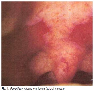
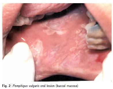
The most common sites of oral PV lesions found in the present study were the lip, buccal mucosa, palatal, ventral surface of tongue, and gingival. Examples of palatal mucosa and buccal mucosa with oral lesions in patients with PV are shown in Figures 1 and 2, respectively.
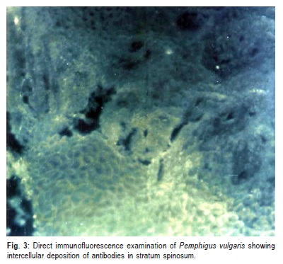
]]> Direct immunofluorescence testing revealed circulating pemphigus antibodies in all patients with oral PV. Figure 3 shows an example of PV with intercellular deposition of antibodies in stratum spinosum.There was no statistically significant difference (p>0.05) between patients with oral PV and controls with respect to the mean age.
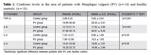
Table 1 shows serum cytokines levels (pg/mL) of TNFα, IL-2, IL-4, and IL-6 in patients with oral PV and controls. In comparison with controls, patients with oral PV had 640% and 179% higher mean TNF-α and IL-6 levels (p<0.001 and p<0.001), respectively. On the other hand, there were no significant differences in the levels of IL-2 and IL-4 of patients with oral PV compared with controls (p>0.05 and p>0.05), showing only 1% and 4% increase, respectively.
]]>
]]> Discussion
PV is a chronic, antibody mediated autoimmune disease that affects the skin and oral mucous membrane with distinct clinical, histopathological, and immunological features16. The exact cause of oral PV is unknown. Several studies support the immunological basis for the disease. Of importance to dentist is the frequency with which oral lesions were the initial presenting feature of oral PV as they often precede the skin lesions by several months or may be the major, if not the only, manifestation in some patients8.
Serum cytokine levels were recently investigated in many autoimmune disorders such as Lichen planus (LP) and rheumatoid arthritis6, and a relationship between serum cytokine levels and the clinical appearance of these diseases has been reported. Previous studies6,17 have reported that, the increase of some cytokines levels, such as IL-1, IL-6, IL- 8 and TNF-α in the serum of patients with inflammatory and autoimmune diseases is well known, and these elevated cytokine levels seem to be important mediators restricting each disease6,17. In the present study, the serum levels of some proinflammatory cytokines (IL-2, IL-6, TNF-α) and the antiinflammatory cytokine IL-4 were measured in Egyptian patients with oral PV and compared with healthy controls.
Cytokine generation is regulated by other cytokines and also by itself6. In cytokine network with autocrine and paracrine control, cytokine actions are usually balanced; however, cytokine imbalance occurs under pathological conditions and large amounts of cytokines are generated, which may be beneficial or harmful to the body17. Unbalanced cytokine actions are considered to be one of the immunopathogenesis mechanisms of autoimmune disorders6. Elevation of cytokine activities and unbalanced cytokine network may induce oral mucosal lesions6. The identification of cytokine activities in patient's tissues and sera seems to be advantageous for pathological analysis of oral diseases6.
The present study revealed a 640% increase in serum levels TNF-α of patients with oral PV compared to controls. Increased serum levels of TNF-α were reported by many authors. Thus, Alecu et al.18 found increased TNF-α levels in the sera and blister fluid obtained from patients with PV. Similar results were obtained by D'Auria et al.1,13 who showed increased levels and in situ expression of TNF-α. Also, Narbutt et al.2 showed 72% increase in TNF-α levels in the sera of patients with PV compared to control. This indicates that, TNF-α may play a role in the disease process of PV. Like IL-1, TNF-α is also a key stone in the cytokine network. TNF-α is a cytokine involved in the majority of inflammatory processes, and its increased activity is found in many skin diseases including psoriasis, SLE, or systemic sclerosis19-20. TNF-α is released by cells under various stimuli including bacterial infections or ultraviolet radiation. It plays a role in many biological processes, enhances phagocytosis, cytotoxicity, and modulates activity of other cytokines such as IL-1 and IL-421.
TNF-α is generated from macrophages, T and B cells and endothelial cells17. The important inducers of TNF-α production include viruses, IL-1, immune complex, endotoxin and TNF-α itself6. The TNF-α plays a major role in cell mediated cytotoxicity being able to induce cytotoxic T-cell differentiation, enhances monocyte cytotoxicity, and stimulates lymphokine activated natural killer cells22. TNF-α has marked effects on epithelial cells. It is cytotoxic at high concentration and anti-proliferative at lower concentration22. Prolonged release of TNF-α has been implicated in epithelial cell damage. So in the view of the enhanced cytotoxicity and epithelial cell lysis reported in PV, TNF-α seems to be implicated in the pathologic process of PV.
The increased serum levels of TNF-α may be attributed to the activation of macrophages during recognizing the antigenic epitopes and presenting them to T-lymphocytes. Moreover, mononuclear leukocytes might be stimulated to migrate into the submucosal area to produce cytokines including TNF-α23. Alternatively, the predominant cellular sources of TNF-α are the mast cells, macrophages, monocytes and endothelial cells. These cells can secrete TNF-α into the circulation, thus the TNF-α serum levels reach an appreciable amount that could play a significant role in the immunopathogenesis of PV, and in the same time gets reflected in the patient's serum22. Consequently, it seems that, in PV the antigen-antibody complex formation within the epithelial layer may induce increased cytokine release including TNF-α, which enhances the epithelial cell damage.
The IL-6 is a multifunctional cytokine which is an important mediator in host response to injury and infection. It stimulates the production of acute phase protein by liver cells24. Previous studies24-25, reported that, the production of IL-6 may be a response to many inducers such as TNF-α, IL- 4, IL-3, and some viruses. Moreover, the production of IL-6 by epithelial cells is increased in many inflammatory autoimmune diseases such as Lichen planus and psoriasis19,26. It seems likely therefore that, IL-6 plays an important role in immunopathogenesis of a number of immunoinflammatory skin diseases, and since several inflammatory skin diseases also affect the oral mucous membrane such as LP, SLE, and PV, it is reasonable to detect high serum level of IL-6 in patients with active PV oral lesions24.
The results of the present study revealed a marked increase of IL-6 serum levels (179%) in patients with oral PV compared with those of the control group. As much as 72% increase in serum IL-6 levels was reported by others in active stage of PV2. The elevated IL-6 serum levels in patients with active PV oral lesions seem to be compatible with the high levels of IL-6 generated and released in acute inflammatory conditions. The high IL-6 serum levels in patients with PV may be attributed to the elevated serum levels of TNF-α which is one of the strong inducers for generation and release of IL-624. There are wide variety of cells that are responsible for generation and release of IL-6 including lymphocytes, endothelial cells and epithelial cells. Therefore, IL-6 seems to be generated and released into the circulation in acute and chronic immunoinflammatory as well as autoimmune disorders17,24. Since PV is a chronic inflammatory mucocutaneous disease, the higher IL-6 serum levels in patients with oral PV seem to be a reasonable finding.
The findings of the present study revealed a slight, nonsignificant elevation of IL-4 serum levels in patients with oral PV compared with those of control. However, Satyam et al.9 reported significantly higher levels of serum IL-4 in patients with PV compared to healthy controls and suggested that this increase shows the induction of T(H)2 cells in the pathogenesis of PV. Also, Keskin et al.11 reported elevated levels of IL-4 in serum of patients with PV and showed reduced levels of this cytokine to the control values following treatment with high-dose, long term systemic corticosteroids with or without immunoglobulins. The IL-4 has a wide range of biological functions that include activation of immunoglobulin synthesis, T-cell proliferation, and T-cell adhesion to endothelial cells. Generation of IL-4 in some patients with autoimmune disorders seems to be likely in these diseases.
]]> In the present study, it could not be confirmed any significant changes in IL-2 serum levels in patients with oral PV when compared to the healthy controls. However, Satyam et al.9 reported decreased levels of IL-2 in the sera of patients with PV.In conclusion, the present findings showed that TNF-α and IL-6 levels were significantly increased in the sera of Egyptian patients with oral PV, probably suggesting its role in the pathogenesis of this disease.
Acknowledgements
We acknowledge the help and guidance of Prof Mohmoud Kandeel of Oral Diagnosis, Oral Medicine & Periodontology Department, Faculty of Oral & Dental Medicine, Cairo University, Egypt.
References
1. D'Auria L, Bonifati A, Mussi A, D'Agosto G, De Simone C, Giacalone et al. Cytokines in the sera of patients with pemphigus vulgaris: interleukin-6 and tumour necrosis factor-alpha levels are significantly increased as compared to healthy subjects and correlate with disease activity. Eur Cytok Netw. 1997; 8: 383-7. [ Links ]
2. Narbutt J, Lukamowicz J, Bogaczewicz J, Sysa-Jedrzejowska A, Torzecka JD, Leslak A. Serum concentrations of interleukin-6 is increased both in active and remission stages of Pemphigus vulgaris. Mediators Inflamm. 2008; 2008: 875-94.
]]> 3. Stern JNH, Keski n DB, Barteneva N, Zuniga J, Yunis EJ, Ahmed AR. Possible role of natural killer cells in Pemphigus vulgaris - preliminary observations. Clin Exp Immunol. 2008; 15: 472-81.4. Raychaudhuri SP, Smriti K. Biologics: target-specific treatment of systemic and cutaneous autoimmune diseases. Ind J Dermatol. 2009; 54: 100-9.
5. Femiano F. Pemphigus vulgaris: recent advances in our understanding of its pathogenesis. Minerva Stomatol. 2007; 56: 215-23.
6. Yamamoto T, Yoneda K, Ueta E. Serum cytokine levels in patients with oral mucous membrane disorders. J Oral Pathol Med. 1991; 20: 275-9.
7. Shanmugasekar C, Ram Gaesh VR, Jayaramun A, Srinivas CR. Blister fluid immunofluorescence in case of pemphigus vulgaris. Ind J Dermatol. 2010; 55: 188-9.
8. Mignogna MD, Lorenzolo M, Robert C. Oral pemphigus: Long term behaviour and clinical response to treatment with deflazacort in sixteen cases. J Oral Surg Pathol Med. 2000; 29: 145-52.
9. Satyam A, Khandpur S, Sharma VK, Sharma A. Involvement of T(H)1/ T(H)2 cytokines in the pathogenesis of autoimmune skin disease- Pemphigus vulgaris. Immunol Invest. 2009; 38: 498-509.
10. Baroni A, Perfetto B, Ruocco E, Greco R, Criscuolo D, Ruocco V. Cytokine pattern in blister fluid and sera of patients with pemphigus. Dermatology. 2002; 205: 116-21.
11. Keskin DB, Stern JN, Fridkis-Hareli M, Razzaque Ahme A. Cytokine profiles in pemphi-gus vulgaris patients treated with intravenous Igs as compared to controlled immunosuppressive therapy. Cytokine. 2008; 4: 15-21.
12. Feliciani C, Toto P, Amerio P, Pour SM, Coscione G, Shivji G et al. In vitro and in vivo expression of interleukin-1-alpha and tumor necrosis factor-alpha mRNA in pemphigus vulgaris: interleukin-1-alpha and tumor necrosis factor-alpha are involved in acantholysis. J Invest Dermatol. 2002; 114: 71-7.
]]> 13. Lopez-Robles E, Avalos-Diaz E, Vega-Memije E, et al. TNFá and IL-6 are mediators in the blistering process of pemphigus. Int J Dermatol. 2001; 40: 185-8.14. Ahmed AR, Yunis EJ, Kharti K, Wagner R, Notani G, Awdeh Z et al. Major histocompatibility complexhaplotype in Ashkenazi Jewish patients with pemphigus vulgaris. Proc Natl Acad Sci. 1990; 87: 7658-62.
15. Brenner S, Tur E, Shapio J, Ruocco V, A'avino M, Ruocco E et al. Pemphigus vulgaris: environmental factors, occupational, behavioral, medical and frequently quetionnaire . Int J Dermatol. 2001; 40: 562-9.
16. John C, Robinson MA, Lozada F. Oral pemphigus vulgaris. Oral Surg Oral Med Oral Pathol. 1997; 84: 349-55.
17. Tetsuya Y, Kazunori Y, Osaki T. Serum cytokines, interleukin-2 receptor, and soluble intercellular adhesion molecule-1 in oral disorders. Oral Surg Oral Med Oral Pathol. 1994; 78: 727-35.
18. Alecu M, Alecu S, Coman G, Galatescu E, Ursaciuc C. TNF-á and IL- 6 in serum and blister liquid of pemphigus vulgaris patients. Roumanian. Arch Microbiol Immunol. 1999; 58: 121-30.
19. Gomi T, Shiohara T, Munakata T, Imanishi K, Nagashima M. Interleukin 1á, tumor necrosis factor á, and interferon ã in psoriasis. Arch Dermatol. 1991; 127: 827-30.
20. Kantor TV, Friberg D, Medsger TA Jr, Buckingham RB, Whiteside TL. Cytokine production and serum levels in systemic sclerosis. Clin Immunol Immunopathol. 1992; 65: 278-85.
21. Kock A, Schwarz T, Kimbauer R, Urbanski A, Perry P, Ansel JC et al. Human keratinocytes are a source for tumor necrosis factor á: evidence for synthesis and release upon stimulation with endotoxin or ultraviolet light. J Exp Med. 1990; 172):1609-14.
22. Sugermann NW, Savage GJ, Seymour L. Is there a role for tumor necrosis factor-alpha (TNF-á) in oral lichen planus? J Oral Pathol Med. 1996; 25: 219-24.
]]> 23. Takeuchi Y, Tohani I, Kaneda T. Immuno-histochemical analysis of cells in mucosal lesions of lichen planus. J Oral Pathol Med. 1988; 17: 367-71.24. Farthing PM, Thornhill MH. IL-1 á and IL-6 production by oral and skin keratinocytes: similarities and differences in response to cytokine treatment in vitro. J Oral Pathol Med. 1996; 25: 157-62.
25. Wilson M, Reddi, Henderson B. Cytokine-inducing components of periodonto-pathogenic bacteria. J Priodont Res. 1996; 31: 393-407.
26. Yamamoto T, Osaki T, Ueta K. Cytokine production by keratinocytes and mononuclear infiltrates in oral lichen planus. J Oral Pathol Med. 1994; 23: 309-15.
 Correspondence:
Basiouny El-Gamal
Department of Clinical Biochemistry
College of Medicine, King Khalid University,
Abha, Saudi Arabia Phone: +966 5431757799
E-mail: basiouny_el_gamal@hotmail.com
Received for publication: 01, 2011
Accepted: April 20, 2011