Apical and cervical displacement produced by hand and engine-driven stainless steel and nickel-titanium instruments in simulated curved root canal
Benito André Silveira MiranziI; Gilberto Antônio BorgesI; Almir José Silveira MiranziIV; Fernando Hueb de MenezesII; Mário Alfredo Silveira MiranziIII; Luis Henrique BorgesI
I DDS, PhD, Department of Biomaterials, Dental School, University of Uberaba, Brazil
IIDDS, PhD, Department of Biopathology, Dental School, University of Uberaba, Brazil
IIIDDS, PhD, Department of Community Medicine, Federal University of Triângulo Mineiro Brazil
IVDDS, PhD, Department of Dental Prosthodontics, University of Uberaba, Brazil
]]>
ABSTRACT
Aim: The purpose of this study was evaluate the occurrence of displacement on the apical and cervical thirds of artificial curved root canals with 30° of curvature, comparing the use of stainless steel hand and oscillatory files with nickel-titanium (NI-TI) hand, rotary and oscillatory files. Methods: Seventy artificial curved root canals were divided into 7 groups (n=10). All canals were prepared with sizes 15 to 40 instruments at the working length according to a crown-down technique. The amount of material removed in the curved portion of the simulated canals at 2 mm level outside and 11 mm inside was measured. Data were analyzed statistically by ANOVA and Tukey's test (p<0.05). Results: There was statistically significant differences (p<0.05) at 2 mm level between NI-TI and steel stainless groups. At 11 mm, statistically significant differences (p<0.05) were observed between the groups that used rotary systems (5 and 6) and the remaining groups. Conclusions: The most centered preparations were those belonging to groups 5 and 6, at both levels. Root canal preparation with NI-TI files yielded better results than the stainless steel files especially when mechanically activated.
Keywords: root canal preparation, endodontic instruments, root canal therapy.
Introduction
The proper preparation of curved root canals is directly related to the capacity of providing uniform removal of intracanal dentin without resulting in undesirable canal aberrations. The use of stainless steel files for dilatation of curved canals may cause problems such as zipping, elbow, and perforations especially when the canals have to be enlarged above a size 25 file1.
The introduction of nickel-titanium (NI-TI) files made possible to provide more regular preparations because these instruments have high flexibility, low elasticity modulus and shape memory, which lead these files to producing fewer mistakes during instrumentation of narrow and curved canals1-6. It has been shown that instrumentation with stainless steel hand files yield a larger number of aberrations compared with NI-TI rotary files4-5,7-8. Nevertheless, neither stainless steel files nor NI-TI instruments (either manual or engine-driven) are able to yield complete cleaning and shaping of canal walls9-11.
The purpose of this study was evaluate the occurrence of displacement on the apical and cervical thirds of artificial curved root canals with 30° of curvature, comparing the use of stainless steel hand and oscillatory files with NI-TI hand, rotary and oscillatory files.
]]>Material and methods
Seventy artificial curved root canals (Tecnodon, Uberaba, MG, Brazil) were fabricated following the methodology proposed by Dummer et al .12 with gradual curvatures of about 30º, measured according to the Schneider's method13.
The working length (WL) was determined 1 mm short of the apical portion of the artificial root canals using size 10 K-files (Union Broach, Montgomeryville, PA, USA).
Three reference points were made in the acrylic blocks to allow further precise superimposition of the images obtained before and after preparation of the simulated root canals. The blocks were positioned always in the same direction and photographed with a Nikon F-2 camera (Nikon, Tokyo, Japan) using standard object/film distance. In order to quantify the possible distortions produced by instrumentation, two measured sections were put close to the resin blocks. After preparation, the blocks were photographed again, maintaining the initial position and the previously established object/film distance. After digitization of the photographs with a flatbed scanner Genius HR5 SCSI (KYE DCOM systems, China) images were superimposed using Adobe Photoshop 6.0 image-editing software and Image tool 3.0 image-analysis software for possible provoked changes.
The 70 canal-resin blocks were randomly assigned to 7 groups (n=10) and instrumented by a single operator. In all groups, the straight portion of the canals was prepared with size 1 to 3 Gates-Glidden drills (Union Broach, Montgomeryville, Pennsylvania USA), according to a crown-down instrumentation technique, and root canal preparation was complemented using sizes 15 to 40 files at the WL. In group 1, the canals were manually prepared with Flex-R stainless steel files (Union Broach) using balanced force. In group 2, the canals were manually prepared with Onix-R NI-TI files (Union Broach) as in group 1. In group 3, the simulated canals were prepared with Flex-R stainless steel files activated by Endo-Gripper oscillatory system (Union Broach). In group 4, the canals were prepared with Onix-R NI-TI files (Union Broach) activated by Endo-Gripper oscillatory system. In group 5, the canals were prepared with Pow-R NITI files (Union Broach) activated by Anthogyr (Micro-Mega, France) rotary pneumatic engine using a handpiece with 1:64 reduction. In group 6, the canals were prepared with Pow-R NI-TI files activated by Endo-Plus motor (VK Driller, Brazil) with controlled speed of 250 rpm. In group 7, the canals were prepared with Flex-R files activated by Endo-Plus motor and Endo-Gripper handpiece allowing constant oscillation of 250 rpm.
At each change of instrument, the canals were copiously irrigated with 2 mL of distilled water together with bi-distilled glycerin (Farmax, São Paulo, SP, Brazil) to lubricate the canals and facilitate instrumentation.
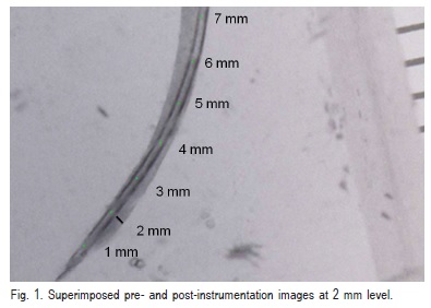
The blocks with root canals were fixed on a mini lathe (Western, L/C,T/T, China) to facilitate instrumentation. The superimposed images were magnified 4 times and evaluated using Image Tool 3.0 software, which allows measuring distances, angles and areas in digitized images. Initially, it was calibrated in millimeters as unit of measure having as reference to calibration the measured sections placed close to the blocks. Then, the software calculated by means of a drawning, the amount of material removed in the curved portion of the simulated canals at 2 mm level outside (Figure 1) and 11 mm inside.
The normality Lilliefors was applied to the two levels analyzed and it showed normal curve which allowed using one-way ANOVA and Tukey's post-hoc test. The significance level was set at 5%.
]]>
Results
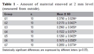
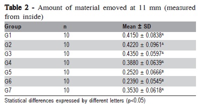
Results are shown in Tables 1 and 2. There were statistically significant differences (p<0.05) at 2 mm level between NI-TI and steel stainless groups (Table 1). On the other level, at 11 mm, statistically significant differences (p<0.05) were observed between groups 5 and 6 (rotary systems) when compared with the remaining groups (Table 2).
In group 1, it was observed loss of the WL and blockage, mainly after the use of size 25 file. Difficulties were found on moving from size 30 to 35 file and from size 35 to 40 file. There was zipping and elbow formation. A size 35 file fractured during instrumentation.
]]>
In group 2, there was difficulty in moving from size 20 to 25 file and from size 30 to 35 file. There was loss of WL but no instrument breakage was observed.
In group 3, files up to size 25 were flexible enough to accompany the curvature without resistance. However, from size 30 on there was loss of the WL and need for using greater force with the handpiece. In spite of zipping and elbow formation, there was neither fracture nor distortion of any instrument.
On the simulated canals in group 4, the file had to be first prepared manually, until reaching the WL and then the mechanical instrumentation system could be used. Sizes 35 and 40 files did not reach the WL. There was neither fracture nor distortion of any instrument.
]]> The files in group 5 had no difficulty in penetrating the canals. The WL was reached without finding resistance. There was not blockage and a size 25 file fractured.Likewise, the canals in group 6 were enlarged up to size 40 files without any difficulty. There was no instrument breakage and only a size 35 file presented tip distortion.
No instrument fracturing or distortion was observed in group 7. It was noticed transport starting from size 30 file with loss of the WL, blockage and zipping and elbow formation. These findings are similar to those of group 3, which used the same hand piece but without speed control.
Discussion
Several methodologies have been used to evaluate root canal preparation7-8,10-11. Simulated canals allow standardization of length, radius, diameter and curvature angle, while natural teeth present variations of canal shape and diameter. Dentin hardness may not be identical to that of the resin used for preparation of the blocks containing the artificial canals7-8,12.
In this study, both hand and engine-driven (oscillatory or rotary) stainless steel and NI-TI files had similar design configuration not to interfere with the results. The use of Pow-R is justified because the study evaluated the most effective preparation techniques and not the file design. Even though they are not used anymore, the Pow-R files are similar to the conventional (stainless steel) instruments used in hand instrumentation. Tan and Messer14 indicate files with small taper to finish the preparation in the work length, to improve the intracanal cleaning and filling.
It was observed that the NI-TI instruments were able to shape the simulated curved canals more appropriately, as observed in previous studies7-8,11. It was also verified that rotary NI-TI instrumentation using pneumatic or electric engines was more effective than manual instrumentation with stainless steel and NI-TI files. These findings are consistent with those of previous studies1,11. However, Rasquin et al.15 found better shaping and cleaning ability for AET (Endo- Eze®) files (oscillatory) compared with RaCe rotary system (FKG® Dentaire) in the cervical third. The authors did not found significant differences in the apical part.
In all groups, it was observed that root canal preparation selected areas on the curved portion of the canal; in the apical region of the curvature the selected areas occurred on the external side while in the cervical region of the curvature they occurred in the internal side. Similar outcomes have been reported2,7,16. Canals prepared with NI-TI instruments driven by rotary or oscillatory systems showed more centered preparations17.
There was greater impaction of resin and, consequently, loss of the WL for the stainless steel instruments used either manually or activated by the Endo-Gripper oscillatory motion. Similar observations with less extrusion and impaction using of NI-TI files coupled to rotary systems have been reported18-19.
]]> Preparation of curved root canals up to size 40 instrument confer a better cleaning and facilitate filling1,20. Therefore, all root canals were prepared up to size 40 files at the WL, aiming to comply with the basic requirements for good root canal shaping. However in the present results, the stainless steel instruments should be used up to file 20, followed by rotary Ni-Ti instruments. The investigation analyzed the hypothesis that the oscillatory instrumentation with controlled speed and stainless steel files would be an alternative to decrease deformations during preparation of curved and strait root canals. The results showed that no significant difference was found for the tested hypothesis, resulting in similar deformation and blockage (Table 1). The crown-down technique combined with apical enlargement determine apical foramen diameter, as well as prevent aberrations of curved canals1,18,21-22. As the instrumentation technique used in all groups was the same, it could be assumed that the type of metal and movement of instruments are crucial to keep the artificial root canal curvature. Further studies should be carried out on natural teeth to assess root canal cleaning because, as previously mentioned, simulated root canal methodology analyzes better other requirements.In conclusion, the most centered preparations were those belonging to groups prepared with NI-TI (groups 5 and 6) files and rotary systems, at both levels analyzed. Mechanical root canal preparation with NI-TI instruments yielded the best results.
References
1. Pécora JD, Capelli A. Shock of Paradigms on the instrumentation of curved root canals. Braz Dent J. 2006; 17: 3-5. [ Links ]
2. Gergi R, Rjeily JA, Sader J, Naaman A. Comparison of canal transportation and centering ability of Twisted Files, Pathfile-ProTaper system, and Stainless Stell Hand K-Files by using computed tomography. J Endod. 2010; 36: 904-7.
3. Sadeghi S. Shaping ability of NiTi rotary versus stainless stell hand instruments in simulated curved canals. Med Oral Patol Oral Cir Bucal. 2010; 15: 505-8.
4. Thompson SA. An overview of nickel-titanium alloys used in dentistry. Int Endod J. 2000; 33: 297-10.
5. Vanni JR, Albuquerque DS, Reiss C, Barato Filho F, Limongi O, Della Bona A. Apical displacement produced by rotary nickel-titanium instruments and stainless steel files. J Appl Oral Sci. 2004; 12: 51-5.
]]> 6. Walia H, Brantley WA, Gerstein H. An initial investigation of the bending and torsional properties of a Nitinol root canal files. J Endod. 1988: 14: 346-51.7. Schäfer E, Lohmann D. Efficiency of rotary nickel-titanium FlexMaster instruments compared with stainless steel hand K-Flexofiles – Part 1. Shaping ability in simulated curved canals. Int Endod J. 2002; 35: 505-13.
8. Schäfer E, Florek H. Efficiency of rotary nickel-titanium K3 instruments compared with stainless steel hand K-Flexofiles. Part 1. Shaping ability in simulated curved canals. Int Endod J. 2003; 36: 199-207.
9. Baumann MA. Nickel-titanium: options and challenges. Dent Clin North Am. 2004; 48: 55-67.
10. Chen JL, Messer HH. A comparison of stainless steel hand and rotary nickel-titanium instrumentation using a silicone impression technique. Aust Dent J. 2002; 47: 12-20.
11. Schirrmeister JF, Strohl C, Altenburger MJ, Wrbas K-T, Hellwig E. Shaping ability and safety of five different rotary nickel-titanium instruments compared with stainless steel hand instrumentation in simulated curved root canals. Oral Surg Oral Med Oral Pathol Oral radiol Endod. 2006; 101: 807-13.
12. Dummer PMH, Alodeh MHA, Al-Omari MAO. A method for the construction of simulated root canals in clear resin blocks. Int Endod J. 1991; 24: 63-6.
13. Schneider SW. A Comparison of canal preparation in straight and curved root canals. Oral Surg Oral Med Oral Pathol. 1971; 32: 271-5.
14. Tan BT, Messer HH. The quality of apicaql canal preparation using hand and rotary instruments with specific criteria for enlargement based on initial apical file size. J Endod. 2002; 28: 658-64.
15. Rasquin LC, Carvalho FB, Lima RKP. In vitro evaluation of root canal preparation using oscillatory and rotary systems in flattened root canals. J Appl Oral Sci. 2007; 15: 65-9.
]]> 16. Miranzi BAS, Miranzi MAS, Costa WF, Rezende LC. Estudo comparativo in vitro da instrumentação de canais artificiais mediante o emprego das limas Flex-R, valendo-se de dois tamanhos de preparos apicais. Rev USF. 1999; 17: 43-9.17. Setzer FC, Kwon TK, KarabucaK B. Comparison of apical transportation between two rotary files systems and two hybrid rotary instrumentation sequences. J Endod. 2010; 36: 1226-9.
18. Al-Omari MAO, Dummer PMH. Canal blockage and debris extrusion with eight preparation techniques. J Endod. 1995; 21: 154-8.
19. Mckendry DJ. Comparison of balanced forces, endosonic, and step-back filing instrumentation techniques: quantification of extruded apical debris. J Endod. 1990; 16: 24-7.
20. Card SJ, Sigurdsson A, Orstavik D, Trope M. The effectiveness of increased apical enlargement in reducing intracanal bacteria. J Endod. 2002; 28: 779-83.
21. Barroso JM, Guerisoli DMZ, Capelli A., Saquy PC, Pécora JD. Influence of cervical preflaring on determination of apical file size in maxillary premolars: SEM analysis. Braz Dent J. 2005; 16: 1-9.
22. Vanni JR, Santos R, Limongi O, Guerisoli DMZ, Capelli A, Pécora JD. Influence of cervical preflaring on determination of apical file size in maxillary molars: SEM analysis. Braz Dent J. 2005; 16: 181-6.
 Correspondence:
Benito André Silveira Miranzi ]]>
Universidade de Uberaba UNIUBE,
Departamento de Endodontia
Av. Nenê Sabino, 1801, Bloco H, Sala nº 207
3055-500 - Uberaba, MG, Brazil. Phone: (34)84069382
E-mail: bmiranzi@mednet.com.br
Received for publication: January 24, 2011
Accepted: May 31, 2011