<?xml version="1.0" encoding="ISO-8859-1"?><article xmlns:mml="http://www.w3.org/1998/Math/MathML" xmlns:xlink="http://www.w3.org/1999/xlink" xmlns:xsi="http://www.w3.org/2001/XMLSchema-instance">
<front>
<journal-meta>
<journal-id>1677-3225</journal-id>
<journal-title><![CDATA[Brazilian Journal of Oral Sciences]]></journal-title>
<abbrev-journal-title><![CDATA[Braz. j. oral sci.]]></abbrev-journal-title>
<issn>1677-3225</issn>
<publisher>
<publisher-name><![CDATA[Faculdade de Odontologia de Piracicaba, UNICAMP]]></publisher-name>
</publisher>
</journal-meta>
<article-meta>
<article-id>S1677-32252013000100011</article-id>
<title-group>
<article-title xml:lang="en"><![CDATA[Conservative treatment of the dentigerous cyst: report of two cases]]></article-title>
</title-group>
<contrib-group>
<contrib contrib-type="author">
<name>
<surname><![CDATA[Carrera]]></surname>
<given-names><![CDATA[Manoela]]></given-names>
</name>
<xref ref-type="aff" rid="A01"/>
</contrib>
<contrib contrib-type="author">
<name>
<surname><![CDATA[Dantas]]></surname>
<given-names><![CDATA[Danilo Borges]]></given-names>
</name>
<xref ref-type="aff" rid="A02"/>
</contrib>
<contrib contrib-type="author">
<name>
<surname><![CDATA[Marchionni]]></surname>
<given-names><![CDATA[Antônio Mareio]]></given-names>
</name>
<xref ref-type="aff" rid="A03"/>
</contrib>
<contrib contrib-type="author">
<name>
<surname><![CDATA[Oliveira]]></surname>
<given-names><![CDATA[Marília Gerhardt de]]></given-names>
</name>
<xref ref-type="aff" rid="A04"/>
</contrib>
<contrib contrib-type="author">
<name>
<surname><![CDATA[Andrade]]></surname>
<given-names><![CDATA[Miguel Gustavo Setúbal]]></given-names>
</name>
<xref ref-type="aff" rid="A03"/>
</contrib>
</contrib-group>
<aff id="A01">
<institution><![CDATA[,UNICAMP Piracicaba Dental School Departament of Oral Diagnosis]]></institution>
<addr-line><![CDATA[Piracicaba SP]]></addr-line>
<country>Brazil</country>
</aff>
<aff id="A02">
<institution><![CDATA[,Bahia Foundation for Developement of Science Area of Oral and Maxillofacial Surgery ]]></institution>
<addr-line><![CDATA[Salvador BA]]></addr-line>
<country>Brazil</country>
</aff>
<aff id="A03">
<institution><![CDATA[,Roberto Santos General Hospital and Child Hospital Area of Oral and Maxillofacial Surgeon ]]></institution>
<addr-line><![CDATA[Salvador BA]]></addr-line>
<country>Brazil</country>
</aff>
<aff id="A04">
<institution><![CDATA[,Catholic University of Rio Grande do Sul Areas of Oral and Maxillofacial Surgery and Pathology ]]></institution>
<addr-line><![CDATA[ ]]></addr-line>
</aff>
<pub-date pub-type="pub">
<day>00</day>
<month>03</month>
<year>2013</year>
</pub-date>
<pub-date pub-type="epub">
<day>00</day>
<month>03</month>
<year>2013</year>
</pub-date>
<volume>12</volume>
<numero>1</numero>
<fpage>52</fpage>
<lpage>56</lpage>
<copyright-statement/>
<copyright-year/>
<self-uri xlink:href="http://revodonto.bvsalud.org/scielo.php?script=sci_arttext&amp;pid=S1677-32252013000100011&amp;lng=en&amp;nrm=iso"></self-uri><self-uri xlink:href="http://revodonto.bvsalud.org/scielo.php?script=sci_abstract&amp;pid=S1677-32252013000100011&amp;lng=en&amp;nrm=iso"></self-uri><self-uri xlink:href="http://revodonto.bvsalud.org/scielo.php?script=sci_pdf&amp;pid=S1677-32252013000100011&amp;lng=en&amp;nrm=iso"></self-uri><abstract abstract-type="short" xml:lang="en"><p><![CDATA[The purpose of this paper is to present two cases of dentigerous cyst associated to permanent teeth in children treated by conservative techniques. Dentigerous cyst is the most common developmental cysts of the jaws. Conservative treatment is very effective to this entity and aims at eliminating the cystic tissue and preserving the permanent tooth involved in the pathology. Two techniques are described as conservative treatment for these cysts, marsupialization and the decompression. Two children presented with dentigerous cysts. A female child was affected by a large lesion at the right side of the mandible associated to tooth 45. The other lesion arose at the left maxilla associated to tooth 21 of a male child. Each dentigerous cyst promoted severe tooth displacement. The first patient was treated with decompression and the second with marsupialization.]]></p></abstract>
<kwd-group>
<kwd lng="en"><![CDATA[dentigerous cyst]]></kwd>
<kwd lng="en"><![CDATA[marsupializtion]]></kwd>
<kwd lng="en"><![CDATA[decompression]]></kwd>
<kwd lng="en"><![CDATA[conservative treatment]]></kwd>
</kwd-group>
</article-meta>
</front><body><![CDATA[ <p align="right"><font face="Verdana, Arial, Helvetica, sans-serif" size="2"><b>CASE    REPORT</b></font></p>     <p>&nbsp;</p>     <p><font face="Verdana, Arial, Helvetica, sans-serif" size="4"><b><a name="top"/></a>Conservative    treatment of the dentigerous cyst: report of two cases</b></font></p>     <p>&nbsp;</p>     <p>&nbsp;</p>     <p><font face="Verdana, Arial, Helvetica, sans-serif" size="2"><b>Manoela Carrera<sup>I</sup>;    Danilo Borges Dantas<sup>II</sup>; Ant&ocirc;nio Mareio Marchionni<sup>III</sup>;    Mar&iacute;lia Gerhardt de Oliveira<sup>IV</sup>; Miguel Gustavo Set&uacute;bal    Andrade<sup>III</sup></b></font></p>     <p><font face="Verdana, Arial, Helvetica, sans-serif" size="2"><sup>I</sup>Departament    of Oral Diagnosis, Piracicaba Dental School, University of Campinas (UNICAMP),    Piracicaba, SP, Brazil<br/>   <sup>II</sup>Area of Oral and Maxillofacial Surgery, Bahia Foundation for Developement    of Science (FBDC) /SESAB, Salvador, BA, Brazil<br/>   <sup>III</sup>Area of Oral and Maxillofacial Surgeon, Roberto Santos General    Hospital and Child Hospital, Salvador and Feira de Santana, Bahia, BA, Brazil<br/>   <sup>IV</sup>Areas of Oral and Maxillofacial Surgery and Pathology, Catholic    University of Rio Grande do Sul, Porto Alegre, RS, Brazil</font></p>     <p><font face="Verdana, Arial, Helvetica, sans-serif" size="2"><a href="#back">Correspondence</a></font></p>     <p>&nbsp;</p>     <p>&nbsp;</p> <hr noshade size="1">     ]]></body>
<body><![CDATA[<p><font face="Verdana, Arial, Helvetica, sans-serif" size="2"><b>ABSTRACT</b></font></p>     <p><font face="Verdana, Arial, Helvetica, sans-serif" size="2">The purpose of    this paper is to present two cases of dentigerous cyst associated to permanent    teeth in children treated by conservative techniques. Dentigerous cyst is the    most common developmental cysts of the jaws. Conservative treatment is very    effective to this entity and aims at eliminating the cystic tissue and preserving    the permanent tooth involved in the pathology. Two techniques are described    as conservative treatment for these cysts, marsupialization and the decompression.    Two children presented with dentigerous cysts. A female child was affected by    a large lesion at the right side of the mandible associated to tooth 45. The    other lesion arose at the left maxilla associated to tooth 21 of a male child.    Each dentigerous cyst promoted severe tooth displacement. The first patient    was treated with decompression and the second with marsupialization.</font></p>     <p><font face="Verdana, Arial, Helvetica, sans-serif" size="2"><b>Keywords:</b>    dentigerous cyst, marsupializtion, decompression, conservative treatment.</font></p> <hr noshade size="1">     <p>&nbsp;</p>     <p>&nbsp;</p>     <p><font face="Verdana, Arial, Helvetica, sans-serif" size="3"><b>Introduction</b></font></p>     <p><font face="Verdana, Arial, Helvetica, sans-serif" size="2">Dentigerous cyst    is the most common odontogenic development cyst. It can involve any included    tooth, although molars and canines are the most affected ones. Cystic formation    involving the crown of premolars and incisives is rare<sup>1</sup>.</font></p>     <p><font face="Verdana, Arial, Helvetica, sans-serif" size="2">The classic treatment    for dentigerous cysts is enucleation and extraction of the involved tooth. In    large cysts, an initial marsupialization can reduce the size of the bone defect    before definitive enucleation<sup>2</sup>. Since this cyst has a large prevalence    in children, marsupialization can be advisable as definitive treatment to allow    the eruption of the permanent tooth involved. Children have a great regenerative    potential and tooth with incomplete root development maintain the eruptive strenght<sup>3</sup>.    In such circunstances, therefore, marsupialization or descompression should    be tried as a major therapy<sup>1,4</sup>.</font></p>     <p><font face="Verdana, Arial, Helvetica, sans-serif" size="2">The aim of this    article is to present the cases of two children who presented large dentigerous    cyst associated to teeth where the incidence of this lesion is not so common.    Each cyst was treated by different surgical techniques, marsupialization and    decompression. These children had their permanent teeth erupted and they were    functional in the dental arch in just six months. This paper also illustrates    how these easy therapies are considered important and conservative modalities    to approach this pathology.</font></p>     <p>&nbsp;</p>     ]]></body>
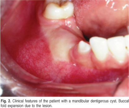
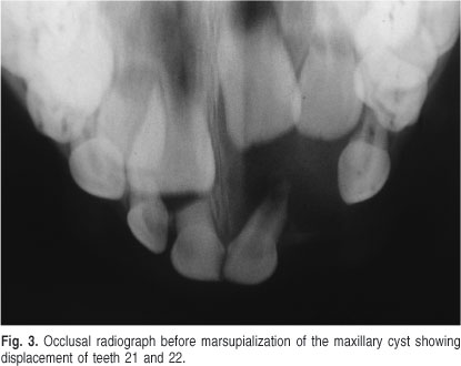
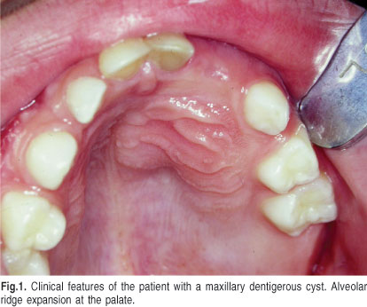
<body><![CDATA[<p><font face="Verdana, Arial, Helvetica, sans-serif" size="3"><b>Clinical Cases</b></font></p>     <p><font face="Verdana, Arial, Helvetica, sans-serif" size="2">This work was conducted    in accordance with bioethical concerns. Written informed consents were obtained    from the children's parents for all imaging exams, for the treatment modality    adopted, and for the publication of the data presented in this paper.</font></p>     <p><font face="Verdana, Arial, Helvetica, sans-serif" size="2">Two patients, a    boy aged 7 years and a girl aged 11 years, were referred to the Oral Surgery    Clinic of the Oral and Maxillofacial Surgery Master's Program of the Catholic    University of Rio Grande do Sul, Brazil. They were accompanied by their respective    parents who complained about facial asymmetry and delay of permanent tooth eruption    in both children. No history of systemic pathologies or previous trauma in the    affected area were reported.</font></p>     <p><font face="Verdana, Arial, Helvetica, sans-serif" size="2">The boy presented    an important facial asymmetry with expansion of paranasal region and upper lip    on the left side that caused elevation of the nasal wing. Intraoral exam revealed    compressible and painless expansion of buccal and palatal cortical plates of    the alveolar ridge at the region of teeth 61, 62, 63 (<a href="#f1">Figure 1</a>).    Dentition was mixed, free from carious lesions and no permanent maxillary incisors    were present in the arch. There was no sensorial or motor deficit at the facial    structures.</font></p>     <p><a name="f1"></a></p>     <p>&nbsp;</p>     <p align="center"><img src="/img/revistas/bjos/v12n1/a11fig01.jpg"></p>     <p>&nbsp;</p>     <p><font face="Verdana, Arial, Helvetica, sans-serif" size="2">The girl presented    a discreet expansion at the right side of the mandibular body resulting in a    mild facial asymmetry. Bone expansion of the buccal cortical plate of the alveolar    ridge caused deletion of the buccal folder (<a href="#f2">Figure 2</a>). Dentition    was also mixed, free from carious lesions and the eruption of teeth 44 and 45    was delayed. There was no complaint of dysesthesia in the face.</font></p>     <p><a name="f2"></a></p>     ]]></body>
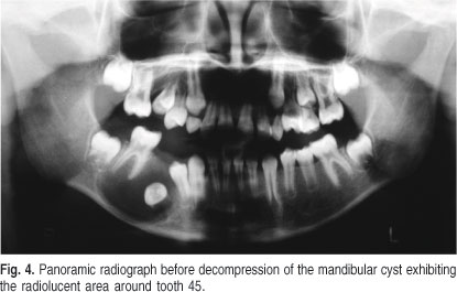
<body><![CDATA[<p>&nbsp;</p>     <p align="center"><img src="/img/revistas/bjos/v12n1/a11fig02.jpg"></p>     <p>&nbsp;</p>     <p><font face="Verdana, Arial, Helvetica, sans-serif" size="2">In both cases,    the radiographic findings revealed a well-delimited radiolucent area, measuring    approximately 20 mm in its largest diameter, with sclerotic margins, completely    associated with the crown of the involved permanent teeth, all in Nolla's stage    8 of tooth development. In the first patient, tooth 21 was within of the radiolucent    image that was also</font></p>     <p><font face="Verdana, Arial, Helvetica, sans-serif" size="2">impacting tooth    22 (<a href="#f3">Figure 3</a>). The other patient had tooth 45 involved with    the radiolucent lesion, and tooth 44 was slightly impacted by the radiolucid    area (<a href="#f4">Figure 4</a>).</font></p>     <p><a name="f3"></a></p>     <p>&nbsp;</p>     <p align="center"><img src="/img/revistas/bjos/v12n1/a11fig03.jpg"></p>     <p>&nbsp;</p>     <p><a name="f4"></a></p>     ]]></body>
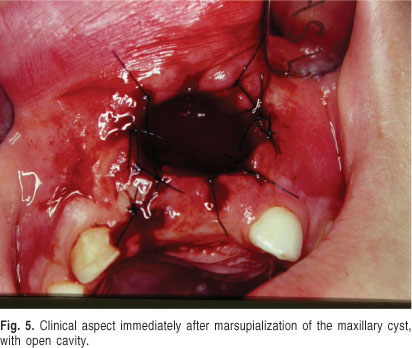
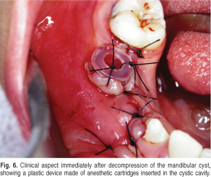
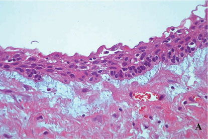
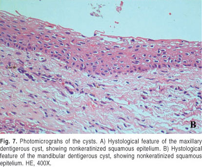
<body><![CDATA[<p>&nbsp;</p>     <p align="center"><img src="/img/revistas/bjos/v12n1/a11fig04.jpg"></p>     <p>&nbsp;</p>     <p><font face="Verdana, Arial, Helvetica, sans-serif" size="2">Aspiration of the    lesion fluid revealed, in both cases, a serous and bloody liquid content. The    clinical diagnosis of dentigerous cyst was concluded allowing the beginning    of an initial treatment with marsupialization in the case of the cyst that involved    the maxillary teeth (<a href="#f5">Figure 5</a>). The lesion at the girl's jaw    was treated by decompression (<a href="#f6">Figure 6</a>). During the surgical    procedures a biopsy was made and the histopathological examination confirmed    the clinical diagnosis of the dentigerous cyst for the lesions of both patients    (<a href="#f7">Figure 7 A</a> and <a href="#f7b">B</a>).</font></p>     <p><a name="f5"></a></p>     <p>&nbsp;</p>     <p align="center"><img src="/img/revistas/bjos/v12n1/a11fig05.jpg"></p>     <p>&nbsp;</p>     <p><a name="f6"></a></p>     <p>&nbsp;</p>     ]]></body>
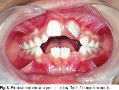
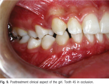
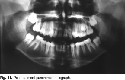
<body><![CDATA[<p align="center"><img src="/img/revistas/bjos/v12n1/a11fig06.jpg"></p>     <p>&nbsp;</p>     <p>&nbsp;</p>     <p><a name="f7"></a></p>     <p>&nbsp;</p>     <p align="center"><img src="/img/revistas/bjos/v12n1/a11fig07a.jpg">    <br>   <a name="f7b"></a><img src="/img/revistas/bjos/v12n1/a11fig07b.jpg"> </p>     <p>&nbsp;</p>     <p><font face="Verdana, Arial, Helvetica, sans-serif" size="2">The follow-up was    accomplished at 7, 30, 60, 120 and 180 days. In both patients, a progressive    improvement was observed in the facial asymmetry. Radiographic decrease of the    lesion diameter and the eruption of the involved teeth were achieved already    in the first 2 months. At 6 months, all teeth had erupted in both cases.</font></p>     <p><font face="Verdana, Arial, Helvetica, sans-serif" size="2">The boy's maxillary    left central incisor erupted with a discrete rotation and the left lateral incisor    was in the course of eruption (<a href="#f8">Figure 8</a>). In the girl, the    two premolars, previously impacted by the cyst, were present in mouth (<a href="#f9">Figure    9</a>). There was no evidence of pathological</font></p>     ]]></body>
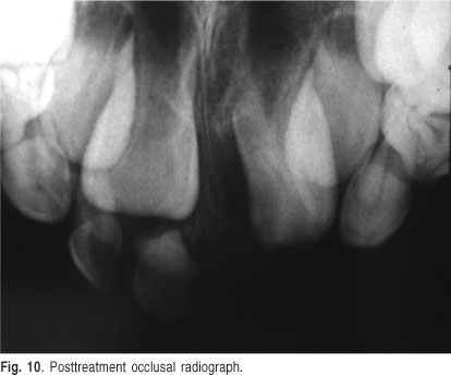
<body><![CDATA[<p><a name="f8"></a></p>     <p>&nbsp;</p>     <p align="center"><img src="/img/revistas/bjos/v12n1/a11fig08.jpg"></p>     <p>&nbsp;</p>     <p><a name="f9"></a></p>     <p>&nbsp;</p>     <p align="center"><img src="/img/revistas/bjos/v12n1/a11fig09.jpg"></p>     <p>&nbsp;</p>     <p><font face="Verdana, Arial, Helvetica, sans-serif" size="2">radiolucency in    both cases (<a href="#f10">Figures 10</a> and <a href="#f11">11</a>).There were    no differences between the two cases with respect to the eruption rate of the    involved teeth nor in the velocity of involution of the radiographic radiolucency.</font></p>     <p><a name="f10"></a></p>     ]]></body>
<body><![CDATA[<p>&nbsp;</p>     <p align="center"><img src="/img/revistas/bjos/v12n1/a11fig10.jpg"></p>     <p>&nbsp;</p>     <p><a name="f11"></a></p>     <p>&nbsp;</p>     <p align="center"><img src="/img/revistas/bjos/v12n1/a11fig11.jpg"></p>     <p>&nbsp;</p>     <p><font face="Verdana, Arial, Helvetica, sans-serif" size="2">It is important    to emphasize that no orthodontic appliance was necessary to assist the eruptive    process. After two years of clinical and radiographic follow-up, there were    no signs of recurrence of the lesion.</font></p>     <p>&nbsp;</p>     <p><font face="Verdana, Arial, Helvetica, sans-serif" size="3"><b>Discussion</b></font></p>     ]]></body>
<body><![CDATA[<p><font face="Verdana, Arial, Helvetica, sans-serif" size="2">The genesis of    the dentigerous cysts has been discussed by Benn and Altini<sup>5</sup>, who    proposed two different processes for cystic degeneration of the reduced epithelium    of enamel organ of an included tooth. The first phenomenon is usually associated    to the compression promoted by tooth eruption at the pericoronary follicle,    which induces fluid accumulation between this tissue and tooth crown.</font></p>     <p><font face="Verdana, Arial, Helvetica, sans-serif" size="2">The second mechanism    is associated to an apical inflammation in the primary predecessor whose cytokines    stimulates cystic degeneration of the permanent tooth follicle. In these cases,    the lesion would be early discovered at the second decade of life during routine    radiographic examinations or even later due to pain and facial swelling<sup>5</sup>.    Both cases described at this paper were probably associated to cystic degeneration    of the permanent teeth follicle since no relationship with inflammatory processes    caused by caries in the primary dentition could be confirmed once both children    had healthy teeth.</font></p>     <p><font face="Verdana, Arial, Helvetica, sans-serif" size="2">Dentigerous cyst    shares clinical and radiographic features with other lesions such as odontogenic    keratocyst, ameloblastoma, ameloblastic fibroma and adenomatoid odontogenic    tumor<sup>5</sup>, which can simultaneously occur with this cyst<sup>6-8</sup>.    Its epitelium can suffer metaplasia into an odontogenic tumor and also into    malign lesions<sup>7</sup>. For this reasons, biopsy is a fundamental diagnostic    element. The conservative techniques to both patients adopted allowed obtaining    parts of the lesion in the same surgical procedure. Since the clinical diagnosis    was confirmed, no other intervention, than follow up, was necessary.</font></p>     <p><font face="Verdana, Arial, Helvetica, sans-serif" size="2">Some different    surgical therapeutic modalities have been applied to treat dentigerous cysts.    Among them, the conservative option is very important in cases of large lesions    and when the permanent teeth involved have eruptive potential<sup>8</sup>. One    possibility is the insertion of a device in cystic lumen in order to keep patent    the communication between the interior of the cyst and the mouth. This procedure    characterizes the decompression and differs from marsupialization because this    technique involves the biopsy of large fragments of tissue<sup>9</sup>.</font></p>     <p><font face="Verdana, Arial, Helvetica, sans-serif" size="2">Factors such as    angulation and position of the tooth associated to the cyst, as well as the    degree of root formation are important requirements for tooth eruption. The    size of the lesion has no influence at eruption. As soon as cyst regression    begins, the involved tooth erupts in the most convenient position depending    on its relationship with the neighboring structures<sup>10</sup>.</font></p>     <p><font face="Verdana, Arial, Helvetica, sans-serif" size="2">The main advantages    of marsupialization or decompression for the treatment of dentigerous cyst are    the stimulus for bone formation after the decrease of cystic pressure. The communication    with oral secretions increases the inflammatory process and both macrophages    and lymphocytes from inflammation release growth factors that enhance bone formation<sup>11-13</sup>.    Preservation of the involved teeth is also another relevant feature of those    modalities<sup>13</sup>.</font></p>     <p><font face="Verdana, Arial, Helvetica, sans-serif" size="2">The largest disadvantage    of the conservative treatment is the preservation of pathological tissue inside    the jaws without histological examination<sup>13</sup>. The lack of patient's    cooperation in cleaning the pathological cavity and the need for periodic follow-up    visits also represent additional disadvantages.</font></p>     <p><font face="Verdana, Arial, Helvetica, sans-serif" size="2">The period of 3    months after marsupialization is suggested as the critical time to decide for    an additional treatment to stimulate tooth eruption if it did not occurred after    cyst regression. After this period, the surgeon should decide between tooth    extraction and orthodontic traction<sup>10</sup>. Other authors share this opinion    and also suggest that after 3 months the size of the cyst decreases and tooth    eruption can already be confirmed radiographically<sup>13</sup>.</font></p>     <p><font face="Verdana, Arial, Helvetica, sans-serif" size="2">The patients reported    in this paper were treated only with decompression or marsupialization. Both    presented cysts with expressive dimensions and two permanent teeth were affected    by the lesion. Since children and their respective parents were very cooperative,    the therapeutic option was curative.</font></p>     <p><font face="Verdana, Arial, Helvetica, sans-serif" size="2">Treatment of dentigerous    cyst through conservative therapy is preferable in children. Marsupialization    and decompression may represent the treatment of choice, but they are also useful    prior to extensive enucleation or curettage. Inherent peculiarities to the dentigerous    cyst regarding size and location, linked to root development and dental positioning,    as well as patient's profile should be strongly considered for a safe and effective    treatment. Marsupialization and decompression are very low invasive techniques    that could easily be conducted by any dentist familiar with basic surgical procedures,    in order to treat the pathology and preserve the tooth or teeth involved with    the cyst.</font></p>     ]]></body>
<body><![CDATA[<p>&nbsp;</p>     <p><font face="Verdana, Arial, Helvetica, sans-serif" size="3"><b>References</b></font></p>     <!-- ref --><p><font face="Verdana, Arial, Helvetica, sans-serif" size="2">1. Kumar R, Singh    RK, Pandey RK, Mohammad S, Ram H. Inflammatory dentigerous cyst in a ten-year-old    child. Natl J Maxillofac Surg. 2012; 3: 80-3.    &nbsp;&nbsp;&nbsp;&nbsp;&nbsp;&nbsp;&nbsp;&nbsp;[&#160;<a href="javascript:void(0);" onclick="javascript: window.open('/scielo.php?script=sci_nlinks&ref=036921&pid=S1677-3225201300010001100001&lng=','','width=640,height=500,resizable=yes,scrollbars=1,menubar=yes,');">Links</a>&#160;]<!-- end-ref --></font></p>     <!-- ref --><p><font face="Verdana, Arial, Helvetica, sans-serif" size="2">2. Pinheiro RS,    Castro GF, Roter M, Netto R, Meirelles V Jr, Janini ME, et al. An unusual dentigerous    cyst in a young child. Gen Dent. 2013; 61: 62-4.    &nbsp;&nbsp;&nbsp;&nbsp;&nbsp;&nbsp;&nbsp;&nbsp;[&#160;<a href="javascript:void(0);" onclick="javascript: window.open('/scielo.php?script=sci_nlinks&ref=036923&pid=S1677-3225201300010001100002&lng=','','width=640,height=500,resizable=yes,scrollbars=1,menubar=yes,');">Links</a>&#160;]<!-- end-ref --></font></p>     <!-- ref --><p><font face="Verdana, Arial, Helvetica, sans-serif" size="2">3. Picciotti M,    Divece L, Parrini S, Pettini M, Lorenzini G. Replantation of tooth involved    in dentigerous cyst: a case report. Eur J Paediatr Dent. 2012; 13: 349-51.    &nbsp;&nbsp;&nbsp;&nbsp;&nbsp;&nbsp;&nbsp;&nbsp;[&#160;<a href="javascript:void(0);" onclick="javascript: window.open('/scielo.php?script=sci_nlinks&ref=036925&pid=S1677-3225201300010001100003&lng=','','width=640,height=500,resizable=yes,scrollbars=1,menubar=yes,');">Links</a>&#160;]<!-- end-ref --></font></p>     <!-- ref --><p><font face="Verdana, Arial, Helvetica, sans-serif" size="2">4. Marwah N, Bishen    KA, Prabha V, Goenka P. A conservative approach in the management of inflammatory    dentigerous cyst in transitional dentition: a case report. Eur J Paediatr Dent.    2012; 13: 349-51.    &nbsp;&nbsp;&nbsp;&nbsp;&nbsp;&nbsp;&nbsp;&nbsp;[&#160;<a href="javascript:void(0);" onclick="javascript: window.open('/scielo.php?script=sci_nlinks&ref=036927&pid=S1677-3225201300010001100004&lng=','','width=640,height=500,resizable=yes,scrollbars=1,menubar=yes,');">Links</a>&#160;]<!-- end-ref --></font></p>     ]]></body>
<body><![CDATA[<!-- ref --><p><font face="Verdana, Arial, Helvetica, sans-serif" size="2">5. Benn A, Altini    M. Dentigerous cysts of inflammatory origin. A clinicopathologic study. Oral    Surg Oral Med Oral Pathol Oral Radiol Endod. 1996; 81: 203-9.    &nbsp;&nbsp;&nbsp;&nbsp;&nbsp;&nbsp;&nbsp;&nbsp;[&#160;<a href="javascript:void(0);" onclick="javascript: window.open('/scielo.php?script=sci_nlinks&ref=036929&pid=S1677-3225201300010001100005&lng=','','width=640,height=500,resizable=yes,scrollbars=1,menubar=yes,');">Links</a>&#160;]<!-- end-ref --></font></p>     <!-- ref --><p><font face="Verdana, Arial, Helvetica, sans-serif" size="2">6. de Matos FR,    Nonaka CF, Pinto LP, de Souza LB, de Almeida Freitas R. Adenomatoid odontogenic    tumor: retrospective study of 15 cases with emphasis on histopathologic features.    Head Neck Pathol. 2012; 6: 430-7.    &nbsp;&nbsp;&nbsp;&nbsp;&nbsp;&nbsp;&nbsp;&nbsp;[&#160;<a href="javascript:void(0);" onclick="javascript: window.open('/scielo.php?script=sci_nlinks&ref=036931&pid=S1677-3225201300010001100006&lng=','','width=640,height=500,resizable=yes,scrollbars=1,menubar=yes,');">Links</a>&#160;]<!-- end-ref --></font></p>     <!-- ref --><p><font face="Verdana, Arial, Helvetica, sans-serif" size="2">7. Lima GS, Fontes    ST, de Ara&uacute;jo LM, Etges A, Tarquinio SB, Gomes AP. A survey of oral and    maxillofacial biopsies in children: a single-center retrospective study of 20    years in Pelotas-Brazil. J Appl Oral Sci. 2008; 16: 397-402.    &nbsp;&nbsp;&nbsp;&nbsp;&nbsp;&nbsp;&nbsp;&nbsp;[&#160;<a href="javascript:void(0);" onclick="javascript: window.open('/scielo.php?script=sci_nlinks&ref=036933&pid=S1677-3225201300010001100007&lng=','','width=640,height=500,resizable=yes,scrollbars=1,menubar=yes,');">Links</a>&#160;]<!-- end-ref --></font></p>     <!-- ref --><p><font face="Verdana, Arial, Helvetica, sans-serif" size="2">8. Hyomoto M, Kawakami    M, Inoue M, Kirita T. Clinical conditions for eruption of maxillary canines    and mandibular premolars associated with dentigerous cysts. Am J Orthod Dentofac    Orthop. 2003; 124: 515-20.    &nbsp;&nbsp;&nbsp;&nbsp;&nbsp;&nbsp;&nbsp;&nbsp;[&#160;<a href="javascript:void(0);" onclick="javascript: window.open('/scielo.php?script=sci_nlinks&ref=036935&pid=S1677-3225201300010001100008&lng=','','width=640,height=500,resizable=yes,scrollbars=1,menubar=yes,');">Links</a>&#160;]<!-- end-ref --></font></p>     <!-- ref --><p><font face="Verdana, Arial, Helvetica, sans-serif" size="2">9. Horowitz RA,    Giannasca PJ, Woodcock CL. Ultrastructural preservation of nuclei and chromatin:    improvement with low-temperature methods. J Microsc. 1990; 157: 205-24.    &nbsp;&nbsp;&nbsp;&nbsp;&nbsp;&nbsp;&nbsp;&nbsp;[&#160;<a href="javascript:void(0);" onclick="javascript: window.open('/scielo.php?script=sci_nlinks&ref=036937&pid=S1677-3225201300010001100009&lng=','','width=640,height=500,resizable=yes,scrollbars=1,menubar=yes,');">Links</a>&#160;]<!-- end-ref --></font></p>     ]]></body>
<body><![CDATA[<!-- ref --><p><font face="Verdana, Arial, Helvetica, sans-serif" size="2">10. Benichou G,    Tonsho M, Tocco G, Nadazdin O, Madsen JC. Innate immunity and resistance to    tolerogenesis in allotransplantation. Front Immunol. 2012; 3: 73.    &nbsp;&nbsp;&nbsp;&nbsp;&nbsp;&nbsp;&nbsp;&nbsp;[&#160;<a href="javascript:void(0);" onclick="javascript: window.open('/scielo.php?script=sci_nlinks&ref=036939&pid=S1677-3225201300010001100010&lng=','','width=640,height=500,resizable=yes,scrollbars=1,menubar=yes,');">Links</a>&#160;]<!-- end-ref --></font></p>     <!-- ref --><p><font face="Verdana, Arial, Helvetica, sans-serif" size="2">11. Gervasio AM,    Silva DA, Taketomi EA, Souza CJ, Sung SS, Loyola AM. Levels of GM-CSF, IL-3,    and IL-6 in fluid and tissue from human radicular cysts. J Dent Res. 2005; 81:    64-8.    &nbsp;&nbsp;&nbsp;&nbsp;&nbsp;&nbsp;&nbsp;&nbsp;[&#160;<a href="javascript:void(0);" onclick="javascript: window.open('/scielo.php?script=sci_nlinks&ref=036941&pid=S1677-3225201300010001100011&lng=','','width=640,height=500,resizable=yes,scrollbars=1,menubar=yes,');">Links</a>&#160;]<!-- end-ref --></font></p>     <!-- ref --><p><font face="Verdana, Arial, Helvetica, sans-serif" size="2">12. Takagi S, Koyama    S. Guided eruption of an impacted second premolar associated with a dentigerous    cyst in the maxillary sinus of a 6-year-old child. J Oral Maxillofac Surg. 1999;    56: 237-45.    &nbsp;&nbsp;&nbsp;&nbsp;&nbsp;&nbsp;&nbsp;&nbsp;[&#160;<a href="javascript:void(0);" onclick="javascript: window.open('/scielo.php?script=sci_nlinks&ref=036943&pid=S1677-3225201300010001100012&lng=','','width=640,height=500,resizable=yes,scrollbars=1,menubar=yes,');">Links</a>&#160;]<!-- end-ref --></font></p>     <!-- ref --><p><font face="Verdana, Arial, Helvetica, sans-serif" size="2">13. Miyawaki S,    Hyomoto M, Tsubauchi J. Eruption speed and rate of angulation change of a cyst-associated    mandibular second premolar after marsupialization of a dentigerous cyst. Am    J Orthod Dentofac Orthop. 1999; 116: 578-84.    &nbsp;&nbsp;&nbsp;&nbsp;&nbsp;&nbsp;&nbsp;&nbsp;[&#160;<a href="javascript:void(0);" onclick="javascript: window.open('/scielo.php?script=sci_nlinks&ref=036945&pid=S1677-3225201300010001100013&lng=','','width=640,height=500,resizable=yes,scrollbars=1,menubar=yes,');">Links</a>&#160;]<!-- end-ref --></font></p>     <p>&nbsp;</p>     <p>&nbsp;</p>     ]]></body>
<body><![CDATA[<p><font face="Verdana, Arial, Helvetica, sans-serif" size="2"><a name="back"/></a><a href="#top"><img src="/img/revistas/bjos/v12n1/seta.jpg" border="0" align="absmiddle"/></a>    <b>Correspondence:</b> <br/>   Miguel Gustavo Set&uacute;bal Andrade<br/>   Oral and Maxillofacial and Tramatology Division<br/>   Roberto Santos General Hospital<br/>   Avenida Silveira Martins, 3386, Cabula<br/>   CEP: 41180-780, Salvador, BA, Brasil<br/>   Phone/Fax: +55 71 33722999<br/>   E-mail: <a href="mailto:miguelsetubal@hotmail.com">miguelsetubal@hotmail.com</a></font></p>     <p><font face="Verdana, Arial, Helvetica, sans-serif" size="2"><b>Received for    publication:</b> November 08, 2012<br/>   <b>Accepted:</b> February 11, 2013</font></p>      ]]></body>
<back>
<ref-list>
<ref id="B1">
<label>1</label><nlm-citation citation-type="journal">
<person-group person-group-type="author">
<name>
<surname><![CDATA[Kumar]]></surname>
<given-names><![CDATA[R]]></given-names>
</name>
<name>
<surname><![CDATA[Singh]]></surname>
<given-names><![CDATA[RK]]></given-names>
</name>
<name>
<surname><![CDATA[Pandey]]></surname>
<given-names><![CDATA[RK]]></given-names>
</name>
<name>
<surname><![CDATA[Mohammad]]></surname>
<given-names><![CDATA[S]]></given-names>
</name>
<name>
<surname><![CDATA[Ram]]></surname>
<given-names><![CDATA[H]]></given-names>
</name>
</person-group>
<article-title xml:lang="en"><![CDATA[Inflammatory dentigerous cyst in a ten-year-old child]]></article-title>
<source><![CDATA[Natl J Maxillofac Surg]]></source>
<year>2012</year>
<volume>3</volume>
<page-range>80-3</page-range></nlm-citation>
</ref>
<ref id="B2">
<label>2</label><nlm-citation citation-type="journal">
<person-group person-group-type="author">
<name>
<surname><![CDATA[Pinheiro]]></surname>
<given-names><![CDATA[RS]]></given-names>
</name>
<name>
<surname><![CDATA[Castro]]></surname>
<given-names><![CDATA[GF]]></given-names>
</name>
<name>
<surname><![CDATA[Roter]]></surname>
<given-names><![CDATA[M]]></given-names>
</name>
<name>
<surname><![CDATA[Netto]]></surname>
<given-names><![CDATA[R]]></given-names>
</name>
<name>
<surname><![CDATA[Meirelles]]></surname>
<given-names><![CDATA[V Jr]]></given-names>
</name>
<name>
<surname><![CDATA[Janini]]></surname>
<given-names><![CDATA[ME]]></given-names>
</name>
</person-group>
<article-title xml:lang="en"><![CDATA[An unusual dentigerous cyst in a young child]]></article-title>
<source><![CDATA[Gen Dent]]></source>
<year>2013</year>
<volume>61</volume>
<page-range>62-4</page-range></nlm-citation>
</ref>
<ref id="B3">
<label>3</label><nlm-citation citation-type="journal">
<person-group person-group-type="author">
<name>
<surname><![CDATA[Picciotti]]></surname>
<given-names><![CDATA[M]]></given-names>
</name>
<name>
<surname><![CDATA[Divece]]></surname>
<given-names><![CDATA[L]]></given-names>
</name>
<name>
<surname><![CDATA[Parrini]]></surname>
<given-names><![CDATA[S]]></given-names>
</name>
<name>
<surname><![CDATA[Pettini]]></surname>
<given-names><![CDATA[M]]></given-names>
</name>
<name>
<surname><![CDATA[Lorenzini]]></surname>
<given-names><![CDATA[G]]></given-names>
</name>
</person-group>
<article-title xml:lang="en"><![CDATA[Replantation of tooth involved in dentigerous cyst: a case report]]></article-title>
<source><![CDATA[Eur J Paediatr Dent]]></source>
<year>2012</year>
<volume>13</volume>
<page-range>349-51</page-range></nlm-citation>
</ref>
<ref id="B4">
<label>4</label><nlm-citation citation-type="journal">
<person-group person-group-type="author">
<name>
<surname><![CDATA[Marwah]]></surname>
<given-names><![CDATA[N]]></given-names>
</name>
<name>
<surname><![CDATA[Bishen]]></surname>
<given-names><![CDATA[KA]]></given-names>
</name>
<name>
<surname><![CDATA[Prabha]]></surname>
<given-names><![CDATA[V]]></given-names>
</name>
<name>
<surname><![CDATA[Goenka]]></surname>
<given-names><![CDATA[P]]></given-names>
</name>
</person-group>
<article-title xml:lang="en"><![CDATA[A conservative approach in the management of inflammatory dentigerous cyst in transitional dentition: a case report]]></article-title>
<source><![CDATA[Eur J Paediatr Dent]]></source>
<year>2012</year>
<volume>13</volume>
<page-range>349-51</page-range></nlm-citation>
</ref>
<ref id="B5">
<label>5</label><nlm-citation citation-type="journal">
<person-group person-group-type="author">
<name>
<surname><![CDATA[Benn]]></surname>
<given-names><![CDATA[A]]></given-names>
</name>
<name>
<surname><![CDATA[Altini]]></surname>
<given-names><![CDATA[M]]></given-names>
</name>
</person-group>
<article-title xml:lang="en"><![CDATA[Dentigerous cysts of inflammatory origin: A clinicopathologic study]]></article-title>
<source><![CDATA[Oral Surg Oral Med Oral Pathol Oral Radiol Endod]]></source>
<year>1996</year>
<volume>81</volume>
<page-range>203-9</page-range></nlm-citation>
</ref>
<ref id="B6">
<label>6</label><nlm-citation citation-type="journal">
<person-group person-group-type="author">
<name>
<surname><![CDATA[de Matos]]></surname>
<given-names><![CDATA[FR]]></given-names>
</name>
<name>
<surname><![CDATA[Nonaka]]></surname>
<given-names><![CDATA[CF]]></given-names>
</name>
<name>
<surname><![CDATA[Pinto]]></surname>
<given-names><![CDATA[LP]]></given-names>
</name>
<name>
<surname><![CDATA[de Souza]]></surname>
<given-names><![CDATA[LB]]></given-names>
</name>
<name>
<surname><![CDATA[de Almeida Freitas]]></surname>
<given-names><![CDATA[R]]></given-names>
</name>
</person-group>
<article-title xml:lang="en"><![CDATA[Adenomatoid odontogenic tumor: retrospective study of 15 cases with emphasis on histopathologic features]]></article-title>
<source><![CDATA[Head Neck Pathol]]></source>
<year>2012</year>
<volume>6</volume>
<page-range>430-7</page-range></nlm-citation>
</ref>
<ref id="B7">
<label>7</label><nlm-citation citation-type="journal">
<person-group person-group-type="author">
<name>
<surname><![CDATA[Lima]]></surname>
<given-names><![CDATA[GS]]></given-names>
</name>
<name>
<surname><![CDATA[Fontes]]></surname>
<given-names><![CDATA[ST]]></given-names>
</name>
<name>
<surname><![CDATA[de Araújo]]></surname>
<given-names><![CDATA[LM]]></given-names>
</name>
<name>
<surname><![CDATA[Etges]]></surname>
<given-names><![CDATA[A]]></given-names>
</name>
<name>
<surname><![CDATA[Tarquinio]]></surname>
<given-names><![CDATA[SB]]></given-names>
</name>
<name>
<surname><![CDATA[Gomes]]></surname>
<given-names><![CDATA[AP]]></given-names>
</name>
</person-group>
<article-title xml:lang="en"><![CDATA[A survey of oral and maxillofacial biopsies in children: a single-center retrospective study of 20 years in Pelotas-Brazil]]></article-title>
<source><![CDATA[J Appl Oral Sci]]></source>
<year>2008</year>
<volume>16</volume>
<page-range>397-402</page-range></nlm-citation>
</ref>
<ref id="B8">
<label>8</label><nlm-citation citation-type="journal">
<person-group person-group-type="author">
<name>
<surname><![CDATA[Hyomoto]]></surname>
<given-names><![CDATA[M]]></given-names>
</name>
<name>
<surname><![CDATA[Kawakami]]></surname>
<given-names><![CDATA[M]]></given-names>
</name>
<name>
<surname><![CDATA[Inoue]]></surname>
<given-names><![CDATA[M]]></given-names>
</name>
<name>
<surname><![CDATA[Kirita]]></surname>
<given-names><![CDATA[T]]></given-names>
</name>
</person-group>
<article-title xml:lang="en"><![CDATA[Clinical conditions for eruption of maxillary canines and mandibular premolars associated with dentigerous cysts]]></article-title>
<source><![CDATA[Am J Orthod Dentofac Orthop]]></source>
<year>2003</year>
<volume>124</volume>
<page-range>515-20</page-range></nlm-citation>
</ref>
<ref id="B9">
<label>9</label><nlm-citation citation-type="journal">
<person-group person-group-type="author">
<name>
<surname><![CDATA[Horowitz]]></surname>
<given-names><![CDATA[RA]]></given-names>
</name>
<name>
<surname><![CDATA[Giannasca]]></surname>
<given-names><![CDATA[PJ]]></given-names>
</name>
<name>
<surname><![CDATA[Woodcock]]></surname>
<given-names><![CDATA[CL]]></given-names>
</name>
</person-group>
<article-title xml:lang="en"><![CDATA[Ultrastructural preservation of nuclei and chromatin: improvement with low-temperature methods]]></article-title>
<source><![CDATA[J Microsc]]></source>
<year>1990</year>
<volume>157</volume>
<page-range>205-24</page-range></nlm-citation>
</ref>
<ref id="B10">
<label>10</label><nlm-citation citation-type="journal">
<person-group person-group-type="author">
<name>
<surname><![CDATA[Benichou]]></surname>
<given-names><![CDATA[G]]></given-names>
</name>
<name>
<surname><![CDATA[Tonsho]]></surname>
<given-names><![CDATA[M]]></given-names>
</name>
<name>
<surname><![CDATA[Tocco]]></surname>
<given-names><![CDATA[G]]></given-names>
</name>
<name>
<surname><![CDATA[Nadazdin]]></surname>
<given-names><![CDATA[O]]></given-names>
</name>
<name>
<surname><![CDATA[Madsen]]></surname>
<given-names><![CDATA[JC]]></given-names>
</name>
</person-group>
<article-title xml:lang="en"><![CDATA[Innate immunity and resistance to tolerogenesis in allotransplantation]]></article-title>
<source><![CDATA[Front Immunol]]></source>
<year>2012</year>
<volume>3</volume>
<page-range>73</page-range></nlm-citation>
</ref>
<ref id="B11">
<label>11</label><nlm-citation citation-type="journal">
<person-group person-group-type="author">
<name>
<surname><![CDATA[Gervasio]]></surname>
<given-names><![CDATA[AM]]></given-names>
</name>
<name>
<surname><![CDATA[Silva]]></surname>
<given-names><![CDATA[DA]]></given-names>
</name>
<name>
<surname><![CDATA[Taketomi]]></surname>
<given-names><![CDATA[EA]]></given-names>
</name>
<name>
<surname><![CDATA[Souza]]></surname>
<given-names><![CDATA[CJ]]></given-names>
</name>
<name>
<surname><![CDATA[Sung]]></surname>
<given-names><![CDATA[SS]]></given-names>
</name>
<name>
<surname><![CDATA[Loyola]]></surname>
<given-names><![CDATA[AM]]></given-names>
</name>
</person-group>
<article-title xml:lang="en"><![CDATA[Levels of GM-CSF, IL-3, and IL-6 in fluid and tissue from human radicular cysts]]></article-title>
<source><![CDATA[J Dent Res]]></source>
<year>2005</year>
<volume>81</volume>
<page-range>64-8</page-range></nlm-citation>
</ref>
<ref id="B12">
<label>12</label><nlm-citation citation-type="journal">
<person-group person-group-type="author">
<name>
<surname><![CDATA[Takagi]]></surname>
<given-names><![CDATA[S]]></given-names>
</name>
<name>
<surname><![CDATA[Koyama]]></surname>
<given-names><![CDATA[S]]></given-names>
</name>
</person-group>
<article-title xml:lang="en"><![CDATA[Guided eruption of an impacted second premolar associated with a dentigerous cyst in the maxillary sinus of a 6-year-old child]]></article-title>
<source><![CDATA[J Oral Maxillofac Surg]]></source>
<year>1999</year>
<volume>56</volume>
<page-range>237-45</page-range></nlm-citation>
</ref>
<ref id="B13">
<label>13</label><nlm-citation citation-type="journal">
<person-group person-group-type="author">
<name>
<surname><![CDATA[Miyawaki]]></surname>
<given-names><![CDATA[S]]></given-names>
</name>
<name>
<surname><![CDATA[Hyomoto]]></surname>
<given-names><![CDATA[M]]></given-names>
</name>
<name>
<surname><![CDATA[Tsubauchi]]></surname>
<given-names><![CDATA[J]]></given-names>
</name>
</person-group>
<article-title xml:lang="en"><![CDATA[Eruption speed and rate of angulation change of a cyst-associated mandibular second premolar after marsupialization of a dentigerous cyst]]></article-title>
<source><![CDATA[Am J Orthod Dentofac Orthop]]></source>
<year>1999</year>
<volume>116</volume>
<page-range>578-84</page-range></nlm-citation>
</ref>
</ref-list>
</back>
</article>