Effect of astaxanthin and fish oil on enzymatic antioxidant system and α-amylase activity of salivary glands from rats
Mariana Ferreira LeiteI; Amanda Martins de LimaI; Simone Jee Sun KangI; Maria Teresa Botti Rodrigues dos SantosI; Rosemari OttonII
I Universidade Cruzeiro do Sul - UNICSUL, Biological and Health Sciences, Department of Pediatric Dentistry, São Paulo, SP, Brasil
II Universidade Cruzeiro do Sul - UNICSUL, Biological and Health Sciences, Department of Health Sciences, São Paulo, SP, Brasil
Salivary glands contribute to oral health. It is therefore of interest to study therapies that may favor their function and protection. Aim: To evaluate the effect of astaxanthin, fish oil and association of them on enzymatic antioxidant system and functional parameters of salivary glands. Methods: Healthy rats (n=32) were divided into 4 groups: untreated-control, astaxanthin-treated (1 mg/kg body weight- BW), fish oil-treated (10 mg EPA/kg BW and 7 mg DHA/kg BW), and fish oil plus astaxanthin-treated. A prophylactic dose was administered in each group daily by gavage, for 45 days. Superoxide dismutase (SOD), catalase, glutathione peroxidase, reductase, and α-amylase activities were determined in salivary glands and compared by ANOVA and Tukey post-test (p<0.05). Results: Parotid gland presented increased catalase and glutathione system and unaffected SOD activity after astaxanthin and astaxanthin plus fish oil treatment (p<0.05). Fish oil stimulated only glutathione peroxidase activity of parotid gland (p<0.05). Submandibular gland presented stimulated SOD and catalase, and reduced glutathione reductase activities after fish oil and fish oil plus astaxanthin treatment (p<0.05). SOD and glutathione reductase activities were reduced by astaxanthin treatment in submandibular gland (p<0.05). Parotid gland presented increased α-amylase activity in all groups supplemented and submandibular glands presented no changes (p<0.05). Conclusions: Astaxanthin, fish oil and combination of them stimulated the antioxidant system and functional parameter of salivary glands, which could be beneficial to oral health.
Keywords: astaxanthin; fish oil; antioxidant system; α-amylase; salivary glands.
Introduction
The salivary glands produce saliva, which contributes to maintaining oral health. The parotid gland has serous cells in abundance, which produce a salivary secretion with abundant water and electrolytes responsible for the buffering capacity and protection of dental surface1. The proteins synthesized by parotid acinar cells are stored in large secretory granules whose composition includes α-amylase, leucine-rich parotid secretory protein (PSP), and proline-rich proteins (PRPs), in addition to multiple minor components2-3 related to digestive and protective functions. The major regulated secretory pathway involves large granules that are exocytosed in response to autonomic stimulation4. In some diseases, for example Sjögren's syndrome (SS), there is a hyposalivation related to organic disorders of salivary glandular tissue. Increased oxidative stress due to high production of reactive oxygen species (ROS) is proposed to be involved in pathogenesis of SS5.
The submandibular gland is composed of a predominance of cells characterized by mucus secretion. Salivary seromucous glands are regulated predominantly by parasympathetic activation of muscarinic receptors, resulting in exocrine secretion of mucins and macroglobulin responsible by lubrication and protection of oral mucosa3-4. In addition to the autonomic innervation, neuropeptides and hormones can influence the secretion and vascularization of submandibular6. Some pathological conditions related to symptoms of xerostomia, such as radiotherapy, diabetes and SS, can alter the secretion of both parotid and submandibular glands5-7-8.
A number of endogenous systems, such as the aerobic metabolism and electron transport chains, generate highly reactive molecules with important biological functions known as reactive oxygen species (ROS), including superoxide and hydrogen peroxide (H2O2). In order to prevent oxidative damage, the antioxidant system presents a group of cellular enzymes (SOD, catalase and glutathione system) responsible for the control of free radicals. While SOD catalyzes the dismutation of superoxide anion (O2") to H2O2, catalase and glutathione (peroxidase, reductase) system reduce cellular toxicity degrading peroxides into oxygen and water9. Nonenzymatic antioxidant system also maintains the balance of reactive oxygen species (ROS), including vitamin C, carotenoids and fish oil.
Antioxidants are expected to serve as potentially therapeutic agents for oxidative stress-related diseases. Astaxanthin (AST) is a xanthophyll carotenoid and current human dietary intake is almost exclusively from seafood. AST has been used with efficacy and safety due to its biological properties, such as antioxidant, antiinflammatory and immunemodulatory properties and cardioprotective effect in therapies for aging, diabetes, cardiovascular disease and other systemic disease10-11. The antioxidant action of AST is mainly due to the presence of oxygenated groups contained in each additional ring structure of the molecule, which reduces the effect of peroxyl, superoxide radicals and singlet oxygen10-11.
]]> Fish oil is a compound rich in polyunsaturated fatty acids (PUFAs) mainly represented by eicosapentaenoic acid (EPA) and docosahexaenoic acid (DHA) that regulates a wide range of functions in the body including blood pressure, blood clotting, modulation of inflammatory response, and correct development and functioning of brain and nervous systems12. Epidemiological studies suggest that among populations ingesting large amounts of PUFAs, mainly present in fish oil, there are reduced risk of neurodegenerative disorders such as Alzheimer's disease, lower incidence of acute myocardial infarction and chronic inflammatory diseases such as rheumatoid arthritis, ulcerative colitis, psoriasis, among other inflammatory diseases13.Recently, our research group published a study evaluating the effect of AST administration on antioxidant parameters of salivary glands in diabetic rats, showing a positive effect after supplementation14. However, AST presented a modest antioxidant effect on salivary glands from healthy rats. For this purpose we measured the enzymatic antioxidant system of parotid and submandibular gland of healthy rats to evaluate whether the combination of AST with fish oil could be more effective than AST and fish oil alone. Moreover, it was also evaluated the α-amylase activity as a functional parameter of salivary gland.
Material and methods
Chemicals and natural products
All purified chemicals were purchased from Sigma- Aldrich Chemical Company (St. Louis, MO, USA), except for common laboratory solutions and buffers, which were obtained from Labsynth (Diadema, São Paulo, SP, Brazil). Fish oil (FO) capsules were purchased from Pharmanostra (São Paulo, SP, Brazil). Each FO capsule of 500 mL contains 9 kcal (38 kJ), 2.0 mg of mixed tocopherols, and 1.0 g of total fat, out of which 30% are from saturated fats, 20% from monounsaturated fats (mostly palmitoleic and oleic acids), and 50% of polyunsaturated fatty acids (300 mg EPA and 200 mg DHA). Natural ASTA supplements (AstaREAL A1010) were obtained as a donation BioReal AB (Gustavsberg, Sweden). AstaREAL A1010 is an astaxanthinrich natural microalgae Haematococcus pluvialis product that contains 5.2-5.8% of total carotenoids, whereas 5.0-5.6% are purely astaxanthin (3.9% as monoesters, 0.9% diesters, and 0.1% in free form). Based on that composition, we calculated the AstaREAL A1010 biomass per gavage volume (of 10% Tween-80 aqueous solution, v/v) and animal body weight (BW) to reach the aforementioned mg ASTA/ Kg BW.
Animals
Adult Wistar male rats (225.6±17.1g) were housed in Plexiglas cages (4 rats/cage) under standard laboratory conditions: 12 h light/dark cycle; lights on at 7:00 a.m.; 22±2°C and ad libitum access to water and Purina rat chow. The animals used were handled in accordance with guidelines of the committee on care and use of laboratory animals resources. The Research Ethics Committee of the Federal University of São Paulo approved the experimental protocol (Protocol number 1938/09).
Supplementation protocols
Four experimental groups of 8 animals each were formed: control (fed with 400 μL of 10% Tween-80 aqueous solution (v/v)); ASTA (fed with 1 mg ASTA/kg body weight (BW)); Fish oil (fed with 10 mg EPA/kg BW and 7 mg DHA/ kg BW) and FO+ASTA (fed with 1 mg ASTA/kg BW, 10 mg EPA/kg BW and 7 mg DHA/kg BW). The animals were treated orally by gavage in a constant volume of 1 mL/kg, 5 days a week, for 45 days. A maximum volume of 400 μL was established in order to prevent regurgitation or stomach discomfort of the animals.
]]> Fish oil content of capsules was diluted in 10% Tween-80 aqueous solution (v/v) to reach final n-3 PUFAs concentrations of 10 mg EPA/kg BW and 7 mg DHA/kg BW. An identical procedure was conducted for animal supplementation with 1 mg ASTA/kg BW. For combined FO and ASTA treatments (FO+ASTA), both components were diluted in the same stock 10% Tween-80 aqueous solution (v/v) to reach previously described concentrations.Experimental procedure and preparation of homogenates
After forty-five days of treatment, fed rats were killed by decapitation. The salivary glands were immediately removed, weighed (50mg), homogenized on ice-cold condition at 10%, with 0.5 mL of 50 mM sodium phosphate buffer, pH 7.4, vortexed briefly and broken down by ultrasonication in a Vibra-Cell Ultrasonic Liquid Processing Equipment (Sonics & Materials, Inc. Newtown, CT USA). A refrigerated centrifugation step was included (10000 x g for 10 min at 4oC) and supernatant was then used for further analysis.
Measurement of Antioxidant Enzymes
• Assay of superoxide dismutase activity (SOD)
The activity of superoxide dismutase (SOD) was measured according to Ewing and Janero15. The complete reaction buffer included 50 mM sodium phosphate buffer, pH 7.4, 0.1 mM EDTA, 50 μM nitrobluetetrazolium (NBT), 78 μM NADH, and 3.3 μM phenazine methosulphate (PMS) used as an O2 - generator. The kinetic absorbance variation at 560 nm was monitored for 2 min to evaluate O2 - dependent reduction of NBT. A control system lacking PMS revealed negligible change in absorbance at 560 nm with an Ultrospec 3000 spectrophotometer (Pharmacia Biotech, Little Chalfont, UK).
• Assay of catalase activity
The decomposition of H2O2 can be followed directly by the decrease in absorbance at 240 nm (ε240 = 0.0394 ± 0.0002 L.mM-1cm-1). One catalase unit is defined as the enzyme concentration required for the decomposition of 1 ìmol of H2O2 per min at 25oC, as described by Aebi16. The complete reaction system for catalase consisted of 0.1 mM phosphate buffer, pH 7.4 and 10 mM H2O2. The reaction was initiated by the addition of 10 mM H2O2 and absorbance was monitored for 2 min at 240 nm with the Ultrospec 3000 spectrophotometer (Pharmacia Biotech, Little Chalfont, UK).
• Assay of Glutathione Peroxidase (GPx) and reductase activities (GR)
GPx activity was measured according to the method described by Mannervik17. Enzyme activity was determined using 2.5 U/mL of glutathione reductase (GR), 10 mM reduced glutathione (GSH), 250 μM sodium azide (as a catalase inhibitor) and 1.2 mM NADPH in the presence of 4.8 mM tert-buthyl hydroperoxide used as substrate. The oxidation of NADPH was monitored at 340 nm for 2 min in 0.2 M phosphate buffer (pH 7.4) in the Ultrospec 3000 spectrophotometer (Pharmacia Biotech). Glutathione reductase (GR) activity was measured using the same methodology described by Mannervik17. Alternatively, GR activity was determined using 3.6 mMNADPH and 10 mM oxidized glutathione (GSSG). Again, the NADPH oxidation was monitored in 0.2 M phosphate buffer, pH 7.4, at 340 nm for 2 min with the Ultrospec 3000 spectrophotometer (Pharmacia Biotech).
]]> • Assay of α-Amylase activity and total protein measurementα-Amylase activity was determined by the method described by Fisher and Stein18, using maltose as standard. The samples were incubated with 1% starch solution in 20 mM phosphate buffer pH 7.0 for 5 min at 30oC. The reaction was interrupted by the addition of an alkaline solution of dinitrosalicylic acid and the mixture was maintained in boiling water for 5 min. The absorbance was determined at 530 nm with the Ultrospec 3000 spectrophotometer (Pharmacia Biotech). Specific enzyme activities were all related to protein concentrations, which were estimated by Bradford19 using bovine serum albumin as a standard.
Statistical analysis
To increase data reliability, each sample was evaluated in duplicate. The data are presented as mean ± standard deviation (SD). The Anderson-Darling test was applied for the evaluation of the frequency distribution of the data. After confirming the normality and homogeneity of data distribution, the biochemical parameters of the groups studied were compared by analysis of variance and Tukey's multiplecomparison test, using the GraphPad InStat Software (GraphPad Software, Inc., San Diego, CA, USA). The level of significance adopted was 5%.
Results
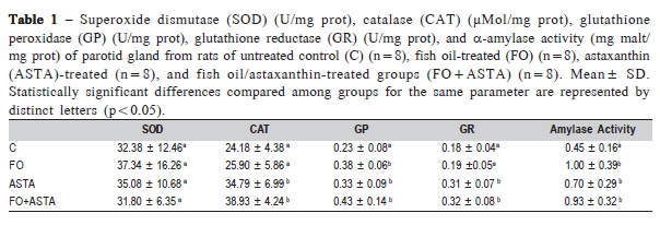
Table 1 and 2 show the enzymatic activities of antioxidant system and α-amylase of parotid and submandibular, respectively. The results of superoxide dismutase (U/mg protein), catalase (μMol/mg protein), glutathione peroxidase (U/mg protein), glutathione reductase (U/mg protein), and α-amylase activities (mg malt/mg protein) are presented as mean and standard deviation.
In the parotid gland (Table 1), fish oil treatment (FO) stimulated the glutathione peroxidase activity (65%) compared to the control group. Astaxanthin treatment (ASTA) and its association with fish oil (FO+ASTA) also increased the catalase (43 and 61 %, respectively), glutathione peroxidase (43 and 86%, respectively), and glutathione reductase activities (72 and 73%, respectively) as compared to the control group (p<0.05). There was no significant effect on the SOD activity in all groups.
]]>
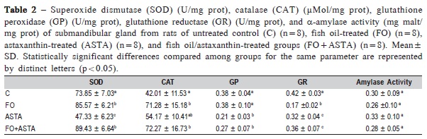
In the submandibular gland (Table 2), fish oil treatment (FO) stimulated the SOD (16%) and catalase activities (70%), whereas it reduced glutathione reductase activity (60%) as compared to the control group (p<0.05). Submandibular gland from rats treated with ASTA presented a reduction of SOD (36%), glutathione peroxidase (45%), and glutathione reductase (24%) activities and an increase in the catalase activity (29%) compared to the control group (p<0.05). Association of fish oil and astaxanthin (FO+ASTA) promoted a stimulating effect on the SOD and catalase activities in the submandibular and an inhibitory effect on glutathione system (peroxidase and reductase, 29 and 15%, respectively) (p<0.05).
Fish oil-treated, ASTA-treated and fish oil plus ASTAtreated groups presented increased α-amylase activity in the parotid gland (120, 55, and 106%, respectively) compared to the control group (p<0.05) (Table 1). No changes were observed in the submandibular gland (Table 2).
]]> Discussion
Our research group has developed scientific studies evaluating the actions of different elements present in the diet on salivary glands and dental pulp14,20-21,with positive effects on oral healthy. In the present study, we evaluated the effect of astaxanthin, fish oil and association of them on antioxidant system and amylase activity of salivary glands from healthy rats. We observed a stimulating effect of parameters studied after supplementation.
The impact of nutritional changes on the oral cavity of human individuals is a topic of interest in dentistry22-23. Dental caries is considered a public health problem that has the diet as one of the main etiological factors, with increased risk by association between inadequate intake of fruits and vegetables and excessive consumption of sugar sweetened beverages and foods22.The effect of diet has also been investigated on periodontal disease and individuals who had a poor diet presented higher number of missing teeth, higher average clinical attachment loss, which were significantly associated with increased odds of periodontitis23.
Considering the contribution of salivary glands in maintaining oral health, it seems of interest to study elements that can act therapeutically in salivary glands, such as vitamins, carotenoids, and minerals. Some studies have shown that administration of vitamins are very beneficial to the salivary glands for modulating the quality and quantity of saliva production and promoting tissue protection in cases of systemic diseases24-25. ASTA, fish oil and the combination of them presented a different pattern of stimulation on antioxidant system according to the type of salivary gland. Studies have shown that the parotid and submandibular glands have different responses to oxidative challenge such as diabetes and exposure to fluorides, particularly with reduced activity of some antioxidant enzymes and increased lipid peroxidation and oxidative damage14,26-27 in the submandibular gland, and greater stability and regenerative ability of parotid gland7,14.When submitted to antioxidant therapies, the salivary glands also exhibit different behavior from each other14, which agrees with our results. It is known that the parotid can be more prepared to oxidative damage by reactive oxygen species by presenting a predominantly aerobic metabolism28, however there are no reports in the literature to clarify the specific mechanism of action of parotid and submandibular when exposed to oxidative or antioxidants conditions. Further studies are required to evaluate the signaling pathways and expression of antioxidants proteins in salivary glands.
The ASTA action on the antioxidant system of salivary glands from healthy rats has been previously evaluated by our research group, presenting mild antioxidant effects14. For this reason, we decide to combine another element, fish oil, which also has a potential antioxidant role. The antioxidant effects observed after combination with fish oil were markedly improved and could be beneficial to salivary glands by enhancing the protective action of enzymatic antioxidant system of parotid and submandibular glands. In addition, we demonstrated that astaxanthin administered in smaller doses (1mg/kg BW) for a longer time (45 days) stimulates enzymatic antioxidant system of parotid gland compared to the protocol used before (20mg/kg BW for 30 days).These results show that a diverse diet may protect salivary glands14.
Some agents have oxidative property, but at low concentrations stimulate cellular enzymes of antioxidant system, acting as pro-oxidant29. Among all biomolecules, lipids are the most sensitive to free radicals. Double bonds in fatty acids form peroxide products by reacting with free radicals and lipid radicals can be formed subsequently upon removal of electrons. Excessive consumption of lipids, including polyunsaturated fatty acid (PUFA), increases lipid peroxidation significantly and may raise the susceptibility of tissues to free radical oxidative damage30. A previous published study showed that submandibular gland presents increased resistance against oxidative damage depends on the source of dietary PUFA31. In the present study, the fish oil administered in low concentration stimulated the enzymatic antioxidant system of submandibular gland, probably acting as pro-oxidant agent.
On the other hand, parotid gland also responded positively to treatment with antioxidants showing increased amylase activity. The response of salivary cells in the secretion process depends on the subtype of autonomic receptors that appears to be different on each gland32, submandibular and parotid glands have a predominance of muscarinic and adrenergic receptors, respectively. Some study showed that the beta-adrenergic agonist induced an increase of cAMP in both salivary glands, but while in the parotid it triggered amylase release, in the submandibular it was unable to increase α-amylase secretion32. Parotid α-amylase release was dependent on adenylate cyclase activation32.Dietary omega 3 fatty acids change the fatty-acid composition of the membrane phospholipids of submandibular salivary glands, accompanied by higher adenylate-cyclase activity33. The increased α-amylase activity in parotid gland could be related to adenylate-cyclase activity stimulation, particularly in the groups that received the fish oil supplementation that showed a more expressive increase of amylase activity. Moreover, further studies are required in order to explain the stimulatory effect of astaxanthin in the α-amylase activity of parotid gland.
α-Amylase is highly abundant salivary protein responsible for the initial digestion of starch, favoring the formation of the food bolus. Its main function is to split the α-1,4-glicosidic bindings of several glycans, such as starch (amylopectin), producing oligosaccharides (dextrin) disaccharides (maltose, isomaltose) and monosaccharide glucose. Its action is inactivated in the acid portions of the gastrointestinal tract and is consequently limited to the mouth34.α-Amylasehas been studied as a biomarker for sympathetic nervous system and functional capacity of salivary glands7,35. If by one side α-amylase provides substrate for bacteria, favoring the formation of dental biofilm36, in contrast, it presents specific binding sites with affinity for microorganisms (cariogenic and periodontopathogenic), forming bacterial agglomerates diluted in saliva that are easily eliminated by swallowing and consequently suffer acid digestion by the stomach37. The stimulation of α-amylase activity in parotid gland of rats supplemented with astaxanthin, fish oil and the combination of them was an interesting result, which could express an improved functional capacity of salivary gland and defense properties of the enzyme.
Further studies are needed to assess the impact of supplementation with antioxidant agents on oral cavity of human healthy individuals, using salivary and clinical parameters that represent the oral immunity. Moreover, there is a lack of studies that assess the expression of antioxidant proteins or key enzymes of pathway signaling of salivary glands from rats subjected to treatment with antioxidants such as ASTA and fish oil, which justifies further studies in this research line.
In conclusion, our results showed that antioxidant therapy could stimulate parotid gland, by increasing the α-amylase activity. FO and ASTA as well as the combination had some antioxidant effect, especially on parotid glands (increase of glutathione and catalase activity) and partially on submandibular gland (increase of catalase activity for all treatments).
]]>Acknowledgements
This research was supported by Research Foundation of the State of São Paulo (FAPESP 2009/12342-8). The authors are grateful to Dra. Rita Mattei from São Paulo University and Dr. Marcelo Paes de Barros, for providing supplemented rats.
References
1. Lee MG, Ohana E, Park HW, Yang D, Muallem S. Molecular mechanism of pancreatic and salivary gland fluid and HCO3 secretion. Physiol Rev. 2012; 92: 39-74. [ Links ]
2. Gorr SU, Venkatesh SG, Darling DS. Parotid secretory granules: crossroads of secretory pathways and protein storage. J Dent Res. 2005; 84: 500-9.
3. Nashida T, Sato R, Imai A, Shimomura H. Gene expression profiles of the three major salivary glands in rats. Biomed Res. 2010; 31: 387-99.
4. Proctor GB, Carpenter GH. Regulation of salivary gland function by autonomic nerves. Auton Neurosci. 2007; 133: 3-18.
]]> 5. Yamada T, Ryo K, Tai Y, Tamaki Y, Inoue H, Mishima K, et at.Evaluation of therapeutic effects of astaxanthin on impairments in salivary secretion. J Clin Biochem Nutr. 2010; 47: 130-7.6. Smith J, Lindsay M, Rahimian R, Anderson L. The influence of estrogen and progesterone on parasympathetic vasodilatation in the rat submandibular gland. Auton Neurosci. 2009; 146: 87-94.
7. Leite MF, Nicolau J.Sodium tungstate on some biochemical parameters of the parotid salivary gland of streptozotocin-induced diabetic rats: a shortterm study. Biol Trace Elem Res. 2009; 127: 154-63.
8. Vieira ACF, Rodrigues ASL, Cruz MCFN, Lopes FF. Profile of salivary gland flow dysfunctions in patients with differentiated thyroid carcinoma submitted to radioiodine therapy. Braz J Oral Sci. 2013; 12: 169-72.
9. Davies MJ. The oxidative environment and protein damage. Biochim Biophys Acta. 2005; 1703: 93-109.
10. Higuera-ciapara I, Felix-Valenzuela L, Goycoolea FM. Astaxanthin: a review of its chemistry and applications. Crit Rev Food Sci Nutr. 2006; 46: 185-96.
11. Fassett RG, Coombes JS. Astaxanthin: a potential therapeutic agent in cardiovascular disease. Mar Drugs. 2011; 9: 447-65.
12. Lordan S, Ross RP, Stanton C. Marine bioactives as functional food ingredients: potential to reduce the incidence of chronic diseases. Mar Drugs. 2011; 9: 1056-100.
13. Calder PC. n-3 Polyunsaturated fatty acids, inflammation, and inflammatory diseases. Am J Clin Nutr. 2006; 83(6 Suppl): 1505S-19S.
14. Leite MF, Lima AM, Massuyama MM, Otton R. Astaxanthin restores the enzymatic antioxidant profile in salivary gland of alloxan-induced diabetic rats. Arch Oral Biol. 2010; 55: 479-85.
]]> 15. Ewing JF, Janero DR. Microplate superoxide dismutase assay employing a nonenzymatic superoxide generator. Anal Biochem. 1995; 232: 243-8.16. Aebi H. Catalase in vitro. Methods Enzymol. 1984; 105: 121-6. 17. Mannervik B. Glutathione peroxidase. Methods Enzymol. 1985; 113: 490-5.
18. Fisher E, Stein EA. Alpha amylase from human saliva. Biochem Prepar. 1961; 8: 27-33.
19. Bradford MM. A rapid and sensitive method for the quantitation of microgram quantities of protein utilizing the principle of protein-dye binding. Anal Biochem. 1976; 72: 248-54.
20. Leite MF, De Lima A, Massuyama MM, Otton R. In vivo astaxanthin treatment partially prevents antioxidant alterations in dental pulp from alloxaninduced diabetic rats. Int Endod J. 2010; 43: 959-67.
21. Leite MF, Lima AM, Otton R. Combination of astaxanthin and fish oil supplementation alters antioxidant enzyme profile of dental pulp tissue. Int Endod J. 2012; 45: 1109-15.
22. Sharma A, Hegde AM. Relationship between body mass index, caries experience and dietary preferences in children. J Clin Pediatr Dent. 2009; 34: 49-52.
23. Bawadi HA, Khader YS, Haroun TF, Al-Omari M, Tayyem RF. The association between periodontal disease, physical activity and healthy diet among adults in Jordan. J Periodontal Res. 2011; 46: 74-81.
24. Ramos FM, Pontual ML, De Almeida SM, Bóscolo FN, Tabchoury CP, et at. Evaluation of radioprotective effect of vitamin E in salivary dysfunction in irradiated rats. Arch Oral Biol. 2006; 51: 96-101.
25. Stumpf WE, Hayakawa N. Salivary glands epithelial and myoepithelial cells are major vitamin D targets. Eur J Drug Metab Pharmacokinet. 2007; 32: 123-9.
]]> 26. Nogueira FN, Carvalho AM, Yamaguti PM, Nicolau J. Antioxidant parameters and lipid peroxidation in salivary glands of streptozotocin-induced diabetic rats. Clin Chim Acta. 2005; 353: 133-9.27. Yamaguti PM, Simões A, Ganzerla E, Souza DN, Nogueira FN, Nicolau J. Effects of single exposure of sodium fluoride on lipid peroxidation and antioxidant enzymes in salivary glands of rats. Oxid Med Cell Longev. 2013; 2013: 674593. doi: 10.1155/2013/674593. Epub 2013 Apr 27.
28. Nicolau J, Sassaki KT. Metabolism of carbohydrate in the major salivary glands of the rats. Arch Oral Biol. 1976; 21: 659-61.
29. Ivanova IP, Trofimova SV, Piskarev IM. Evaluation of prooxidant properties of ascorbic acid. Biofizika. 2013; 58: 582-6.
30. Haggag MS, Elsanhoty RM, Ramadan MF. Impact of dietary oils and fats on lipid peroxidation in liver and blood of albino rats. Asian Pac J Trop Biomed. 2014; 4: 52-8.
31. Avula CP, Fernandes G. Modulation of lipid peroxidation and antioxidant enzymes in murine salivary gland by dietary fatty acid ethyl esters. Life Sci. 1999; 65: 2373-83.
32. Busch L, Sterin-Borda L, Borda E. Difference in the regulatory mechanism of amylase release by rat parotid and submandibular glands. Arch Oral Biol. 2002; 47: 717-22.
33. Alam SQ, Alam BS. In-vivo incorporation of omega 3 fatty acids into membrane lipids of rat salivary glands and changes in adenylate-cyclase activity. Arch Oral Biol. 1988; 33: 295-9.
34. de Almeida PV, Grégio AM, Machado MA, de Lima AA, Azevedo LR. Saliva composition and functions: a comprehensive review. J Contemp Dent Pract. 2008; 9: 72-80.
35. Nater UM, Rohleder N. Salivary alpha-α-amylase as a non-invasive biomarker for the sympathetic nervous system: current state of research. Psychoneuroendocrinol. 2009; 34: 486-96.
]]> 36. Scannapieco FA, Torres G, Levine MJ. Salivary α-amylase: Role in dental plaque and caries formation. Crit Rev Oral Biol Med. 1993; 4: 301-7.37. Choi S, Baik JE, Jeon JH, Cho K, Seo DG, Kum KY, et at. Identification of Porphyromonas gingivalis lipopolysaccharide-binding proteins in human saliva. Mol Immunol. 2011; 48: 2207-13.
 Correspondence:
Mariana Ferreira Leite
Universidade Cruzeiro do Sul
Avenida Ussiel Cirilo 225
CEP: 08060-070 São Miguel Paulista
São Paulo - SP – Brasil
E-mail: mariana.leite@cruzeirodosul.edu.br
]]>
Received for publication: January 29, 2014 A
Accepted: March 20, 2014