<?xml version="1.0" encoding="ISO-8859-1"?><article xmlns:mml="http://www.w3.org/1998/Math/MathML" xmlns:xlink="http://www.w3.org/1999/xlink" xmlns:xsi="http://www.w3.org/2001/XMLSchema-instance">
<front>
<journal-meta>
<journal-id>1806-146X</journal-id>
<journal-title><![CDATA[IJD. International Journal of Dentistry]]></journal-title>
<abbrev-journal-title><![CDATA[IJD, Int. j. dent.]]></abbrev-journal-title>
<issn>1806-146X</issn>
<publisher>
<publisher-name><![CDATA[Universidade Federal de Pernambuco]]></publisher-name>
</publisher>
</journal-meta>
<article-meta>
<article-id>S1806-146X2010000100004</article-id>
<title-group>
<article-title xml:lang="en"><![CDATA[Erosion-like lesions progression in human and bovine enamel]]></article-title>
<article-title xml:lang="pt"><![CDATA[Progessão de lesões erosivas em substrato humano e bovino]]></article-title>
</title-group>
<contrib-group>
<contrib contrib-type="author">
<name>
<surname><![CDATA[Rehder Neto]]></surname>
<given-names><![CDATA[Francisco Carlos]]></given-names>
</name>
<xref ref-type="aff" rid="A01"/>
</contrib>
<contrib contrib-type="author">
<name>
<surname><![CDATA[Turssi]]></surname>
<given-names><![CDATA[Cecília Pedroso]]></given-names>
</name>
<xref ref-type="aff" rid="A02"/>
</contrib>
<contrib contrib-type="author">
<name>
<surname><![CDATA[Serra]]></surname>
<given-names><![CDATA[Mônica Campos]]></given-names>
</name>
<xref ref-type="aff" rid="A01"/>
</contrib>
</contrib-group>
<aff id="A01">
<institution><![CDATA[,University of São Paulo School of Dentistry of Ribeirão Preto Department of Restorative Dentistry]]></institution>
<addr-line><![CDATA[Ribeirão Preto São Paulo]]></addr-line>
<country>Brazil</country>
</aff>
<aff id="A02">
<institution><![CDATA[,University of Uberaba School of Dentistry Laboratory of Biomaterials Research]]></institution>
<addr-line><![CDATA[Uberaba Minas Gerais]]></addr-line>
<country>Brazil</country>
</aff>
<pub-date pub-type="pub">
<day>00</day>
<month>03</month>
<year>2010</year>
</pub-date>
<pub-date pub-type="epub">
<day>00</day>
<month>03</month>
<year>2010</year>
</pub-date>
<volume>9</volume>
<numero>1</numero>
<fpage>16</fpage>
<lpage>20</lpage>
<copyright-statement/>
<copyright-year/>
<self-uri xlink:href="http://revodonto.bvsalud.org/scielo.php?script=sci_arttext&amp;pid=S1806-146X2010000100004&amp;lng=en&amp;nrm=iso"></self-uri><self-uri xlink:href="http://revodonto.bvsalud.org/scielo.php?script=sci_abstract&amp;pid=S1806-146X2010000100004&amp;lng=en&amp;nrm=iso"></self-uri><self-uri xlink:href="http://revodonto.bvsalud.org/scielo.php?script=sci_pdf&amp;pid=S1806-146X2010000100004&amp;lng=en&amp;nrm=iso"></self-uri><abstract abstract-type="short" xml:lang="en"><p><![CDATA[Although on dental erosion studies bovine enamel has been considered a feasible option to human enamel, time dependence related to the erosive challenge remains uncertain. Thus, in this study the following null hypothesis was tested: 1) independent of the number of erosive challenges, no microhardness (SMH) differences exist between human and bovine enamel and 2) the behavior of bovine substrate along the erosive challenges do not differ from the human substrate. Human and bovine enamel slabs were embedded in epoxy resin and serially polished and flattened. Initial SMH tests were measured. The erosive challenge consists of an immersion of the specimens in continuously stirred orange juice (20ml) for 5 minutes at room temperature twice a day, for 5 days. Between the immersions the specimens were washed with deionized water and stored in artificial saliva (20ml). SMH tests were daily measured at the end of 2,4,6,8 and 10 challenges. Data were analyzed by two-way ANOVA as a split-plot design in time and Tukey's test. Regression analyses were used to model the SMH of human and bovine enamel over time. The first null hypothesis was rejected. Regression analysis exhibits a quadratic polynomial reduction of SMH in function of the number of erosive challenges for both substrates, revealing the admission of the second null hypothesis. Bovine substrate showed the most susceptible substrate to erosive challenge, but after several days of erosive challenge this difference disappears. Thus, the use of bovine tooth instead of human tooth in studies of erosive lesions formation seems to be feasible.]]></p></abstract>
<abstract abstract-type="short" xml:lang="pt"><p><![CDATA[Embora em estudos de erosão, o emprego do esmalte bovino seja considerado uma opção viável ao esmalte humano, a comparação do comportamento da indução das lesões erosivas nos diferentes substratos em relação à progressão/perda mineral permanece incerto. O objetivo deste estudo foi definir in vitro, a partir de um protocolo de indução de desgaste corrosivo, o tempo para formação de lesões cujo padrão, avaliado através de microdureza Knoop, seja semelhante em substratos bovinos e humanos. Fragmentos esmalte humano e bovino foram incluídos em resina epóxica, planificados, polidos. Um protocolo para o desenvolvimento de lesões erosivas foi instituído e cada ciclo erosivo consistiu da imersão dos espécimes em 20 ml de suco de laranja, em temperatura ambiente, por 5 minutos, sob agitação, duas vezes ao dia, durante 5 dias. No período entre as exposições e durante a noite, os espécimes foram mantidos em 20 ml de saliva artificial. Após o desafio erosivo leituras de microdureza foram efetuadas para avaliar a perda de conteúdo mineral. A analise de variância mostrou efeito significativo na interação substrato -tempo (p,0096). Ao longo dos episódios erosivos, os substratos dentais - humano e bovino - apresentaram um comportamento semelhante. A microdureza para ambos os substratos variou de acordo com uma função polinomial de segundo grau onde apesar de inicialmente não existir diferença entre os valores de microdureza dos substratos, o esmalte bovino se mostrou mais susceptível aos episódios erosivos; porém com o aumento do número de desafios, a diferença entre os substratos desapareceu.]]></p></abstract>
<kwd-group>
<kwd lng="en"><![CDATA[Erosion]]></kwd>
<kwd lng="en"><![CDATA[microhardness]]></kwd>
<kwd lng="en"><![CDATA[human enamel]]></kwd>
<kwd lng="en"><![CDATA[bovine enamel]]></kwd>
<kwd lng="pt"><![CDATA[Erosão dental]]></kwd>
<kwd lng="pt"><![CDATA[microdureza]]></kwd>
<kwd lng="pt"><![CDATA[esmalte humano]]></kwd>
<kwd lng="pt"><![CDATA[esmalte bovino]]></kwd>
</kwd-group>
</article-meta>
</front><body><![CDATA[ <p align="right"><font face="Verdana, Arial, Helvetica, sans-serif" size="2"><b>ORIGINAL    ARTICLE</b> ARTIGO ORIGINAL</font></p>     <p>&nbsp;</p>     <p><a name="top"></a><font face="Verdana, Arial, Helvetica, sans-serif" size="4"><b>Erosion-like    lesions progression in human and bovine enamel</b></font></p>     <p>&nbsp;</p>     <p><font face="Verdana, Arial, Helvetica, sans-serif" size="3"><b>Progess&atilde;o    de les&otilde;es erosivas em substrato humano e bovino</b></font></p>     <p>&nbsp;</p>     <p>&nbsp;</p>     <p><font face="Verdana, Arial, Helvetica, sans-serif" size="2"><b>Francisco Carlos    Rehder Neto<sup>I</sup>; Cec&iacute;lia Pedroso Turssi<sup>II</sup>; M&ocirc;nica    Campos Serra<sup>III</sup></b></font></p>     <p><font face="Verdana, Arial, Helvetica, sans-serif" size="2"><sup>I</sup>DDS,    MS, doctoral student, Department of Restorative Dentistry, School of Dentistry    of Ribeir&atilde;o Preto, University of S&atilde;o Paulo, Ribeir&atilde;o Preto,    S&atilde;o Paulo, Brazil    <br>   <sup>II</sup>DDS, MS, PhD, Laboratory of Biomaterials Research, School of Dentistry,    University of Uberaba (UNIUBE), Uberaba, Minas Gerais, Brazil    ]]></body>
<body><![CDATA[<br>   <sup>III</sup>DDS, MS, PhD, associate professor, Department of Restorative Dentistry,    School of Dentistry of Ribeir&atilde;o Preto, University of S&atilde;o Paulo,    Ribeir&atilde;o Preto, S&atilde;o Paulo, Brazil</font></p>     <p><font face="Verdana, Arial, Helvetica, sans-serif" size="2"><a href="#back">Correspondence</a></font></p>     <p>&nbsp;</p>     <p>&nbsp;</p> <hr size="1" noshade>     <p><font face="Verdana, Arial, Helvetica, sans-serif" size="2"><b>ABSTRACT</b></font></p>     <p><font face="Verdana, Arial, Helvetica, sans-serif" size="2">Although on dental    erosion studies bovine enamel has been considered a feasible option to human    enamel, time dependence related to the erosive challenge remains uncertain.    Thus, in this study the following null hypothesis was tested: 1) independent    of the number of erosive challenges, no microhardness (SMH) differences exist    between human and bovine enamel and 2) the behavior of bovine substrate along    the erosive challenges do not differ from the human substrate. Human and bovine    enamel slabs were embedded in epoxy resin and serially polished and flattened.    Initial SMH tests were measured. The erosive challenge consists of an immersion    of the specimens in continuously stirred orange juice (20ml) for 5 minutes at    room temperature twice a day, for 5 days. Between the immersions the specimens    were washed with deionized water and stored in artificial saliva (20ml). SMH    tests were daily measured at the end of 2,4,6,8 and 10 challenges. Data were    analyzed by two-way ANOVA as a split-plot design in time and Tukey's test. Regression    analyses were used to model the SMH of human and bovine enamel over time. The    first null hypothesis was rejected. Regression analysis exhibits a quadratic    polynomial reduction of SMH in function of the number of erosive challenges    for both substrates, revealing the admission of the second null hypothesis.    Bovine substrate showed the most susceptible substrate to erosive challenge,    but after several days of erosive challenge this difference disappears. Thus,    the use of bovine tooth instead of human tooth in studies of erosive lesions    formation seems to be feasible.</font></p>     <p><font face="Verdana, Arial, Helvetica, sans-serif" size="2"><b>Key words:</b>    Erosion; microhardness; human enamel; bovine enamel.</font></p> <hr size="1" noshade>     <p><font face="Verdana, Arial, Helvetica, sans-serif" size="2"><b>RESUMO</b></font></p>     <p><font face="Verdana, Arial, Helvetica, sans-serif" size="2">Embora em estudos    de eros&atilde;o, o emprego do esmalte bovino seja considerado uma op&ccedil;&atilde;o    vi&aacute;vel ao esmalte humano, a compara&ccedil;&atilde;o do comportamento    da indu&ccedil;&atilde;o das les&otilde;es erosivas nos diferentes substratos    em rela&ccedil;&atilde;o &agrave; progress&atilde;o/perda mineral permanece    incerto. O objetivo deste estudo foi definir <i>in vitro,</i> a partir de um    protocolo de indu&ccedil;&atilde;o de desgaste corrosivo, o tempo para forma&ccedil;&atilde;o    de les&otilde;es cujo padr&atilde;o, avaliado atrav&eacute;s de microdureza    Knoop, seja semelhante em substratos bovinos e humanos. Fragmentos esmalte humano    e bovino foram inclu&iacute;dos em resina ep&oacute;xica, planificados, polidos.    Um protocolo para o desenvolvimento de les&otilde;es erosivas foi institu&iacute;do    e cada ciclo erosivo consistiu da imers&atilde;o dos esp&eacute;cimes em 20    ml de suco de laranja, em temperatura ambiente, por 5 minutos, sob agita&ccedil;&atilde;o,    duas vezes ao dia, durante 5 dias. No per&iacute;odo entre as exposi&ccedil;&otilde;es    e durante a noite, os esp&eacute;cimes foram mantidos em 20 ml de saliva artificial.    Ap&oacute;s o desafio erosivo leituras de microdureza foram efetuadas para avaliar    a perda de conte&uacute;do mineral. A analise de vari&acirc;ncia mostrou efeito    significativo na intera&ccedil;&atilde;o substrato -tempo (p,0096). Ao longo    dos epis&oacute;dios erosivos, os substratos dentais - humano e bovino - apresentaram    um comportamento semelhante. A microdureza para ambos os substratos variou de    acordo com uma fun&ccedil;&atilde;o polinomial de segundo grau onde apesar de    inicialmente n&atilde;o existir diferen&ccedil;a entre os valores de microdureza    dos substratos, o esmalte bovino se mostrou mais suscept&iacute;vel aos epis&oacute;dios    erosivos; por&eacute;m com o aumento do n&uacute;mero de desafios, a diferen&ccedil;a    entre os substratos desapareceu.</font></p>     <p><font face="Verdana, Arial, Helvetica, sans-serif" size="2"><b>Palavras-chave:</b>    Eros&atilde;o dental; microdureza; esmalte humano; esmalte bovino</font></p> <hr size="1" noshade>     ]]></body>
<body><![CDATA[<p>&nbsp;</p>     <p>&nbsp;</p>     <p><font face="Verdana, Arial, Helvetica, sans-serif" size="3"><b>INTRODUCTION</b></font></p>     <p><font face="Verdana, Arial, Helvetica, sans-serif" size="2">There have been    substantial advances in understanding the pathophysiology of dental erosion,    especially through controlled <i>in vitro</i> studies. Regarding the tooth wear,    the use of <i>in vitro</i> studies showed some advantages, like the high level    of scientific control, ethical aspects related to certain experiments, reduction    in experimental variability, the use of highly sensitive assessment methods,    and the possibility of separate the components of tooth wear and investigate    them separately.</font></p>     <p><font face="Verdana, Arial, Helvetica, sans-serif" size="2">Erosion-like lesions    have been produced through different models, using both human<sup>1-5</sup>    and bovine teeth <sup>1,5,f6,7</sup>. Bovine substrate is widely used in dental    research and presents similarities with human enamel such as the chemical composition<sup>8</sup>,    crystal orientations<sup>9</sup> and microhardness<sup>10,11</sup>. Bovine teeth    have some advantages in relation to the ethical aspects, being easier to obtain    and manipulate than human teeth<sup>10</sup>. In fact, ethics committees are    encouraging their use as an alternative for human substrate<sup>12</sup>. However,    bovine enamel cannot be replaced for human enamel without validation<sup>8</sup>.</font></p>     <p><font face="Verdana, Arial, Helvetica, sans-serif" size="2">Although bovine    enamel has been considered a feasible option to human enamel, few studies have    compared artificial erosion lesions progression in both substrates<sup>1,5,6</sup>.    The aim of this study was to evaluate whether an erosive challenge protocol    that simulates acidic drinks intake is able to induce similar erosion-like lesions    in human and bovine enamel.</font></p>     <p>&nbsp;</p>     <p><font face="Verdana, Arial, Helvetica, sans-serif" size="3"><b>MATERIAL AND    METHODS</b></font></p>     <p><font face="Verdana, Arial, Helvetica, sans-serif" size="2"><i>Experimental    design</i></font></p>     <p><font face="Verdana, Arial, Helvetica, sans-serif" size="2">This study was    designed as a randomized complete block with 15 replications and a split-plot    factorial arrangement. The factors under study were: <i>dental substrate</i>    at two levels (bovine and human enamel) and <i>number of erosive challenges,    at six levels,</i> repeatedly measured on the same specimen, at different times    (T0, T1, T2, T3, T4, and T5). The levels of the factor <i>dental substrate</i>    were assigned to the main plot and the factor <i>number of erosive challenges</i>    were assigned to the subplots. The following null hypothesis were tested: 1)    independent of the number of erosive challenges, no microhardness (SMH) differences    exist between human and bovine enamel and 2) the behavior of bovine substrate    along the erosive challenges do not differ from human substrate. The response    variable was surface microhardness (SMH).</font></p>     ]]></body>
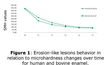
<body><![CDATA[<p><font face="Verdana, Arial, Helvetica, sans-serif" size="2"><i>Preparation    of specimens</i></font></p>     <p><font face="Verdana, Arial, Helvetica, sans-serif" size="2">Sound third human    molars and bovine incisors were cleaned to remove tissue remnants and stored    in a 0.1% thymol solution, at 4&deg;C. Each tooth was sectioned using a low-speed    water-cooled diamond saw (Isomet 1000, Buehler, Lake Bluff, IL, USA) in order    to obtain enamel slabs (3 x 2 x 2 mm) that were inspected for defects under    a stereomicroscope at 40x magnification and discarded if pitted or cracked.    The preselected slabs were embedded in epoxy resin (Epoxicure Resin, Buehler)    with enamel surface facing up and flattened in a water-cooled polishing machine    (Beta Grinder-Polisher, Buehler) using 400, 600 and 1200-grit Al<sub>2</sub>O<sub>3</sub>    papers, and polished on cloths with a 0.3 m.m alumina suspension (Alpha and    Gamma Micropolish, Buehler, Lake Bluff, IL, USA). Specimens were ultrasonically    cleaned in deionized water (T1440D, Odontobr&aacute;s, Ribeir&atilde;o Preto,    SP, Brazil) for 10 minutes and stored at 37º C in 100% relative humidity.</font></p>     <p><font face="Verdana, Arial, Helvetica, sans-serif" size="2"><i>Baseline microhardness    measurements</i></font></p>     <p><font face="Verdana, Arial, Helvetica, sans-serif" size="2">Specimens of human    and bovine enamel were tested for surface Knoop microhardness (HMV-2, Shimadzu,    Kyoto, Japan), under an load of 25 g, applied for 30 s. Indentations were performed    in each specimen at 150, 500 and 1500 u.m from superior margin, and at 100 u.m    to the lateral margin. The microhardness was measured with the aid of dedicated    software (New Age, Software Cams, South Ampton, PA, USA) and the average of    surface microhardness (SMH) values was calculated. Specimens with microhardness    values 20% above or 20% below the average value were discarded.</font></p>     <p><font face="Verdana, Arial, Helvetica, sans-serif" size="2"><i>Erosive Challenges</i></font></p>     <p><font face="Verdana, Arial, Helvetica, sans-serif" size="2">The erosive challenge    was based on that proposed by Amaechi et al, (1999). Briefly, the protocol consisted    of immersing the specimens in 20 mL of pure ready to drink orange juice (pH    - 3.74) (Fazenda Bela Vista, Tapiratiba, SP, Brazil) in an erlenmeyer flask,    which was then placed in an orbital shaker (CT155, Cientec, Piracicaba, SP,    Brazil), with stirring velocity of 100 rpm for 5 minutes, at the room temperature    (25&deg;C), twice daily, for 5 days. From one immersion to the other, the specimens    were washed with deionized water and stored in 20 mL of the artificial saliva    (pH of 6.75), described by McKnight-Hanes and Whitford<sup>13</sup> and modified    by Amaechi et al.,<sup>2</sup>, at 37&deg;C until the following erosive episode.    Saliva was daily changed and the orange juice at each immersion.</font></p>     <p><font face="Verdana, Arial, Helvetica, sans-serif" size="2"><i>Microhardness    analysis along the erosive challenges</i></font></p>     <p><font face="Verdana, Arial, Helvetica, sans-serif" size="2">Surface microhardness    measurements (SMH) were performed following the lesion progression at 250 um    left from the measurements made at the post-indentation. SMH analyses were assessed    under the same load and time described above for the baseline measurements.    The analysis was carried out at the end of each day, after the two daily challenges,    and to clarify this, six points in time were set (T0, T1, T2, T3, T4, T5) (<a href="#f1">Figure    1</a>). The times are in equivalency with levels of the factor <i>number of    challenges,</i> thus, T0 means the initial condition, while the time T5 refers    to the end of the five day, which means, 10 erosive challenges.</font></p>     <p><a name="f1"></a></p>     <p>&nbsp;</p>     ]]></body>
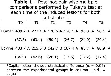
<body><![CDATA[<p align="center"><img src="/img/revistas/ijd/v9n1/04f01.jpg"></p>     <p>&nbsp;</p>     <p><font face="Verdana, Arial, Helvetica, sans-serif" size="2"><i>Statistical    Analysis</i></font></p>     <p><font face="Verdana, Arial, Helvetica, sans-serif" size="2">Data analysis was    carried out using Statgraphics Centurion XV (Manugistics, Rockville, MD, USA)    at a significance level of a = 0.05. After assumptions of normality and homogeneity    of variances had been verified, SMH data were analyzed by two-way ANOVA as a    split-plot design. Post-hoc pair wise multiple comparisons were performed using    the Tukey's test applied at each time (T0, T1, T2, T3, T4 and T5) to identify    differences in microhardness values between human and bovine substrate (L.S.D    - 22.44). Regression analyses were used to model the SMH of human and bovine    enamel, individually, over time.</font></p>     <p>&nbsp;</p>     <p><font face="Verdana, Arial, Helvetica, sans-serif" size="3"><b>RESULTS</b></font></p>     <p><font face="Verdana, Arial, Helvetica, sans-serif" size="2">The first null    hypothesis was rejected, since human and bovine substrate showed differences    regarding the SMH measurements. However, the second null hypothesis was accepted,    regression analysis exhibits a quadratic polynomial reduction of SMH in function    of the number of erosive challenges for both substrates. A two-way ANOVA according    to a split-plot design revealed significant interaction between <i>dental substrate</i>    and <i>number of erosive challenges</i> (p = 0.0096). At the times T0, T3, T4    and T5, SMH values of human and bovine enamel did not differ from each other,    as shown in <a href="#t1">Table 1</a>. After 2 and 4 erosive challenges (T1    and T2), human enamel showed higher SMH than the bovine substrate. The SMH values    for both human and bovine enamel decreased SMH, according to the following models:</font></p>     <p><font face="Verdana, Arial, Helvetica, sans-serif" size="2"><i>For human enamel:</i></font></p>     <p><font face="Verdana, Arial, Helvetica, sans-serif" size="2">SMH = 429,375-159,839*Time    + 18,7089*Time<sup>2</sup> (r<sup>2</sup> = 90,3%)</font></p>     <p><font face="Verdana, Arial, Helvetica, sans-serif" size="2"><i>For bovine enamel:</i></font></p>     ]]></body>
<body><![CDATA[<p><font face="Verdana, Arial, Helvetica, sans-serif" size="2">SMH = 409,545-175,832*Time    + 22,6783*Time<sup>2</sup> (r<sup>2</sup> = 92,0%)</font></p>     <p><a name="t1"></a></p>     <p>&nbsp;</p>     <p align="center"><img src="/img/revistas/ijd/v9n1/04t01.jpg"></p>     <p>&nbsp;</p>     <p><font face="Verdana, Arial, Helvetica, sans-serif" size="3"><b>DISCUSSION</b></font></p>     <p><font face="Verdana, Arial, Helvetica, sans-serif" size="2">Erosive lesions    involve enamel apatite crystals dissolution, which is associated with a surface    softening caused by the frequent contact with acids. This process might weaken    the enamel structure, and when the softened enamel is not re-hardened, they    become more vulnerable to further dissolutions or physical insult, leading to    an erosion lesion formation<sup>14,15,16</sup>.</font></p>     <p><font face="Verdana, Arial, Helvetica, sans-serif" size="2">Equivalency with    levels of the factor <i>number of challenges</i> T0 = 0 pre challenge condition;    T1 = after 2 challenges, or even, at the end of first day; T2 = after 4 challenges    or even, at the end of second day; T3 = after 6 challenges, or even, at the    end of third day; T4 = after 8 challenges or even, at the end of fourth day;    T5 = after 10 challenges, or even, at the end of fifth day.</font></p>     <p><font face="Verdana, Arial, Helvetica, sans-serif" size="2">Orange juices may    cause an appreciable level of erosion on enamel substrate<sup>2,6</sup> and    to simulate the acidic drinks intake, the model used to induce erosion like    lesions was based on ready to drink orange juices. To induce erosion-like lesions,    the chosen method was originally proposed for microradiographic assessments<sup>2</sup>,    and as the study showed, the prolonged intermittent exposure of enamel slabs    to orange juice increases progressively the depth of erosion lesion and the    loss of mineral content, measured by microradiography<sup>2</sup>. However,    although microradiography and even profilometry are accepted methods to assess    erosion lesions<sup>11</sup>, it was not included in this study.</font></p>     <p><font face="Verdana, Arial, Helvetica, sans-serif" size="2">Since this study    was designed to follow the initial stages of erosive lesions formation, preliminary    tests showed that the protocol proposed by Amaechi et al., <sup>2</sup> was    aggressive, thus, instead of six times a day for 24 days, the protocol was modified    for twice daily challenges for five days. Regarding the response variable, as    erosion has been considered a surface phenomenon<sup>17</sup>, to adapt the    methodology, following the study objective, surface microhardness was chosen    as evaluation method.</font></p>     ]]></body>
<body><![CDATA[<p><font face="Verdana, Arial, Helvetica, sans-serif" size="2">As a simple non-destructive    method to assess the early stages of enamel erosion, indentation techniques    have been extensively used to investigate enamel erosion by measuring the hardness    of the enamel surface<sup>7,11,18</sup>. Surface Knoop microhardness was chosen,    since this method allowed measurements along the time in the same specimen<sup>19</sup>,    an aspect taken into account regarding the hypothesis initially proposed.</font></p>     <p><font face="Verdana, Arial, Helvetica, sans-serif" size="2">The SMH values    throughout the elapsing process of lesions formation for both, human and bovine    enamel showed a similar behavior (<a href="#f1">Figure 1</a>). The adopted split    plot design allow to obtain the equation of the fitted model for human and bovine    enamel, and for both substrates the r<sup>2</sup> observed has a value higher    than 90%, which means that the data fitted tightly to the estimated model, and    for both substrates, quadratic polynomial equations were obtained modeling the    decrease of SMH values over time.</font></p>     <p><font face="Verdana, Arial, Helvetica, sans-serif" size="2">Considering that    the microhardness of both substrates was assumed to be the same, previously    to erosive challenge (T0) (<a href="#t1">Table 1</a>), the SMH values of human    and bovine enamel were similar; in fact, it has been showed that the mineral    contents of both substrates are not significantly different<sup>1,5,8,10,20</sup>.</font></p>     <p><font face="Verdana, Arial, Helvetica, sans-serif" size="2">After the times    T1 and T2 (<a href="#t1">Table 1</a>), bovine enamel showed lower SMH values    when compared to human substrate. Considering that bovine enamel had a faster    erosive lesion progression than in human permanent enamel, our results corroborates    with Amaechi et al.,<sup>2</sup> and Attin et al.,<sup>5</sup> , and, this finding    may be related to the higher porosity of bovine enamel<sup>21</sup>. Microscopically,    human and bovine enamel exhibit similar crystals orientations, although bovine    teeth exhibit a wider inter-prismatic region<sup>9</sup>, with bigger crystals    than human enamel,<sup>22</sup> an structural aspect that may enhance the acid    diffusion promoted by the erosive challenge acting on the tissue mechanical    resistance, weakening the enamel structure, leading to lower microhardness values.    Thus, the SMH values obtained in the times T1 and T2 of our study reinforces    this suggestion, since microhardness indentations is a function of the mechanical    properties of the material<sup>11</sup>.</font></p>     <p><font face="Verdana, Arial, Helvetica, sans-serif" size="2">Nevertheless, as    the erosive process continues, in the times T3, T4 and T5, the difference between    both substrates was no longer observed. The increase of erosive challenges along    the time seems to be decisive to induce similar lesions in both human and bovine    enamel. Despite the faster demineralization<sup>23</sup> of bovine enamel due    to its porosity<sup>21</sup>; with the high dissolution of apatite crystals    caused by the acidic contact<sup>1</sup>, when hardness or even mineral content    are reduced, the differences seems to be irrelevant. These results suggest that    in the dependence of erosive challenges, the bovine enamel represents a viable    alternative to human substrate.</font></p>     <p>&nbsp;</p>     <p><font face="Verdana, Arial, Helvetica, sans-serif" size="3"><b>CONCLUSION</b></font></p>     <p><font face="Verdana, Arial, Helvetica, sans-serif" size="2">Bovine substrate    showed the most susceptible substrate to erosive challenge, but after several    days of erosive challenge this difference disappears. On erosion research, the    use of bovine tooth instead of human tooth seems to be a feasible alternative.</font></p>     <p><font face="Verdana, Arial, Helvetica, sans-serif" size="2"><b>Acknowledgements</b></font></p>     <p><font face="Verdana, Arial, Helvetica, sans-serif" size="2">The authors are    indebted to Patr&iacute;cia Marchi for her technical assistance. This research    was supported by CNPQ, process 111925/2004-2005 and his conduction was approved    by the National Ethic Research Committee (CONEP), CAAE n&deg; 0071.0.138.000-06.</font></p>     ]]></body>
<body><![CDATA[<p>&nbsp;</p>     <p><font face="Verdana, Arial, Helvetica, sans-serif" size="3"><b>REFERENCES</b></font></p>     <!-- ref --><p><font face="Verdana, Arial, Helvetica, sans-serif" size="2">1. Meurman, R H,    Frank, R M. Progression and surface ultrastructure of in vitro caused erosive    lesions in human and bovine enamel. Caries Res 1991; 25(2): 81-87.    &nbsp;&nbsp;&nbsp;&nbsp;&nbsp;&nbsp;&nbsp;&nbsp;[&#160;<a href="javascript:void(0);" onclick="javascript: window.open('/scielo.php?script=sci_nlinks&ref=079417&pid=S1806-146X201000010000400001&lng=','','width=640,height=500,resizable=yes,scrollbars=1,menubar=yes,');">Links</a>&#160;]<!-- end-ref --></font></p>     <!-- ref --><p><font face="Verdana, Arial, Helvetica, sans-serif" size="2">2. Amaechi BT,    Higham SM, Edgar WM. Factors influencing the development of dental erosion in    vitro: enamel type, temperature and exposure time. J Oral Rehabil 1999, 26:    624-30.    &nbsp;&nbsp;&nbsp;&nbsp;&nbsp;&nbsp;&nbsp;&nbsp;[&#160;<a href="javascript:void(0);" onclick="javascript: window.open('/scielo.php?script=sci_nlinks&ref=079419&pid=S1806-146X201000010000400002&lng=','','width=640,height=500,resizable=yes,scrollbars=1,menubar=yes,');">Links</a>&#160;]<!-- end-ref --></font></p>     <!-- ref --><p><font face="Verdana, Arial, Helvetica, sans-serif" size="2">3. Ganss C, Klimek    J, Schwarz N. A comparative profilometric in vitro study of the susceptibility    of polished and natural human enamel and dentine surfaces to erosive demineralization    Arch Oral Biol 2000; 45: 897-902.    &nbsp;&nbsp;&nbsp;&nbsp;&nbsp;&nbsp;&nbsp;&nbsp;[&#160;<a href="javascript:void(0);" onclick="javascript: window.open('/scielo.php?script=sci_nlinks&ref=079421&pid=S1806-146X201000010000400003&lng=','','width=640,height=500,resizable=yes,scrollbars=1,menubar=yes,');">Links</a>&#160;]<!-- end-ref --></font></p>     <!-- ref --><p><font face="Verdana, Arial, Helvetica, sans-serif" size="2">4. Lussi A, Kohler    N, Zero D, Schaffner M, Megert B. A comparison of the erosive potential of different    beverages in primary and permanent teeth using an in vitro model. Eur J Oral    Sci 2000; 108: 110-114.    &nbsp;&nbsp;&nbsp;&nbsp;&nbsp;&nbsp;&nbsp;&nbsp;[&#160;<a href="javascript:void(0);" onclick="javascript: window.open('/scielo.php?script=sci_nlinks&ref=079423&pid=S1806-146X201000010000400004&lng=','','width=640,height=500,resizable=yes,scrollbars=1,menubar=yes,');">Links</a>&#160;]<!-- end-ref --></font></p>     ]]></body>
<body><![CDATA[<!-- ref --><p><font face="Verdana, Arial, Helvetica, sans-serif" size="2">5. Attin T, Wegehaupt    F, Gries D, Wiegand A. The potential of deciduous and permanent bovine enamel    as substitute for deciduous and permanent human enamel: Erosion-abrasion experiments.    J Dent 2007; 35(10): 773-777.    &nbsp;&nbsp;&nbsp;&nbsp;&nbsp;&nbsp;&nbsp;&nbsp;[&#160;<a href="javascript:void(0);" onclick="javascript: window.open('/scielo.php?script=sci_nlinks&ref=079425&pid=S1806-146X201000010000400005&lng=','','width=640,height=500,resizable=yes,scrollbars=1,menubar=yes,');">Links</a>&#160;]<!-- end-ref --></font></p>     <!-- ref --><p><font face="Verdana, Arial, Helvetica, sans-serif" size="2">6. Amaechi BT,    Higham SM, Edgar WM. Techniques for the production of eroded lesions in vitro.    J Oral Rehabil 1999b, 26: 97-102.    &nbsp;&nbsp;&nbsp;&nbsp;&nbsp;&nbsp;&nbsp;&nbsp;[&#160;<a href="javascript:void(0);" onclick="javascript: window.open('/scielo.php?script=sci_nlinks&ref=079427&pid=S1806-146X201000010000400006&lng=','','width=640,height=500,resizable=yes,scrollbars=1,menubar=yes,');">Links</a>&#160;]<!-- end-ref --></font></p>     <!-- ref --><p><font face="Verdana, Arial, Helvetica, sans-serif" size="2">7. Attin T, Meyer    K, Hellwig E, Buchalla W, Lennon AM. Effect of mineral supplements to citric    acid on enamel erosion. Arch Oral Biol 2003; 48: 753-759.    &nbsp;&nbsp;&nbsp;&nbsp;&nbsp;&nbsp;&nbsp;&nbsp;[&#160;<a href="javascript:void(0);" onclick="javascript: window.open('/scielo.php?script=sci_nlinks&ref=079429&pid=S1806-146X201000010000400007&lng=','','width=640,height=500,resizable=yes,scrollbars=1,menubar=yes,');">Links</a>&#160;]<!-- end-ref --></font></p>     <!-- ref --><p><font face="Verdana, Arial, Helvetica, sans-serif" size="2">8. Reeh ES, Douglas    WH, Levine MJ. Lubrication of human and bovine enamel compared in an artificial    mouth. Arch Oral Biol 1995; 40(11): 1063-1072.    &nbsp;&nbsp;&nbsp;&nbsp;&nbsp;&nbsp;&nbsp;&nbsp;[&#160;<a href="javascript:void(0);" onclick="javascript: window.open('/scielo.php?script=sci_nlinks&ref=079431&pid=S1806-146X201000010000400008&lng=','','width=640,height=500,resizable=yes,scrollbars=1,menubar=yes,');">Links</a>&#160;]<!-- end-ref --></font></p>     <!-- ref --><p><font face="Verdana, Arial, Helvetica, sans-serif" size="2">9. Boyde A. Dental    morphology and evolution. 1971, ed. Dahlberg A. A; Chicago University Press,    Chicago, 81-94.    &nbsp;&nbsp;&nbsp;&nbsp;&nbsp;&nbsp;&nbsp;&nbsp;[&#160;<a href="javascript:void(0);" onclick="javascript: window.open('/scielo.php?script=sci_nlinks&ref=079433&pid=S1806-146X201000010000400009&lng=','','width=640,height=500,resizable=yes,scrollbars=1,menubar=yes,');">Links</a>&#160;]<!-- end-ref --></font></p>     ]]></body>
<body><![CDATA[<!-- ref --><p><font face="Verdana, Arial, Helvetica, sans-serif" size="2">10. Mellberg JR:    Hard-tissue substrates for evaluation of cariogenic and anti-cariogenic activity    in situ. J Dent Res 1992 71 (special issue):913-19.    &nbsp;&nbsp;&nbsp;&nbsp;&nbsp;&nbsp;&nbsp;&nbsp;[&#160;<a href="javascript:void(0);" onclick="javascript: window.open('/scielo.php?script=sci_nlinks&ref=079435&pid=S1806-146X201000010000400010&lng=','','width=640,height=500,resizable=yes,scrollbars=1,menubar=yes,');">Links</a>&#160;]<!-- end-ref --></font></p>     <!-- ref --><p><font face="Verdana, Arial, Helvetica, sans-serif" size="2">11. Barbour ME,    Rees JS. The laboratory assessment of enamel erosion: a review. J Dent 2004;    32(8): 591602.    &nbsp;&nbsp;&nbsp;&nbsp;&nbsp;&nbsp;&nbsp;&nbsp;[&#160;<a href="javascript:void(0);" onclick="javascript: window.open('/scielo.php?script=sci_nlinks&ref=079437&pid=S1806-146X201000010000400011&lng=','','width=640,height=500,resizable=yes,scrollbars=1,menubar=yes,');">Links</a>&#160;]<!-- end-ref --></font></p>     <!-- ref --><p><font face="Verdana, Arial, Helvetica, sans-serif" size="2">12. Fonseca RB,    Haiter-Neto F, Fernandes-Neto AJ, Barbosa GAS, Soares CJ. Radiodensity of enamel    and dentin of human, bovine and swine teeth. Arch Oral Biol. 2004; 49(11): 919-22.    &nbsp;&nbsp;&nbsp;&nbsp;&nbsp;&nbsp;&nbsp;&nbsp;[&#160;<a href="javascript:void(0);" onclick="javascript: window.open('/scielo.php?script=sci_nlinks&ref=079439&pid=S1806-146X201000010000400012&lng=','','width=640,height=500,resizable=yes,scrollbars=1,menubar=yes,');">Links</a>&#160;]<!-- end-ref --></font></p>     <!-- ref --><p><font face="Verdana, Arial, Helvetica, sans-serif" size="2">13. Mcknight-Hanes    C, Whitford GM. Fluoride release from three glass ionomer materials and the    effects of varnishing with or without finishing. Caries Res 1992; 26: 345-50.    &nbsp;&nbsp;&nbsp;&nbsp;&nbsp;&nbsp;&nbsp;&nbsp;[&#160;<a href="javascript:void(0);" onclick="javascript: window.open('/scielo.php?script=sci_nlinks&ref=079441&pid=S1806-146X201000010000400013&lng=','','width=640,height=500,resizable=yes,scrollbars=1,menubar=yes,');">Links</a>&#160;]<!-- end-ref --></font></p>     <!-- ref --><p><font face="Verdana, Arial, Helvetica, sans-serif" size="2">14. Imfeld T. Dental    erosion. Definition, classification and links. Eur J Oral Sci 1996; 104: 151-155.    &nbsp;&nbsp;&nbsp;&nbsp;&nbsp;&nbsp;&nbsp;&nbsp;[&#160;<a href="javascript:void(0);" onclick="javascript: window.open('/scielo.php?script=sci_nlinks&ref=079443&pid=S1806-146X201000010000400014&lng=','','width=640,height=500,resizable=yes,scrollbars=1,menubar=yes,');">Links</a>&#160;]<!-- end-ref --></font></p>     ]]></body>
<body><![CDATA[<!-- ref --><p><font face="Verdana, Arial, Helvetica, sans-serif" size="2">15. Zero DT. Etiology    of dental erosion-extrinsic factors. Eur J Oral Sci 1996; 104: 162-77.    &nbsp;&nbsp;&nbsp;&nbsp;&nbsp;&nbsp;&nbsp;&nbsp;[&#160;<a href="javascript:void(0);" onclick="javascript: window.open('/scielo.php?script=sci_nlinks&ref=079445&pid=S1806-146X201000010000400015&lng=','','width=640,height=500,resizable=yes,scrollbars=1,menubar=yes,');">Links</a>&#160;]<!-- end-ref --></font></p>     <!-- ref --><p><font face="Verdana, Arial, Helvetica, sans-serif" size="2">16. Barbour ME,    Rees GD. The role of erosion, abrasion and attrition in tooth wear. J Clin Dent    2006; 17(4): 88-93.    &nbsp;&nbsp;&nbsp;&nbsp;&nbsp;&nbsp;&nbsp;&nbsp;[&#160;<a href="javascript:void(0);" onclick="javascript: window.open('/scielo.php?script=sci_nlinks&ref=079447&pid=S1806-146X201000010000400016&lng=','','width=640,height=500,resizable=yes,scrollbars=1,menubar=yes,');">Links</a>&#160;]<!-- end-ref --></font></p>     <!-- ref --><p><font face="Verdana, Arial, Helvetica, sans-serif" size="2">17. Lussi A. Erosive    tooth wear: a multifactorial condition of growing concern and increasing knowledge.    Monograf Oral Sci 2006; 20:1-8.    &nbsp;&nbsp;&nbsp;&nbsp;&nbsp;&nbsp;&nbsp;&nbsp;[&#160;<a href="javascript:void(0);" onclick="javascript: window.open('/scielo.php?script=sci_nlinks&ref=079449&pid=S1806-146X201000010000400017&lng=','','width=640,height=500,resizable=yes,scrollbars=1,menubar=yes,');">Links</a>&#160;]<!-- end-ref --></font></p>     <!-- ref --><p><font face="Verdana, Arial, Helvetica, sans-serif" size="2">18. Messias DC,    Martins ME, Serra MC, Turssi CP. Feasibility of Using Sodium Bicarbonate Solution    as a Damage-limiting Strategy for Erosion Lesions. Oral Health Prev Dent 2008;    6: 155-158.    &nbsp;&nbsp;&nbsp;&nbsp;&nbsp;&nbsp;&nbsp;&nbsp;[&#160;<a href="javascript:void(0);" onclick="javascript: window.open('/scielo.php?script=sci_nlinks&ref=079451&pid=S1806-146X201000010000400018&lng=','','width=640,height=500,resizable=yes,scrollbars=1,menubar=yes,');">Links</a>&#160;]<!-- end-ref --></font></p>     <!-- ref --><p><font face="Verdana, Arial, Helvetica, sans-serif" size="2">19. Argenta RM,    Tabchoury CP, Cury JA. A modified pH-cycling model to evaluate fluoride effect    on enamel demineralization. Braz Oral Res. 2003 17(3): 241-6.    &nbsp;&nbsp;&nbsp;&nbsp;&nbsp;&nbsp;&nbsp;&nbsp;[&#160;<a href="javascript:void(0);" onclick="javascript: window.open('/scielo.php?script=sci_nlinks&ref=079453&pid=S1806-146X201000010000400019&lng=','','width=640,height=500,resizable=yes,scrollbars=1,menubar=yes,');">Links</a>&#160;]<!-- end-ref --></font></p>     ]]></body>
<body><![CDATA[<!-- ref --><p><font face="Verdana, Arial, Helvetica, sans-serif" size="2">20. Edmunds DH,    Whittaker DK, Green RM: Suitability of human, bovine, equine, and ovine tooth    enamel for studies of artificial bacterial carious lesions. Caries Res 1988    22(6): 327-36.    &nbsp;&nbsp;&nbsp;&nbsp;&nbsp;&nbsp;&nbsp;&nbsp;[&#160;<a href="javascript:void(0);" onclick="javascript: window.open('/scielo.php?script=sci_nlinks&ref=079455&pid=S1806-146X201000010000400020&lng=','','width=640,height=500,resizable=yes,scrollbars=1,menubar=yes,');">Links</a>&#160;]<!-- end-ref --></font></p>     <!-- ref --><p><font face="Verdana, Arial, Helvetica, sans-serif" size="2">21. Arends, J,    Christoffersen, J, Ruben, J, Jongebloed, WL. Remineralization of bovine dentine    <i>in vitro.</i> The influence of the F contents in solution on mineral distribution.    Caries Res 1989; 23(5): 309-314.    &nbsp;&nbsp;&nbsp;&nbsp;&nbsp;&nbsp;&nbsp;&nbsp;[&#160;<a href="javascript:void(0);" onclick="javascript: window.open('/scielo.php?script=sci_nlinks&ref=079457&pid=S1806-146X201000010000400021&lng=','','width=640,height=500,resizable=yes,scrollbars=1,menubar=yes,');">Links</a>&#160;]<!-- end-ref --></font></p>     <!-- ref --><p><font face="Verdana, Arial, Helvetica, sans-serif" size="2">22. Arends J, Jongebloed    WL. Crystallites dimensions of enamel. J. Biol Bucca, 1978; 6:161-71.    &nbsp;&nbsp;&nbsp;&nbsp;&nbsp;&nbsp;&nbsp;&nbsp;[&#160;<a href="javascript:void(0);" onclick="javascript: window.open('/scielo.php?script=sci_nlinks&ref=079459&pid=S1806-146X201000010000400022&lng=','','width=640,height=500,resizable=yes,scrollbars=1,menubar=yes,');">Links</a>&#160;]<!-- end-ref --></font></p>     <!-- ref --><p><font face="Verdana, Arial, Helvetica, sans-serif" size="2">23. Featherstone,    JDB, Mellberg, JR. Relative rates of progress of artificial carious lesions    in bovine, ovine, and human enamel. Caries Res 1981; 15(1); 109114.    &nbsp;&nbsp;&nbsp;&nbsp;&nbsp;&nbsp;&nbsp;&nbsp;[&#160;<a href="javascript:void(0);" onclick="javascript: window.open('/scielo.php?script=sci_nlinks&ref=079461&pid=S1806-146X201000010000400023&lng=','','width=640,height=500,resizable=yes,scrollbars=1,menubar=yes,');">Links</a>&#160;]<!-- end-ref --></font></p>     <p>&nbsp;</p>     <p>&nbsp;</p>     ]]></body>
<body><![CDATA[<p><font face="Verdana, Arial, Helvetica, sans-serif" size="2"><a name="back"></a><a href="#top"><img src="/img/revistas/ijd/v9n1/seta.jpg" border="0"></a>    <b> Correspondence:</b>     <br>   Francisco Carlos Rehder Neto    <br>   Departamento de Odontologia Restauradora    <br>   Faculdade de Odontologia de Ribeir&atilde;o Preto - USP    <br>   Av. do Caf&eacute;, s/n Monte Alegre    <br>   Cep: 14040-904 Ribeir&atilde;o Preto - SP, Brazil    <br>   e-mail: <a href="mailto:fcrehder@usp.br">fcrehder@usp.br</a>    <br>   phone: 55-16-3602-4075    <br>   fax: 55-16-3633-4187</font></p>     <p><font face="Verdana, Arial, Helvetica, sans-serif" size="2">Recebido em 07/01/2010    ]]></body>
<body><![CDATA[<br>   Aprovado em 18/03/2010</font></p>      ]]></body>
<back>
<ref-list>
<ref id="B1">
<label>1</label><nlm-citation citation-type="journal">
<person-group person-group-type="author">
<name>
<surname><![CDATA[Meurman]]></surname>
<given-names><![CDATA[R H]]></given-names>
</name>
<name>
<surname><![CDATA[Frank]]></surname>
<given-names><![CDATA[R M]]></given-names>
</name>
</person-group>
<article-title xml:lang="en"><![CDATA[Progression and surface ultrastructure of in vitro caused erosive lesions in human and bovine enamel]]></article-title>
<source><![CDATA[Caries Res]]></source>
<year>1991</year>
<volume>25</volume>
<numero>2</numero>
<issue>2</issue>
<page-range>81-87</page-range></nlm-citation>
</ref>
<ref id="B2">
<label>2</label><nlm-citation citation-type="journal">
<person-group person-group-type="author">
<name>
<surname><![CDATA[Amaechi]]></surname>
<given-names><![CDATA[BT]]></given-names>
</name>
<name>
<surname><![CDATA[Higham]]></surname>
<given-names><![CDATA[SM]]></given-names>
</name>
<name>
<surname><![CDATA[Edgar]]></surname>
<given-names><![CDATA[WM]]></given-names>
</name>
</person-group>
<article-title xml:lang="en"><![CDATA[Factors influencing the development of dental erosion in vitro: enamel type, temperature and exposure time]]></article-title>
<source><![CDATA[J Oral Rehabil]]></source>
<year>1999</year>
<volume>26</volume>
<page-range>624-30</page-range></nlm-citation>
</ref>
<ref id="B3">
<label>3</label><nlm-citation citation-type="journal">
<person-group person-group-type="author">
<name>
<surname><![CDATA[Ganss]]></surname>
<given-names><![CDATA[C]]></given-names>
</name>
<name>
<surname><![CDATA[Klimek]]></surname>
<given-names><![CDATA[J]]></given-names>
</name>
<name>
<surname><![CDATA[Schwarz]]></surname>
<given-names><![CDATA[N]]></given-names>
</name>
</person-group>
<article-title xml:lang="en"><![CDATA[A comparative profilometric in vitro study of the susceptibility of polished and natural human enamel and dentine surfaces to erosive demineralization]]></article-title>
<source><![CDATA[Arch Oral Biol]]></source>
<year>2000</year>
<volume>45</volume>
<page-range>897-902</page-range></nlm-citation>
</ref>
<ref id="B4">
<label>4</label><nlm-citation citation-type="journal">
<person-group person-group-type="author">
<name>
<surname><![CDATA[Lussi]]></surname>
<given-names><![CDATA[A]]></given-names>
</name>
<name>
<surname><![CDATA[Kohler]]></surname>
<given-names><![CDATA[N]]></given-names>
</name>
<name>
<surname><![CDATA[Zero]]></surname>
<given-names><![CDATA[D]]></given-names>
</name>
<name>
<surname><![CDATA[Schaffner]]></surname>
<given-names><![CDATA[M]]></given-names>
</name>
<name>
<surname><![CDATA[Megert]]></surname>
<given-names><![CDATA[B]]></given-names>
</name>
</person-group>
<article-title xml:lang="en"><![CDATA[A comparison of the erosive potential of different beverages in primary and permanent teeth using an in vitro model]]></article-title>
<source><![CDATA[Eur J Oral Sci]]></source>
<year>2000</year>
<volume>108</volume>
<page-range>110-114</page-range></nlm-citation>
</ref>
<ref id="B5">
<label>5</label><nlm-citation citation-type="journal">
<person-group person-group-type="author">
<name>
<surname><![CDATA[Attin]]></surname>
<given-names><![CDATA[T]]></given-names>
</name>
<name>
<surname><![CDATA[Wegehaupt]]></surname>
<given-names><![CDATA[F]]></given-names>
</name>
<name>
<surname><![CDATA[Gries]]></surname>
<given-names><![CDATA[D]]></given-names>
</name>
<name>
<surname><![CDATA[Wiegand]]></surname>
<given-names><![CDATA[A]]></given-names>
</name>
</person-group>
<article-title xml:lang="en"><![CDATA[The potential of deciduous and permanent bovine enamel as substitute for deciduous and permanent human enamel: Erosion-abrasion experiments]]></article-title>
<source><![CDATA[J Dent]]></source>
<year>2007</year>
<volume>35</volume>
<numero>10</numero>
<issue>10</issue>
<page-range>773-777</page-range></nlm-citation>
</ref>
<ref id="B6">
<label>6</label><nlm-citation citation-type="journal">
<person-group person-group-type="author">
<name>
<surname><![CDATA[Amaechi]]></surname>
<given-names><![CDATA[BT]]></given-names>
</name>
<name>
<surname><![CDATA[Higham]]></surname>
<given-names><![CDATA[SM]]></given-names>
</name>
<name>
<surname><![CDATA[Edgar]]></surname>
<given-names><![CDATA[WM]]></given-names>
</name>
</person-group>
<article-title xml:lang="en"><![CDATA[Techniques for the production of eroded lesions in vitro]]></article-title>
<source><![CDATA[J Oral Rehabil]]></source>
<year>1999</year>
<volume>26</volume>
<page-range>97-102</page-range></nlm-citation>
</ref>
<ref id="B7">
<label>7</label><nlm-citation citation-type="journal">
<person-group person-group-type="author">
<name>
<surname><![CDATA[Attin]]></surname>
<given-names><![CDATA[T]]></given-names>
</name>
<name>
<surname><![CDATA[Meyer]]></surname>
<given-names><![CDATA[K]]></given-names>
</name>
<name>
<surname><![CDATA[Hellwig]]></surname>
<given-names><![CDATA[E]]></given-names>
</name>
<name>
<surname><![CDATA[Buchalla]]></surname>
<given-names><![CDATA[W]]></given-names>
</name>
<name>
<surname><![CDATA[Lennon]]></surname>
<given-names><![CDATA[AM]]></given-names>
</name>
</person-group>
<article-title xml:lang="en"><![CDATA[Effect of mineral supplements to citric acid on enamel erosion]]></article-title>
<source><![CDATA[Arch Oral Biol]]></source>
<year>2003</year>
<volume>48</volume>
<page-range>753-759</page-range></nlm-citation>
</ref>
<ref id="B8">
<label>8</label><nlm-citation citation-type="journal">
<person-group person-group-type="author">
<name>
<surname><![CDATA[Reeh]]></surname>
<given-names><![CDATA[ES]]></given-names>
</name>
<name>
<surname><![CDATA[Douglas]]></surname>
<given-names><![CDATA[WH]]></given-names>
</name>
<name>
<surname><![CDATA[Levine]]></surname>
<given-names><![CDATA[MJ]]></given-names>
</name>
</person-group>
<article-title xml:lang="en"><![CDATA[Lubrication of human and bovine enamel compared in an artificial mouth]]></article-title>
<source><![CDATA[Arch Oral Biol]]></source>
<year>1995</year>
<volume>40</volume>
<numero>11</numero>
<issue>11</issue>
<page-range>1063-1072</page-range></nlm-citation>
</ref>
<ref id="B9">
<label>9</label><nlm-citation citation-type="book">
<person-group person-group-type="author">
<name>
<surname><![CDATA[Boyde]]></surname>
<given-names><![CDATA[A]]></given-names>
</name>
</person-group>
<source><![CDATA[Dental morphology and evolution]]></source>
<year>1971</year>
<page-range>81-94</page-range><publisher-loc><![CDATA[Chicago ]]></publisher-loc>
<publisher-name><![CDATA[Chicago University Press]]></publisher-name>
</nlm-citation>
</ref>
<ref id="B10">
<label>10</label><nlm-citation citation-type="journal">
<person-group person-group-type="author">
<name>
<surname><![CDATA[Mellberg]]></surname>
<given-names><![CDATA[JR]]></given-names>
</name>
</person-group>
<article-title xml:lang="en"><![CDATA[Hard-tissue substrates for evaluation of cariogenic and anti-cariogenic activity in situ]]></article-title>
<source><![CDATA[J Dent Res]]></source>
<year>1992</year>
<volume>71</volume>
<numero>special issue</numero>
<issue>special issue</issue>
<page-range>913-19</page-range></nlm-citation>
</ref>
<ref id="B11">
<label>11</label><nlm-citation citation-type="journal">
<person-group person-group-type="author">
<name>
<surname><![CDATA[Barbour]]></surname>
<given-names><![CDATA[ME]]></given-names>
</name>
<name>
<surname><![CDATA[Rees]]></surname>
<given-names><![CDATA[JS]]></given-names>
</name>
</person-group>
<article-title xml:lang="en"><![CDATA[The laboratory assessment of enamel erosion: a review]]></article-title>
<source><![CDATA[J Dent]]></source>
<year>2004</year>
<volume>32</volume>
<numero>8</numero>
<issue>8</issue>
<page-range>591602</page-range></nlm-citation>
</ref>
<ref id="B12">
<label>12</label><nlm-citation citation-type="journal">
<person-group person-group-type="author">
<name>
<surname><![CDATA[Fonseca]]></surname>
<given-names><![CDATA[RB]]></given-names>
</name>
<name>
<surname><![CDATA[Haiter-Neto]]></surname>
<given-names><![CDATA[F]]></given-names>
</name>
<name>
<surname><![CDATA[Fernandes-Neto]]></surname>
<given-names><![CDATA[AJ]]></given-names>
</name>
<name>
<surname><![CDATA[Barbosa]]></surname>
<given-names><![CDATA[GAS]]></given-names>
</name>
<name>
<surname><![CDATA[Soares]]></surname>
<given-names><![CDATA[CJ]]></given-names>
</name>
</person-group>
<article-title xml:lang="en"><![CDATA[Radiodensity of enamel and dentin of human, bovine and swine teeth]]></article-title>
<source><![CDATA[Arch Oral Biol]]></source>
<year>2004</year>
<volume>49</volume>
<numero>11</numero>
<issue>11</issue>
<page-range>919-22</page-range></nlm-citation>
</ref>
<ref id="B13">
<label>13</label><nlm-citation citation-type="journal">
<person-group person-group-type="author">
<name>
<surname><![CDATA[Mcknight-Hanes]]></surname>
<given-names><![CDATA[C]]></given-names>
</name>
<name>
<surname><![CDATA[Whitford]]></surname>
<given-names><![CDATA[GM]]></given-names>
</name>
</person-group>
<article-title xml:lang="en"><![CDATA[Fluoride release from three glass ionomer materials and the effects of varnishing with or without finishing]]></article-title>
<source><![CDATA[Caries Res]]></source>
<year>1992</year>
<volume>26</volume>
<page-range>345-50</page-range></nlm-citation>
</ref>
<ref id="B14">
<label>14</label><nlm-citation citation-type="journal">
<person-group person-group-type="author">
<name>
<surname><![CDATA[Imfeld]]></surname>
<given-names><![CDATA[T]]></given-names>
</name>
</person-group>
<article-title xml:lang="en"><![CDATA[Dental erosion: Definition, classification and links]]></article-title>
<source><![CDATA[Eur J Oral Sci]]></source>
<year>1996</year>
<volume>104</volume>
<page-range>151-155</page-range></nlm-citation>
</ref>
<ref id="B15">
<label>15</label><nlm-citation citation-type="journal">
<person-group person-group-type="author">
<name>
<surname><![CDATA[Zero]]></surname>
<given-names><![CDATA[DT]]></given-names>
</name>
</person-group>
<article-title xml:lang="en"><![CDATA[Etiology of dental erosion-extrinsic factors]]></article-title>
<source><![CDATA[Eur J Oral Sci]]></source>
<year>1996</year>
<volume>104</volume>
<page-range>162-77</page-range></nlm-citation>
</ref>
<ref id="B16">
<label>16</label><nlm-citation citation-type="journal">
<person-group person-group-type="author">
<name>
<surname><![CDATA[Barbour]]></surname>
<given-names><![CDATA[ME]]></given-names>
</name>
<name>
<surname><![CDATA[Rees]]></surname>
<given-names><![CDATA[GD]]></given-names>
</name>
</person-group>
<article-title xml:lang="en"><![CDATA[The role of erosion, abrasion and attrition in tooth wear]]></article-title>
<source><![CDATA[J Clin Dent]]></source>
<year>2006</year>
<volume>17</volume>
<numero>4</numero>
<issue>4</issue>
<page-range>88-93</page-range></nlm-citation>
</ref>
<ref id="B17">
<label>17</label><nlm-citation citation-type="journal">
<person-group person-group-type="author">
<name>
<surname><![CDATA[Lussi]]></surname>
<given-names><![CDATA[A]]></given-names>
</name>
</person-group>
<article-title xml:lang="en"><![CDATA[Erosive tooth wear: a multifactorial condition of growing concern and increasing knowledge]]></article-title>
<source><![CDATA[Monograf Oral Sci]]></source>
<year>2006</year>
<volume>20</volume>
<page-range>1-8</page-range></nlm-citation>
</ref>
<ref id="B18">
<label>18</label><nlm-citation citation-type="journal">
<person-group person-group-type="author">
<name>
<surname><![CDATA[Messias]]></surname>
<given-names><![CDATA[DC]]></given-names>
</name>
<name>
<surname><![CDATA[Martins]]></surname>
<given-names><![CDATA[ME]]></given-names>
</name>
<name>
<surname><![CDATA[Serra]]></surname>
<given-names><![CDATA[MC]]></given-names>
</name>
<name>
<surname><![CDATA[Turssi]]></surname>
<given-names><![CDATA[CP]]></given-names>
</name>
</person-group>
<article-title xml:lang="en"><![CDATA[Feasibility of Using Sodium Bicarbonate Solution as a Damage-limiting Strategy for Erosion Lesions]]></article-title>
<source><![CDATA[Oral Health Prev Dent]]></source>
<year>2008</year>
<volume>6</volume>
<page-range>155-158</page-range></nlm-citation>
</ref>
<ref id="B19">
<label>19</label><nlm-citation citation-type="journal">
<person-group person-group-type="author">
<name>
<surname><![CDATA[Argenta]]></surname>
<given-names><![CDATA[RM]]></given-names>
</name>
<name>
<surname><![CDATA[Tabchoury]]></surname>
<given-names><![CDATA[CP]]></given-names>
</name>
<name>
<surname><![CDATA[Cury]]></surname>
<given-names><![CDATA[JA]]></given-names>
</name>
</person-group>
<article-title xml:lang="en"><![CDATA[A modified pH-cycling model to evaluate fluoride effect on enamel demineralization]]></article-title>
<source><![CDATA[Braz Oral Res]]></source>
<year>2003</year>
<volume>17</volume>
<numero>3</numero>
<issue>3</issue>
<page-range>241-6</page-range></nlm-citation>
</ref>
<ref id="B20">
<label>20</label><nlm-citation citation-type="journal">
<person-group person-group-type="author">
<name>
<surname><![CDATA[Edmunds]]></surname>
<given-names><![CDATA[DH]]></given-names>
</name>
<name>
<surname><![CDATA[Whittaker]]></surname>
<given-names><![CDATA[DK]]></given-names>
</name>
<name>
<surname><![CDATA[Green]]></surname>
<given-names><![CDATA[RM]]></given-names>
</name>
</person-group>
<article-title xml:lang="en"><![CDATA[Suitability of human, bovine, equine, and ovine tooth enamel for studies of artificial bacterial carious lesions]]></article-title>
<source><![CDATA[Caries Res]]></source>
<year>1988</year>
<volume>22</volume>
<numero>6</numero>
<issue>6</issue>
<page-range>327-36</page-range></nlm-citation>
</ref>
<ref id="B21">
<label>21</label><nlm-citation citation-type="journal">
<person-group person-group-type="author">
<name>
<surname><![CDATA[Arends]]></surname>
<given-names><![CDATA[J]]></given-names>
</name>
<name>
<surname><![CDATA[Christoffersen]]></surname>
<given-names><![CDATA[J]]></given-names>
</name>
<name>
<surname><![CDATA[Ruben]]></surname>
<given-names><![CDATA[J]]></given-names>
</name>
<name>
<surname><![CDATA[Jongebloed]]></surname>
<given-names><![CDATA[WL]]></given-names>
</name>
</person-group>
<article-title xml:lang="en"><![CDATA[Remineralization of bovine dentine in vitro: The influence of the F contents in solution on mineral distribution]]></article-title>
<source><![CDATA[Caries Res]]></source>
<year>1989</year>
<volume>23</volume>
<numero>5</numero>
<issue>5</issue>
<page-range>309-314</page-range></nlm-citation>
</ref>
<ref id="B22">
<label>22</label><nlm-citation citation-type="journal">
<person-group person-group-type="author">
<name>
<surname><![CDATA[Arends]]></surname>
<given-names><![CDATA[J]]></given-names>
</name>
<name>
<surname><![CDATA[Jongebloed]]></surname>
<given-names><![CDATA[WL]]></given-names>
</name>
</person-group>
<article-title xml:lang="en"><![CDATA[Crystallites dimensions of enamel]]></article-title>
<source><![CDATA[J. Biol Bucca,]]></source>
<year>1978</year>
<volume>6</volume>
<page-range>161-71</page-range></nlm-citation>
</ref>
<ref id="B23">
<label>23</label><nlm-citation citation-type="journal">
<person-group person-group-type="author">
<name>
<surname><![CDATA[Featherstone]]></surname>
<given-names><![CDATA[JDB]]></given-names>
</name>
<name>
<surname><![CDATA[Mellberg]]></surname>
<given-names><![CDATA[JR]]></given-names>
</name>
</person-group>
<article-title xml:lang="en"><![CDATA[Relative rates of progress of artificial carious lesions in bovine, ovine, and human enamel]]></article-title>
<source><![CDATA[Caries Res]]></source>
<year>1981</year>
<volume>15</volume>
<numero>1</numero>
<issue>1</issue>
<page-range>109114</page-range></nlm-citation>
</ref>
</ref-list>
</back>
</article>