<?xml version="1.0" encoding="ISO-8859-1"?><article xmlns:mml="http://www.w3.org/1998/Math/MathML" xmlns:xlink="http://www.w3.org/1999/xlink" xmlns:xsi="http://www.w3.org/2001/XMLSchema-instance">
<front>
<journal-meta>
<journal-id>1981-8637</journal-id>
<journal-title><![CDATA[RGO.Revista Gaúcha de Odontologia (Online)]]></journal-title>
<abbrev-journal-title><![CDATA[RGO, Rev. gaúch. odontol. (Online)]]></abbrev-journal-title>
<issn>1981-8637</issn>
<publisher>
<publisher-name><![CDATA[Mundi Brasil Gráfica e Editora Ltda.]]></publisher-name>
</publisher>
</journal-meta>
<article-meta>
<article-id>S1981-86372012000200003</article-id>
<title-group>
<article-title xml:lang="en"><![CDATA[Analysis of smear layer removal by different irrigants]]></article-title>
<article-title xml:lang="pt"><![CDATA[Análise da remoção de smear layer por diferentes soluções auxiliares]]></article-title>
</title-group>
<contrib-group>
<contrib contrib-type="author">
<name>
<surname><![CDATA[GONÇALVES]]></surname>
<given-names><![CDATA[Rodrigo Diego]]></given-names>
</name>
<xref ref-type="aff" rid="A01"/>
</contrib>
<contrib contrib-type="author">
<name>
<surname><![CDATA[Fassina]]></surname>
<given-names><![CDATA[Felipe Henrique]]></given-names>
</name>
<xref ref-type="aff" rid="A01"/>
</contrib>
<contrib contrib-type="author">
<name>
<surname><![CDATA[ONODA]]></surname>
<given-names><![CDATA[Hélio Katsuya]]></given-names>
</name>
<xref ref-type="aff" rid="A01"/>
</contrib>
<contrib contrib-type="author">
<name>
<surname><![CDATA[GUERISOLI]]></surname>
<given-names><![CDATA[Danilo Mathias Zanello]]></given-names>
</name>
<xref ref-type="aff" rid="A01"/>
</contrib>
<contrib contrib-type="author">
<name>
<surname><![CDATA[PEREIRA]]></surname>
<given-names><![CDATA[Key Fabiano Souza]]></given-names>
</name>
<xref ref-type="aff" rid="A01"/>
</contrib>
<contrib contrib-type="author">
<name>
<surname><![CDATA[YOSHINARI]]></surname>
<given-names><![CDATA[Gerson Hiroshi]]></given-names>
</name>
<xref ref-type="aff" rid="A01"/>
</contrib>
</contrib-group>
<aff id="A01">
<institution><![CDATA[,Universidade Federal de Mato Grosso do Sul Faculdade de Odontologia ]]></institution>
<addr-line><![CDATA[Campo Grande MS]]></addr-line>
<country>Brasil</country>
</aff>
<pub-date pub-type="pub">
<day>00</day>
<month>06</month>
<year>2012</year>
</pub-date>
<pub-date pub-type="epub">
<day>00</day>
<month>06</month>
<year>2012</year>
</pub-date>
<volume>60</volume>
<numero>2</numero>
<fpage>157</fpage>
<lpage>161</lpage>
<copyright-statement/>
<copyright-year/>
<self-uri xlink:href="http://revodonto.bvsalud.org/scielo.php?script=sci_arttext&amp;pid=S1981-86372012000200003&amp;lng=en&amp;nrm=iso"></self-uri><self-uri xlink:href="http://revodonto.bvsalud.org/scielo.php?script=sci_abstract&amp;pid=S1981-86372012000200003&amp;lng=en&amp;nrm=iso"></self-uri><self-uri xlink:href="http://revodonto.bvsalud.org/scielo.php?script=sci_pdf&amp;pid=S1981-86372012000200003&amp;lng=en&amp;nrm=iso"></self-uri><abstract abstract-type="short" xml:lang="en"><p><![CDATA[Objective This study assessed in vitro the ability of different irrigating solutions to remove the smear layer after root canal instrumentation. Methods Twenty upper human canines were instrumented and irrigated with different irrigating solutions: Group I (positive control) - saline; Group II (negative control) - 17% EDTA in water; Group III - 24% EDTA gel; and Group IV - BioPure MTAD Results Micrographs of the middle and apical thirds were taken for determination of the area percentage covered by smear layer. Analysis of variance and the Tukey test showed that BioPure MTAD and 17% EDTA in water had similar efficacy (p&gt;0.05), producing clean canal walls with open dentinal tubules. Saline and 24% EDTA gel also had similar efficacy, but in this case much of the root canal walls were still covered by the smear layer. The results from Groups I and III were significantly different from those of Groups II and IV (p<0.01). There were no differences between the root thirds of each group (p&gt;0.05). Conclusion None of the irrigating solutions were able to completely remove the smear layer from the root canal walls. Biopure MTAD and 17% EDTA in water proved to be more effective for removing the smear layer than 24% EDTA gel or saline.]]></p></abstract>
<abstract abstract-type="short" xml:lang="pt"><p><![CDATA[Objetivo Avaliar in vitro a ação de diferentes soluções auxiliares quanto à capacidade de remoção da smear layer após o preparo biomecânico. Métodos Vinte caninos superiores humanos foram preparados com instrumentos rotatórios ProTaper® (Dentsply-Maillefer, Ballaigues, Suíça), recebendo diferentes soluções auxiliares de acordo com o grupo experimental. No Grupo I (controle positivo), foi utilizado soro fisiológico; no Grupo II (controle negativo), solução aquosa de EDTA a 17%; o Grupo III recebeu gel de EDTA a 24% e o Grupo IV, Biopure® MTAD (Tulsa-Dentsply, Tulsa, OK, EUA). Resultados Fotomicrografias dos terços médio e apical foram obtidas e analisadas quantitativamente, obtendo-se a porcentagem de áreas cobertas por smear layer. Os resultados obtidos foram submetidos à análise estatística (teste ANOVA), que indicou o BioPure® MTAD (Tulsa-Dentsply, Tulsa, OK, EUA) e a solução de EDTA a 17% como tendo capacidade semelhante de remoção da smear layer no terço médio (p>0,05). O gel de EDTA a 24% apresentou capacidade de remoção de smear layer semelhante ao soro fisiológico, sendo inferior ao Biopure® MTAD (Tulsa-Dentsply, Tulsa, OK, EUA) ou à solução aquosa de EDTA. Não houve diferenças entre os terços radiculares estudados. Conclusão Nenhuma solução irrigadora removeu totalmente a lama dentinária nos terços estudados. O Biopure® MTAD (Tulsa-Dentsply, Tulsa, OK, EUA) mostrou resultados semelhantes à solução aquosa de EDTA a 17%, e o gel de EDTA a 24% não foi capaz de eliminar de forma satisfatória a smear layer das paredes dentinárias.]]></p></abstract>
<kwd-group>
<kwd lng="en"><![CDATA[Chelating agents.]]></kwd>
<kwd lng="en"><![CDATA[Endodontics.]]></kwd>
<kwd lng="en"><![CDATA[Root canal preparation.]]></kwd>
<kwd lng="en"><![CDATA[Smear layer.]]></kwd>
<kwd lng="pt"><![CDATA[Quelantes.]]></kwd>
<kwd lng="pt"><![CDATA[Endodontia.]]></kwd>
<kwd lng="pt"><![CDATA[Preparo de canal radicular.]]></kwd>
<kwd lng="pt"><![CDATA[Camada de esfregaço.]]></kwd>
</kwd-group>
</article-meta>
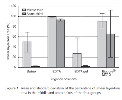
</front><body><![CDATA[ <p align="right"><font size="2" face="Verdana, Arial, Helvetica, sans-serif"><b>ORIGINAL</b> / ORIGINAL</font></p>     <p>&nbsp;</p>     <p><font size="4" face="Verdana, Arial, Helvetica, sans-serif"><a name="top"/></a><B>Analysis of smear layer removal by different irrigants</B></font></p>     <p>&nbsp;</p>     <p><font size="3" face="Verdana, Arial, Helvetica, sans-serif"><b>An&aacute;lise da remo&ccedil;&atilde;o de smear layer por diferentes solu&ccedil;&otilde;es auxiliares</b> </font></p>     <p>&nbsp;</p>     <p>&nbsp;</p>     <p><font size="2" face="Verdana, Arial, Helvetica, sans-serif"><b>Rodrigo Diego GON&Ccedil;ALVES <sup>I</sup>; Felipe Henrique Fassina DOMINGUES <sup>II</sup>; H&eacute;lio Katsuya ONODA <sup>II</sup>; Danilo Mathias Zanello GUERISOLI <sup>II</sup>; Key Fabiano Souza PEREIRA <sup>II</sup>; Gerson Hiroshi YOSHINARI <sup>II</sup></b></font></p>     <p><font size="2" face="Verdana, Arial, Helvetica, sans-serif"><sup>I</sup> Universidade Federal de Mato Grosso do Sul, Faculdade de Odontologia. Av. Senador Filinto M&uuml;ller, 1, 79080-190, Campo Grande, MS, Brasil     <br> <sup>II</sup> Universidade Federal de Mato Grosso do Sul, Faculdade de Odontologia. Campo Grande, MS, Brasil</font></p>     ]]></body>
<body><![CDATA[<p><font size="2" face="Verdana, Arial, Helvetica, sans-serif"><a href="#back">Correspondence:</a></font></p>     <p>&nbsp;</p>     <p>&nbsp;</p> <hr size="1" noshade>     <p><font size="3" face="Verdana, Arial, Helvetica, sans-serif"><b>ABSTRACT</b> </font></p>     <p><font size="2" face="Verdana, Arial, Helvetica, sans-serif"><b>Objective</b>    <br>    </font><font size="2" face="Verdana, Arial, Helvetica, sans-serif">This study assessed in vitro the ability of different irrigating solutions to remove the smear layer after root canal instrumentation. </font></p>     <p><font size="2" face="Verdana, Arial, Helvetica, sans-serif"><b>Methods</b>    <br>    </font><font size="2" face="Verdana, Arial, Helvetica, sans-serif">Twenty upper human canines were instrumented and irrigated with different irrigating solutions: Group I (positive control) - saline; Group II (negative control) - 17% EDTA in water; Group III - 24% EDTA gel; and Group IV - BioPure MTAD </font></p>     <p><font size="2" face="Verdana, Arial, Helvetica, sans-serif"><b>Results</b>    <br>    </font><font size="2" face="Verdana, Arial, Helvetica, sans-serif">Micrographs of the middle and apical thirds were taken for determination of the area percentage covered by smear layer. Analysis of variance and the Tukey test showed that BioPure MTAD and 17% EDTA in water had similar efficacy (p&gt;0.05), producing clean canal walls with open dentinal tubules. Saline and 24% EDTA gel also had similar efficacy, but in this case much of the root canal walls were still covered by the smear layer. The results from Groups I and III were significantly different from those of Groups II and IV (p&lt;0.01). There were no differences between the root thirds of each group (p&gt;0.05). </font></p>     ]]></body>
<body><![CDATA[<p><font size="2" face="Verdana, Arial, Helvetica, sans-serif"><b>Conclusion</b>    <br>    </font><font size="2" face="Verdana, Arial, Helvetica, sans-serif">None of the irrigating solutions were able to completely remove the smear layer from the root canal walls. Biopure MTAD and 17% EDTA in water proved to be more effective for removing the smear layer than 24% EDTA gel or saline. </font></p>     <p><font size="2" face="Verdana, Arial, Helvetica, sans-serif"><B>Indexing terms: </B>Chelating agents. Endodontics. Root canal preparation. Smear layer.</font></p> <hr size="1" noshade>     <p><font size="3" face="Verdana, Arial, Helvetica, sans-serif"><B>RESUMO</B> </font></p>     <p><font size="2" face="Verdana, Arial, Helvetica, sans-serif"><b>Objetivo</b>    <br>  </font><font size="2" face="Verdana, Arial, Helvetica, sans-serif">Avaliar in vitro a a&ccedil;&atilde;o de diferentes solu&ccedil;&otilde;es auxiliares quanto &agrave; capacidade de remo&ccedil;&atilde;o da smear layer ap&oacute;s o preparo biomec&acirc;nico. </font></p>     <p><font size="2" face="Verdana, Arial, Helvetica, sans-serif"><b>M&eacute;todos</b>    <br>  </font><font size="2" face="Verdana, Arial, Helvetica, sans-serif">Vinte caninos superiores humanos foram preparados com instrumentos rotat&oacute;rios ProTaper&reg; (Dentsply-Maillefer, Ballaigues, Su&iacute;&ccedil;a), recebendo diferentes solu&ccedil;&otilde;es auxiliares de acordo com o grupo experimental. No Grupo I (controle positivo), foi utilizado soro fisiol&oacute;gico; no Grupo II (controle negativo), solu&ccedil;&atilde;o aquosa de EDTA a 17%; o Grupo III recebeu gel de EDTA a 24% e o Grupo IV, Biopure&reg; MTAD (Tulsa-Dentsply, Tulsa, OK, EUA).  </font></p>     <p><font size="2" face="Verdana, Arial, Helvetica, sans-serif"><b>Resultados</b>    <br>  </font><font size="2" face="Verdana, Arial, Helvetica, sans-serif">Fotomicrografias dos ter&ccedil;os m&eacute;dio e apical foram obtidas e analisadas quantitativamente, obtendo-se a porcentagem de &aacute;reas cobertas por smear layer. Os resultados obtidos foram submetidos &agrave; an&aacute;lise estat&iacute;stica (teste ANOVA), que indicou o BioPure&reg; MTAD (Tulsa-Dentsply, Tulsa, OK, EUA) e a solu&ccedil;&atilde;o de EDTA a 17% como tendo capacidade semelhante de remo&ccedil;&atilde;o da smear layer no ter&ccedil;o m&eacute;dio (p&gt;0,05). O gel de EDTA a 24% apresentou capacidade de remo&ccedil;&atilde;o de smear layer semelhante ao soro fisiol&oacute;gico, sendo inferior ao Biopure&reg; MTAD (Tulsa-Dentsply, Tulsa, OK, EUA) ou &agrave; solu&ccedil;&atilde;o aquosa de EDTA. N&atilde;o houve diferen&ccedil;as entre os ter&ccedil;os radiculares estudados. </font></p>     ]]></body>
<body><![CDATA[<p><font size="2" face="Verdana, Arial, Helvetica, sans-serif"><b>Conclus&atilde;o</b>    <br>  </font><font size="2" face="Verdana, Arial, Helvetica, sans-serif">Nenhuma solu&ccedil;&atilde;o irrigadora removeu totalmente a lama dentin&aacute;ria nos ter&ccedil;os estudados. O Biopure&reg; MTAD (Tulsa-Dentsply, Tulsa, OK, EUA) mostrou resultados semelhantes &agrave; solu&ccedil;&atilde;o aquosa de EDTA a 17%, e o gel de EDTA a 24% n&atilde;o foi capaz de eliminar de forma satisfat&oacute;ria a smear layer das paredes dentin&aacute;rias. </font></p>     <p><font size="2" face="Verdana, Arial, Helvetica, sans-serif"><B>Termos de indexa&ccedil;&atilde;o: </B>Quelantes. Endodontia. Preparo de canal radicular. Camada de esfrega&ccedil;o.</font></p> <hr noshade size="1">     <p>&nbsp;</p>     <p>&nbsp;</p>     <p><font size="3" face="Verdana, Arial, Helvetica, sans-serif"><B> INTRODUCTION</B></font></p>     <p><font size="2" face="Verdana, Arial, Helvetica, sans-serif">In addition to endodontic instrumentation, the root canal system is also cleaned and disinfected by supplementary solutions that will perform the critical sanitation of the root canal<sup>1</sup>.</font></p>     <p><font size="2" face="Verdana, Arial, Helvetica, sans-serif"> The smear layer, a dentinal mud or magma can be defined as an amorphous, irregular and granulose substrate composed of two distinct layers: a 1 to 2 &mu;m superficial layer consisting of organic matter and a second, 6 to 40 &mu;m, more mineralized and deeper layer consisting mainly of dentinal debris. It is produced by the cutting action of endodontic instruments on moist dentin and consists of inorganic matter, odontoblast process fragments, microorganisms and necrotic pulp remnants<sup>2-3</sup>. </font></p>     <p><font size="2" face="Verdana, Arial, Helvetica, sans-serif">The smear layer can function as an infection route and substrate for bacterial growth<sup>4</sup>, and negatively affect the penetration and adhesion of obturation materials on the dentinal tubules<sup>5</sup>. </font></p>     <p><font size="2" face="Verdana, Arial, Helvetica, sans-serif">Ethylenediaminetetraacetic acid (EDTA) has proven to be the best supplementary solution for the removal of the smear layer from root canals<sup>5-8</sup>. On the other hand, the few published studies on EDTA gel show that its ability to remove the smear layer is different<sup>9-11</sup>. </font></p>     ]]></body>
<body><![CDATA[<p><font size="2" face="Verdana, Arial, Helvetica, sans-serif">Biopure MTAD is marketed as a bactericidal solution for root canal irrigation capable of removing the smear layer. It consists of a tetracycline isomer (doxycycline), 10% citric acid and polysorbate 80. It has been shown to remove the smear layer better than EDTA<sup>12</sup>. </font></p>     <p><font size="2" face="Verdana, Arial, Helvetica, sans-serif">The objective of the present study was to use scanning electron microscopy (SEM) to determine the ability of different supplementary solutions to remove the smear layer from the walls of the middle and apical thirds of root canals.</font></p>     <p>&nbsp;</p>     <p><font size="3" face="Verdana, Arial, Helvetica, sans-serif"><B> METHODS</B></font></p>     <p><font size="2" face="Verdana, Arial, Helvetica, sans-serif">Twenty upper human canines extracted for different reasons were obtained at the Federal University of Mato Grosso do Sul School of Dentistry tooth bank. All specimens were stored in a refrigerated, 2% glutaraldehyde aqueous solution until ready for use.</font></p>     <p><font size="2" face="Verdana, Arial, Helvetica, sans-serif"> The crowns of the teeth were removed by a double-sided diamond disc #7020 (KG Sorensen, S&atilde;o Paulo, Brazil). Canal length was determined by the manual file Flexofile ISO 10 (Dentsply-Maillefer, Ballaigues, Switzerland) and a dental microscope with a magnification of 8x was used to see when the file crossed the apical foramen (DF Vasconcellos, S&atilde;o Paulo, Brazil). The working length was calculated by subtracting 1mm from the canal length. </font></p>     <p><font size="2" face="Verdana, Arial, Helvetica, sans-serif">The canals were instrumented by rotary nickeltitanium ProTaper&reg; files (Dentsply-Maillefer, Ballaigues, Switzerland) as instructed by the manufacturer. At each change of instrument, the canals were irrigated with 3ml of a 5.25% sodium hypochlorite solution (Pharmac&ecirc;utica, Campo Grande, Brazil). </font></p>     <p><font size="2" face="Verdana, Arial, Helvetica, sans-serif">The specimens were randomly divided into four groups according to the solution used in the last irrigation: Group I &ndash; saline (Pharmac&ecirc;utica, Campo Grande, Brazil) as positive control for the smear layer. Group II &ndash; 17% EDTA aqueous solution (Pharmac&ecirc;utica, Campo Grande, Brazil) as negative control; Group III &ndash; 24% EDTA gel (Biodin&acirc;mica, Ibipor&atilde;, Brazil); and Group IV - Biopure MTAD (Tulsa- Dentsply, Tulsa, OK, USA). </font></p>     <p><font size="2" face="Verdana, Arial, Helvetica, sans-serif">Each supplementary solution stayed inside the canals for 5 minutes. The canals were then rinsed with saline and blotted (Tanari, Manaus, Brazil). The canals in Group IV were irrigated as recommended by the manufacturer, that is, irrigation with MTAD for 5 minutes and no subsequent saline rinsing. </font></p>     <p><font size="2" face="Verdana, Arial, Helvetica, sans-serif">A longitudinal groove was made on the vestibular and lingual surfaces of the roots and a surgical hammer and bibeveled chisel were used to cleave the specimens into two parts. One of these parts was randomly selected and prepared for SEM (JEOL JSM-5410, Tokyo, Japan). </font></p>     ]]></body>
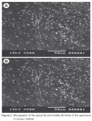
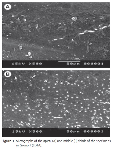
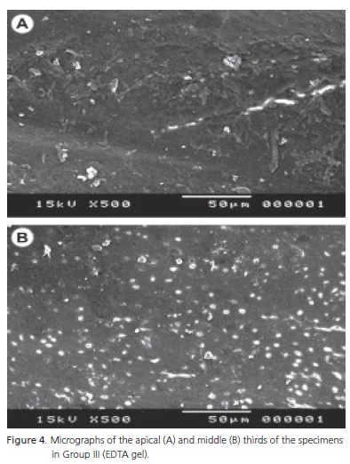
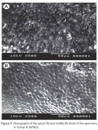
<body><![CDATA[<p><font size="2" face="Verdana, Arial, Helvetica, sans-serif">The middle and apical thirds of each hemisection were magnified 500 times and representative micrographs were taken from each area. The images were analyzed quantitatively by the software ImageTool version 3.0 (University of San Antonio, Texas, USA), which superposed the images with a grid containing 300 cells. All cells containing smear layer were marked. This cells were analyzed by a previously calibrated doctor in dentistry (emphasis on endodontics), who routinely analyzes micrographs. The results were expressed as percentage of the middle and apical third areas of each group covered by smear layer, calculated by the software GraphPad Prism v5.0 (GraphPad Software Inc., USA). The groups were then compared by two-way analysis of variance (two-way ANOVA) complemented by the Tukey. </font></p>     <p><font size="2" face="Verdana, Arial, Helvetica, sans-serif">The study was approved by the Research Ethics Committee of the Federal University of Mato Grosso do Sul, protocol number 954.</font><font size="2" face="Verdana, Arial, Helvetica, sans-serif"> </font></p>     <p>&nbsp;</p>     <p><font size="3" face="Verdana, Arial, Helvetica, sans-serif"><B> RESULTS</B></font></p>     <p><font size="2" face="Verdana, Arial, Helvetica, sans-serif">The results after quantitative analysis of the micrographs are shown in <a href="#fig01">Figure 1</a>. </font></p>     <p><font size="2" face="Verdana, Arial, Helvetica, sans-serif">One of the specimens in Group IV (BioPure MTAD) was lost because of processing error. Figures <a href="#fig02">2</a> to <a href="#fig05">5</a> show the micrographs of the most representative areas of each experimental group. </font></p>     <p><font size="2" face="Verdana, Arial, Helvetica, sans-serif">The efficacy of the study solutions differed significantly (p&lt;0.001). More smear layer remained in the groups irrigated with saline and EDTA gel, which did not differ from each other (p&gt;0.05). The groups irrigated with 17% EDTA in water or BioPure MTAD had more dentinal tubules exposed, indicating that these substances removed the smear layer more effectively (p&gt;0.05). </font></p>     <p><font size="2" face="Verdana, Arial, Helvetica, sans-serif">The only group whose middle third differed significantly from its apical third with respect to percentage of area covered by smear layer was Group I (saline) (p&lt;0.01). There were no significant differences between the middle and apical thirds of the other groups (p&gt;0.05).</font></p>     <p>&nbsp;</p>     <p><a name="fig01"></a></p>     ]]></body>
<body><![CDATA[<p>&nbsp; </p>     <p align="center"><img src="/img/revistas/rgo/v60n2/a03fig01.jpg">     <p>&nbsp;</p>     <p><a name="fig02"></a></p>     <p>&nbsp; </p>     <p align="center"><img src="/img/revistas/rgo/v60n2/a03fig02.jpg">     <p>&nbsp;</p>     <p><a name="fig03"></a></p>     <p>&nbsp; </p>     <p align="center"><img src="/img/revistas/rgo/v60n2/a03fig03.jpg">     ]]></body>
<body><![CDATA[<p>&nbsp;</p>     <p><a name="fig04"></a></p>     <p>&nbsp; </p>     <p align="center"><img src="/img/revistas/rgo/v60n2/a03fig04.jpg">     <p>&nbsp;</p>     <p><a name="fig05"></a></p>     <p>&nbsp; </p>     <p align="center"><img src="/img/revistas/rgo/v60n2/a03fig05.jpg">     <p>&nbsp;</p>     <p><font size="3" face="Verdana, Arial, Helvetica, sans-serif"><B> DISCUSSION</B></font></p>     ]]></body>
<body><![CDATA[<p><font size="2" face="Verdana, Arial, Helvetica, sans-serif">Alternating irrigation with sodium hypochlorite and EDTA during instrumentation was proposed by Goldman et al.<sup>13</sup>, and has since been adopted by endodontists worldwide. Hence, 17% EDTA in water was used by the present study for the last irrigation as negative control, while saline was used lastly as positive control. It is known that this solution, free of chelating agents, is unable to remove the smear layer<sup>14</sup>.</font></p>     <p><font size="2" face="Verdana, Arial, Helvetica, sans-serif"> This study found that Biopure MTAD was not significantly different from 17% EDTA in water in its ability to remove the smear layer (p&gt;0.05), confirming the results of other studies<sup>15-16</sup>. On the other hand, Torabinejad et al.<sup>12</sup> reported that MTAD can remove the smear layer more effectively in the apical third than EDTA. </font></p>     <p><font size="2" face="Verdana, Arial, Helvetica, sans-serif">The 24% EDTA gel was not as effective in removing the smear layer as Biopure MTAD or 17% EDTA in water, corroborating the finding. Meanwhile, Dotto et al.<sup>9</sup>, Sampaio et al.<sup>10</sup> and Putzer et al.<sup>11</sup> found that EDTA gel effectively removed the smear layer from dentinal walls.</font></p>     <p><font size="2" face="Verdana, Arial, Helvetica, sans-serif"> In the present study, EDTA gel was not capable of removing the smear layer effectively from any of the study thirds. The results obtained by ETDA gel and saline were similar (p&gt;0.05). </font></p>     <p><font size="2" face="Verdana, Arial, Helvetica, sans-serif">None of the specimens irrigated with Biopure MTAD presented regions of dentinal erosion, contrary to what has been reported by Torabinejad et al.<sup>12</sup>, Tay et al.<sup>15</sup>, Gonzalez-L&oacute;pez et. al.<sup>17</sup> and Grande et. al.<sup>18</sup>. </font></p>     <p><font size="2" face="Verdana, Arial, Helvetica, sans-serif">There were no significant differences between the middle and apical thirds (p&gt;0.05).</font></p>     <p>&nbsp;</p>     <p><font size="3" face="Verdana, Arial, Helvetica, sans-serif"><B> CONCLUSION</B></font></p>     <p><font size="2" face="Verdana, Arial, Helvetica, sans-serif">None of the irrigation solutions tested was capable of fully removing the dentinal mud from the study thirds. The efficacy of Biopure MTAD was similar to that of 17% EDTA in water, both very effective for removing the smear layer. On the other hand, 24% EDTA gel was not capable of removing the smear layer from the dentinal walls effectively, and proved to be similar to saline.</font></p>     <p><font size="2" face="Verdana, Arial, Helvetica, sans-serif"><B> Collaborators</B></font></p>       ]]></body>
<body><![CDATA[<p><font size="2" face="Verdana, Arial, Helvetica, sans-serif">   RD GON&Ccedil;ALVES performed the experimental work which included preparing the specimens and taking the micrographs; he also helped to write the article. FHF DOMINGUES helped to write the article and with the literature search. HK ONODA helped to assess the micrographs using the software ImageTool, tabulate the results and write the article. DMZ GUERISOLI supervised the study and was responsible for the study design, statistical analyses and writing of the article. KFS PEREIRA supervised the methodology and writing of the article. GH YOSHINARI conceived the experiment, coordinated the execution team and helped with the experimental tasks. </font></p>     <p>&nbsp;</p>     <p><font size="3" face="Verdana, Arial, Helvetica, sans-serif"><B>REFERENCES </B></font></p>     <p><font size="2" face="Verdana, Arial, Helvetica, sans-serif">1. Burns RC, Cohen S. Caminhos da polpa. 9&ordf; ed. Rio de Janeiro: Elsivier; 2007. </font></p>     <p><font size="2" face="Verdana, Arial, Helvetica, sans-serif">2. Mader CL, Baumgartner C, Peters DD. Scanning electron microscopic investigation of the smeared layer on root canal walls. J Endod. 1984;10(10):477-83. doi: 10.1016/S0099- 2399(84)80204-6. </font></p>     <p><font size="2" face="Verdana, Arial, Helvetica, sans-serif">3. Goldman M, Goldman LB, Cavaleri R, Borgis J, Lin PS. The efficacy of several endodontic irrigating solutions: a scanning electron microscopic study: part 2. J Endod. 1982;8(11):487-92. doi: 10.1016/S0099-2399(82)80073-3. </font></p>     <p><font size="2" face="Verdana, Arial, Helvetica, sans-serif">4. Pashley DH. Dentin-predentin complex and its permeability: physiological overview. J Dent Res. 198;64(Spec n.):613-20.</font></p>     <p><font size="2" face="Verdana, Arial, Helvetica, sans-serif"> 5. Sen BH, Wesselink PR, T&uuml;rk&uuml;n M. The smear layer. A phenomenon in root canal therapy. Int J Endod. 1995;28(3):141-8. doi: 10.1111/j.1365-2591.1995.tb00289.x. </font></p>     <p><font size="2" face="Verdana, Arial, Helvetica, sans-serif">6. Calt S, Serper A. Time-dependent effects of EDTA on dentin structures. J Endod. 2002;28(1):17-9. doi: 10.1097/00004770- 200201000-00004. </font></p>     <p><font size="2" face="Verdana, Arial, Helvetica, sans-serif">7. Guerisoli DMZ, Marchesan MA, Walmsley AD, Lumley PJ, P&eacute;cora JD. Evaluation of smear layer removal by EDTAC and sodium hypochlorite with ultrasonic agitation. Int Endod J. 2002;35(5):418-21. doi: 10.1046/j.1365-2591.2002.00488.x. </font></p>     ]]></body>
<body><![CDATA[<p><font size="2" face="Verdana, Arial, Helvetica, sans-serif">8. Wu L, Mu Y, Deng X, Zhang S, Zhou D. Comparison of the effect of four decalcifying agents combined with 60&deg;C 3% sodium hypochlorite on smear layer removal. J Endod. 2012;38(3):381- 4. doi: 10.1016/j.joen.2011.11.013. </font></p>     <p><font size="2" face="Verdana, Arial, Helvetica, sans-serif">9. Dotto SR, Travassos RMC, Oliveira EPM, Machado MEL, Martins JL. Evaluation of ethylenediaminetetraacetic acid (EDTA) solution and gel for smear layer removal. Aust Endod J. 2007;33(2):62-5. doi: 10.1111/j.1747-4477.2007.00048.x. </font></p>     <p><font size="2" face="Verdana, Arial, Helvetica, sans-serif">10. Sampaio JEC, Rached RSGA, Pilatti GL, Theodoro LH, Batista LHC. Effectiveness of EDTA and EDTA-T brushing on the removal of root surface smear layer. Pesqui Odontol Bras. 2003;17(4):319- 25. doi: 10.1590/S1517-74912003000400005. </font></p>     <p><font size="2" face="Verdana, Arial, Helvetica, sans-serif">11. Putzer P, Hoy L, G&uuml;nay H. Highly concentrated EDTA gel improves cleaning efficiency of root canal preparation in vitro. Clin Oral Investig. 2008;12(4):319-24. doi: 10.1007/s00784-008-0197-5. </font></p>     <p><font size="2" face="Verdana, Arial, Helvetica, sans-serif">12. Torabinejad M, Khademi AA, Babagoli J, Cho Y, Johnson WB, Bozhilov K, et al. A new solution for the removal of the smear layer. J Endod. 2003;29(3):170-5. doi: 10.1097/00004770- 200303000-00002. </font></p>     <p><font size="2" face="Verdana, Arial, Helvetica, sans-serif">13. Goldman M, Goldman LB, Cavaleri R, Borgis J, Lin PS. The efficacy of several endodontic irrigating solutions: a scanning electron microscopic study: part 2. J Endod. 1982;8(11):487-92. doi: 10.1016/S0099-2399(82)80073-3. </font></p>     <p><font size="2" face="Verdana, Arial, Helvetica, sans-serif">14. Yamada RS, Armas A, Godman M, Lin PS. A scanning microscopic comparison of a high volume final flush with several irrigating solutions: part 3. J Endod. 1983;9(4):137-42. doi: 10.1016/ S0099-2399(83)80032-6. </font></p>     <p><font size="2" face="Verdana, Arial, Helvetica, sans-serif">15. Tay FR, Pashley DH, Loushine RJ, Doyle MD, Gillespie WT, Weller RN, et al. Ultrastructure of smear layer-covered intraradicular dentin after irrigation with BioPure MTAD. J Endod. 2006;32(3):218-21. doi: 10.1016/j.joen.2005.10.035. </font></p>     <p><font size="2" face="Verdana, Arial, Helvetica, sans-serif">16. Tay FR, Hosoya Y, Loushine RJ, Pashley DH, Weller RN, Low DCY. Ultrastructure of intraradicular dentin after irrigation with Biopure MTAD. II. The consequence of obturation with an epoxy resin based sealer. J Endod. 2006;32(5):473-77. doi: 10.1016/j.joen.2005.10.054.</font></p>     <p><font size="2" face="Verdana, Arial, Helvetica, sans-serif"> 17. Gonz&aacute;lez-L&oacute;pez S, Camejo-Aguilar D, Sanchez-Sanchez P, Bola&ntilde;os-Carmona V. Effect of CHX on the decalcifying effect of 10% citric acid, 20% citric acid, or 17% EDTA. J Endod. 2006;32(8):781-4. doi: 10.1016/j.joen.2006.02.006. </font></p>     ]]></body>
<body><![CDATA[<p><font size="2" face="Verdana, Arial, Helvetica, sans-serif">18. Grande NM, Plotino G, Falanga A, Pomponi M, Somma F. Interaction between EDTA and Sodium Hypochlorite: A Nuclear Magnetic Resonance Analysis. J Endod. 2006;32(5):460-4. doi: 10.1016/j.joen.2005.08.007.</font></p>     <p>&nbsp;</p>     <p>&nbsp;</p>     <p><font size="2" face="Verdana, Arial, Helvetica, sans-serif"><a name="back"/></a><a href="#top"><img src="/img/revistas/rgo/v60n2/seta.jpg" border="0" align="absmiddle"/></a><b>Correspondence to:</b>    <br>   </font><font size="2" face="Verdana, Arial, Helvetica, sans-serif">RD GON&Ccedil;ALVES</font><font size="2" face="Verdana, Arial, Helvetica, sans-serif">    <br>   e-mail: </font><font size="2" face="Verdana, Arial, Helvetica, sans-serif"><a href="mailto:rodrigodgd@yahoo.com.br" target="_blank">rodrigodgd@yahoo.com.br</a></font></p>     <p>&nbsp;</p>       <p><font face="Verdana, Arial, Helvetica, sans-serif" size="2"><b>Received on:</b> 5/2/2011<br/> <b>Final version resubmitted on:</b> 11/11/2011<br/> <b>Approved on:</b> 12/1/2012</font></p>     <p>&nbsp;</p>      ]]></body>
<back>
<ref-list>
<ref id="B1">
<label>1</label><nlm-citation citation-type="book">
<person-group person-group-type="author">
<name>
<surname><![CDATA[Burns]]></surname>
<given-names><![CDATA[RC]]></given-names>
</name>
<name>
<surname><![CDATA[Cohen]]></surname>
<given-names><![CDATA[S.]]></given-names>
</name>
</person-group>
<source><![CDATA[Caminhos da polpa.]]></source>
<year>2007</year>
<publisher-loc><![CDATA[Rio de Janeiro ]]></publisher-loc>
<publisher-name><![CDATA[Elsivier]]></publisher-name>
</nlm-citation>
</ref>
</ref-list>
</back>
</article>