Assessment of sodium fluoride 2% as a new method of disinfecting extracted human teeth
Avaliação de fluoreto de sódio 2% como um novo método de desinfecção dentes humanos extraídos
Monize Ferreira Figueiredo de CARVALHOI; Lissandra C. S. SANTOSII; Evandro WATANABEIII; Adriana Maria BOTELHOIV; Karine Taís Aguiar TAVANOIV
I Universidade Federal de Minas Gerais, Faculdade de Ciências da Saúde, Programa de Pós-Graduação em Odontologia ]]>
II Universidade de São Paulo, Programa de Pós-Graduação em Enfermagem. Ribeirão Preto, SP, Brasil
ABSTRACT
Objective
Investigate the action of sodium fluoride 2% as a new method of disinfecting/sterilizing extracted human teeth, using E. faecalis.
Methods ]]> The survival rate of E. faecalis ATCC 29212 was assessed, in terms of absorbance from the culture media, using a spectrophotometer. The sterilization assessment was carried out in the following groups: Group I (GI) - control; GII - autoclave; GIII - sodium fluoride seven days; GIV – sodium fluoride 14 days.
Results
When using the autoclave and sodium fluoride solution (2%), the quantity of bacteria reduced significantly when compared to the control group (p <0.001). Statistically significant differences were also found when the experimental groups were compared with each other (p < 0.001).
Conclusion
The results of the present study suggest that sodium fluoride 2% solution can be considered a new disinfection method based on its capacity to reduce the load of E. faecalis.
Indexing terms: Anti-bacterial agents. Disinfection. Fluoride. Tooth extracted.
RESUMO
Objetivo
Investigar a ação do fluoreto de sódio à 2% como um novo método de desinfecção/esterilização de dentes humanos extraídos, usando E. faecalis.
Métodos ]]> A taxa de sobrevivência do E. faecalis ATCC 29212 foram avaliadas, em termos de absorbância a partir do meio de cultura, usando espectofotômetro. A avaliação de esterilização ocorreu de acordo com os seguintes grupos: Grupo I (GI) - controle; GII - autoclave; GIII – fluoreto de sódio 7 dias; GIV – fluoreto de sódio 14 dias.
Resultados
Quando utilizado autoclave e solução de fluoreto de sódio (2%), a quantidade de bactérias foi reduzida significativamente quando comparado com o grupo de controle (p <0.001). Diferenças estatisticamente significantes também foram encontrados quando os grupos experimentais foram comparados uns com os outros (p < 0.001).
Conclusão
Os resultados do presente estudo sugerem que a solução de fluoreto de sódio à 2% pode ser considerado como um novo método de desinfecção com base na sua capacidade para reduzir a carga de E. faecalis.
Termos de indexação: Antibacterianos. Desinfecção. Flúor. Extração dentária.
INTRODUCTION
The control of crossed infections is a critical aspect of odontology1. Extracted human teeth are often used by research and educational institutions for scientific development and didactic activities, respectively. Thus, there are situations (examinations, preparations or research) in which no acceptable substitute for these teeth exists2-3.
The dental organ is composed of pulp, root tissue and periradicular tissue. This complex structure is one of the main reasons for the transmission of infectious diseases such as the Hepatitis B (HBV) and C (HCV) viruses, the acquired immunodeficiency virus (AIDS) and other pathogens of the blood4. Consequently, the Occupational Safety and Health Administration (OSHA) considers extracted human teeth used for research and education purposes to be a potential source of microorganisms5. Therefore, in an attempt to control crossed infections in the USA, Centers for Disease Control and Prevention (CDC) exposed the need to sterilize extracted human teeth prior to use6-7.
]]> Different methods of disinfecting/sterilizing extracted teeth have been tested, with varying degrees of succes8. Among the effective chemical and physical methods, formalin solution (10%) and autoclave are easy-to-use, inexpensive and adequate sterilizers for routine use9. However, characteristics such as the potential for irritation and highly carcinogenic qualities10 as well as alterations in the structure of dentin11, are disadvantages that have led to the need for an adequate alternative disinfectant for extracted teeth.Sodium fluoride has been used for decades in dental practice as an effective anti-carcinogenic agent12. This inorganic salt also has the advantage of being anti-enzymatic and microbicidal13-14. Fluoride ions are transported by simple diffusion to the interior of the cells and have a deleterious effect on the microorganism and cells.
As yet, no studies have assessed the efficiency of sodium fluoride as a disinfectant for extracted human teeth. The aim of the present study was to determine if sodium fluoride 2% could be used as a new method of disinfecting/ sterilizing extracted teeth, providing an alternative to the autoclave method.
METHODS
Standardization of the specimens
This study received approval from the Human Research Ethics Committee of the Universidade Federal dos Vales do Jequitinhonha e Mucuri (UFVJM). Fiftysix extracted permanent molars were selected for use in the present study. After calculating the sample size for comparing two proportions, a proportion of 100% disinfection/sterilization using the autoclave method, Dominici et al.2, a confidence interval of 95% and a standard deviation of 5% were selected. In an attempt to compensate for eventual losses, the sample was increased by 20%. It was essential that the recently-extracted molars had none of the following: restorations; carious lesions; fractures; abrasion and morphological abnormalities. All of the teeth were cleaned with curettes to remove debris and polished with rubber cups and pumice stone powder at a low rotation. The teeth were stored in sterile distilled water until the time of testing to prevent dehydration.
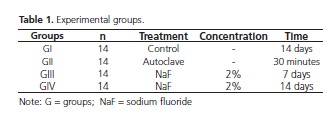
The specimens (n=56) were divided randomly into three experimental groups and one control group (n=14). They were then sterilized with ethylene oxide (ACECIL, Campinas, SP, Brazil) prior to the microbiological analysis.
Formation of biofilm with E. faecalis
All procedures were conducted in a biological safety cabinet (VecoFlow Ltda, Campinas, SP, Brazil). Inoculum standardized from E. faecalis ATCC 29212 was obtained using a spectrophotometer (108 UFC/mL). Aliquots of 1% of the standardized bacterial inoculum were transferred to Tryptic Soy Broth (TSB; Difco, Detroit, USA).
]]> Microplates containing the specimens were maintained in microaerophilic conditions in a bench incubator with orbital agitation (Quimis Aparelhos Científicos Ltda, Diadema, SP, Brazil) at 37° C for 48 hours. After 24 hours, the culture media was renewed without adding the microbial inoculum.Division of the Experimental Groups
Three experimental groups were established to assess the sterilization method (Table 1). The teeth in the autoclave group were submitted to sterilization at 121º C for 30 minutes (15 psi). The groups with sodium fluoride (NaF) 2% were submersed in the solution for seven or 14 days (the solution was renewed after the first week). In the control group, the teeth were maintained in saline solution (0.85%) for 14 days at room temperature.
Samples and microbiological analysis
After each treatment type, the specimens from each group were removed aseptically using a sterilized clinical tweezers and transferred to bottles (200 mL) containing Alternative Thioglycollate Medium (NHI; Himedia). The bottles containing each tooth were incubated at 37° C for 14 days.
]]> There was evidence of turbidity in the samples and analysis of the solution absorbance was carried out in the spectrophotometer. The sterility test was conducted using a thioglycollate medium. Afterwards, the absorbance of the medium was determined by bacterial growth at a wavelength of 625nm.
Data analysis
Analysis of the absorbance values in the groups tested was performed using the Shapiro-Wilk test, which confirmed a normal distribution (p > 0.05). Consequently, the t-test for independent samples was used to compare the values between the groups (control vs. NaF; autoclave vs. NaF), with the results expressed as mean ± standard deviation (SD) values. The level of significance was set at p < 0.05. The statistical analysis was performed using SPSS software (SPSS Inc., Chicago, IL, USA), version 21.0.
RESULTS
Analysis of the turbidity of the broth with bacterial growth was realized for the four groups up to 14 days of incubation. Evidence of turbidity in the broths indicated bacterial growth and consequently, ineffective sterilization.
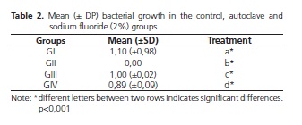
The negative control group, without antimicrobial treatment, exhibited turbidity in the bottles of alternative thioglycollate medium. Conversely, no microbial growth was observed in the autoclave group after incubation (Table 2).
With regards to the NaF 2% solution, the specimens stored in solution for 14 days exhibited a greater reduction of bacterial load than those treated for only seven days (Table 2).
The statistical analysis performed using the t-test for independent samples confirmed statistically significant differences (p <0,05) between the control, autoclave, NaF 2% seven days and NaF 2% 14 days groups (Table 2).
]]>
DISCUSSION
The experimental model adopted in the present study enabled the reinfection of extracted human teeth, which is very similar to what occurs in clinical situations16. The microorganism E. faecalis was used due to its resistance qualities, capacity to form biofilm and ability to survive as a monoculture17. In the present study, the autoclave and NaF 2% experimental groups significantly reduced the presence of E. faecalis, when compared with the control group.
The present study showed that using autoclave at 121º C for 30 minutes (15 psi) was effective in the sterilization of extracted human teeth. The results of this research are in agreement with those obtained in previous studies that indicated the capacity of this method to deactivate different microorganisms, such as viruses, fungi and spores2,8-9. However, a number of previous studies have stated that the method has disadvantages, which are related to the maintenance of the structural properties of extracted teeth11,18-19.
Although the NaF 2% solution did not sterilize the specimens, the microbial load was reduced after seven and 14 days of treatment. Thus, the results of the present study suggest that NaF 2% solution is involved in anti-bacterial activity. This finding corroborates the results of earlier studies, in which fluoride was reported to play an important role in bacterial inhibition20-21. The use of NaF 2% solution for two weeks resulted in the greatest disinfectant capacity. This treatment method caused a statistically significant reduction in the quantity of E. faecalis, when compared to the treatment method that only used NaF 2% solution for seven days. This difference could be explained by the fact that NaF exhibits deleterious effects that are dependent on the concentration used and the duration of exposure15.
The active mechanism in which fluoride induces microbial death has been addressed by several authors at different times. Some have attributed the destruction of the microorganisms to the inhibition of the transport of glucose to the interior of cells as a result of excessive acidification in the cytoplasm22. Other studies have demonstrated the capacity of fluoride to inhibit important enzymes and proteins, including: enolase, adenosine triphosphate, phosphoserine and phosphotyrosyl phosphatase15,21,23. Thus, these alterations negatively affect the metabolism of the microorganisms and may lead to cellular death.
Another important aspect assessed was the stipulation of the concentration of NaF solution needed to promote microbial activity. A previous study indicated that high concentrations of fluoride ions (> 0.12%) would lead to a bactericidal effect24. Chouhan & Flora25 demonstrated that NaF could be biologically active, even in low concentrations. The present study used NaF 2% solution and the results indicated significant bactericidal activity on the microorganism E. faecalis. The results of the present study are in agreement with the work of Tong et al.21, who found that NaF 2% had a strong bactericidal effect on the biofilm of S. mutans. After analysis, the biofilm was found to be irregular and distorted, when compared with its original shape.
]]> However, the fact that the NaF 2% solution was not capable of eliminating all of the microorganisms could be explained by a previous study conducted by Chouhan & Flora25, who reported that higher concentrations of NaF caused a reduced ionic mobility and consequently, a lower availability of fluoride ions, whereas lower concentrations of NaF exhibited greater ion mobility, thereby promoting microbial activity.In the literature, different methods have been suggested for the disinfection of extracted teeth. Recent studies have tested materials such as Gigasept PA (6) and vinegar26, both of which exhibited disinfectant capacity. However, the former is a high level hospital disinfectant and thus, it is difficult to obtain. The latter requires further studies to better understand its active mechanism. NaF 2% solution offers the advantages of being easy to obtain, inexpensive, easy-to-use and fast.
Gamma radiation is known as the best method of sterilizing extracted human teeth. Results of previous studies have indicated that this method does not affect the permeability of dentin or cause structural alterations9,11. However, gamma radiation is an expensive and complex technique that is not easily accessible19. Consequently, it was not used in the present study, which could be considered to be a limitation of the research.
Based on the results of this investigation, NaF 2% solution can be considered as a viable method of disinfection for extracted teeth. The procedure is fast and only requires NaF powder and distilled water. However, further studies should be conducted to assess NaF solution as a possible means of storage, given that disinfection and storage means must be effective and also ensure the structural integrity of the tooth3. Therefore, new concentrations should be tested to assess possible ionic exchanges between the surface of the tooth and the NaF solution, as well as to assess interference in the sterilization process.
CONCLUSION
Based on the results of the present study, NaF 2% solution could be considered an effective method of disinfecting extracted human teeth, due to is capacity to reduce the microbial load of the microorganism E. faecalis. It is important to bear in mind that extracted human teeth should be handled with extreme caution, even after the disinfection process.
Acknowledgements
The study was supported by FAPEMIG.
]]>Collaborators
All authors participated in the study design. MFF CARVALHO and AM BOTELHO writing the manuscript. AM BOTELHO, KTA TAVANO and E WATANABE reviewed the paper. MFF CARVALHO, AM BOTELHO and KTA TAVANO was involved in the data analysis.
REFERENCES
1. Hashemipour MA, Mozafarinia R, Mirzadeh A, Mozafarinia R, Mirzadeh A, Aramon M, et al. Knowledge, attitudes, and performance of dental students in relation to sterilization/ disinfection methods of extracted human teeth. Dent Res J (Isfahan). 2013 Jul-Aug;10(4):482-88. [ Links ]
2. Dominici JT, Eleazer PD, Clark SJ, Staat RH, Scheetz, JP. Disinfection/ sterilization of extracted teeth for dental student use. J Dent Educ. 2001;65:1278-1280.
3. Attam K, Talwar S, Yadav S, Miglani S. Comparative analysis of the effect of autoclaving and 10% formalin storage on extracted teeth: a microleakage evaluation. J Conserv Dent. 2009;12(1):26- 30. doi: 10.4103/0972-0707.53338
4. Tate WH, White RR. Disinfection of human teeth for education purposes. J Dent Educ.1991;55:583-585.
]]> 5. Centers for Disease Control. Recommended Infection Control Practices For Dentistry. MMWR. 1993;42:8-9.6. Hope CK, Griffiths DA, Prior DM. Finding an alternative to formalin forsterilization of extracted teeth for teaching purposes. J Dent Educ. 2013;77: 68-71.
7. Goel K, Gupta R, Solanki J, Nayak M. A comparative study between microwave irradiation and sodium hypochlorite chemical disinfection: a prosthodontic view. J Clin Diagn Res. 2014; 8(4):42- 46. doi: 10.7860/JCDR/2014/8578.4274
8. Lolayekar NV, Bhat VS, Bhat SS. Disinfection methods of extracted human teeth. J Oral Health Comm Dent. 2007;1:27-29.
9. Kumar M, Sequeira PS, Peter S, Bhat GK. Sterilisation of extracted human teeth for educational use. Indian J Med Microbiol. 2005;23:256-258.
10. Cuny E, Carpenter WM. Extracted teeth: decontamination, disposal and use. Dent Assoc J. 1997;25(11):801-804.
11. Dewald JP. The use of extracted teeth for in vitro bonding studies: a review of infection control considerations. Dent Mater. 1997;13(2):74-81.
12. Xu X, Wang Y, Liao S, Wen ZT, Fan Y. Synthesis and characterization of antibacterial dental monomers and composites. J Biomed Mater Res B. 2012;100b: 1151-1162. doi: 10.1002/jbm.b.32683
13. Clarkson, BH. Caries prevention: fluoride. Adv Dent Res. 1991;5:41- 45.
14. Ogard B, Seppa L, Rolla G. Professionals topical fluoride apllications: clinical efficacy and mechanism of action. Adv Dent Res. 1994;8:190-201.
]]> 15. Everett ET. Fluoride's effects on the formation of teeth and bones, and the influence of genetics. J Den Res. 2011;90(5):552-560. doi: 10.1177/002203451038462616. Dornelles-Morgental R, Guerreiro-Tanomaru JM, Faria-Júnior NB, Hungaro-Duarte MA, Kuga MC, Tanomaru-Filho M. Antibacterial efficacy of endodontic irrigating solutions and their combinations in root canals contaminated with enterococcus faecalis. Oral Surg Oral Med Oral Pathol Oral Radiol Endod. 2011;112(3):396-400. doi: 10.1016/j.tripleo.2011.02.004
17. Stuart CH, Schwartz AS, Beeson TJ, Owatz CB. Enterococcus faecalis: its role in root canal treatment failure and current concepts in retreatment. J Endod. 2006;32(2): 93-98. doi: 10.1016/j.joen.2005.10.049
18. Amaechi BT, Higham SM, Edgar WM. The use of gamma irradiation for the sterilization of enamel for intra-oral cariogenicity tests. J Oral Rehabil. 1999 Oct;26(10):809-13.
19. Viana PS, Machado AL, Giampaolo ET, Pavarina AC, Vergani, CE. Disinfection of bovine enamel by microwave irradiation: effect on the surface microhardness and demineralization/ remineralization processes. Caries Res. 2010;44(4):349-357. doi: 10.1159/000318528
20. Hamilton IR. Biochemical effects of fluoride on oral bacteria. J Dent Res. 1990; 69:660-667.
21. Tong Z, Zhoub L, Jianga W, Kuanga R, Lic J, Tao R, et al. An in vitro synergetic evaluation of the use of nisin and sodium fluoride or chlorhexidine against streptococcus mutans. Peptides. 2011;32(10):2021-2026. doi: 10.1016/j.peptides.2011.09.002
22. Sutton SV, Bender GR, Marquis RE. Fluoride inhibition of protontranslocating atpases of oral bacteria. Infect Immun. 1987 Nov;55(11):2597-603.
23. Lussi A, Hellwig E, Klimek J. Fluorides - mode of action and recommendations for use. Schweiz Monatsschr Zahnmed. 2012;122(11):1030-42.
24. Mjor IA, Moorhead JE, Dahl JE. Reasons for replacement of restorations in permanent teeth in general dental practice. Int Dent J. 2000 Dec;50(6):361-6. doi: 10.1111/j.1875-595X.2000. tb00569.x
]]> 25. Chouhan S, Flora SJ. Effects of fluoride on the tissue oxidative stress and apoptosis in rats: biochemical assays supported by ir spectroscopy data. Toxicology. 2008; 254(1-2):61-67. doi: 10.1016/j.tox.2008.09.00826. Tijare M, Smitha D, Kasetty S, Kallianpur S, Gupta S, Amith HV. Vinegar as a disinfectant of extracted human teeth for dental educational use. J Oral Maxil Pathol. 2014;18(1):14-18. doi: 10.4103/0973-029X.131883
 Correspondence to:
MFF CARVALHO
Av. Pres. Antônio Carlos, 6627, Campus Pampulha
31270-901, Belo Horizonte, MG, Brasil
e-mail: monize_c@ hotmail.com
]]> Received on: 19/7/2015
]]>