<?xml version="1.0" encoding="ISO-8859-1"?><article xmlns:mml="http://www.w3.org/1998/Math/MathML" xmlns:xlink="http://www.w3.org/1999/xlink" xmlns:xsi="http://www.w3.org/2001/XMLSchema-instance">
<front>
<journal-meta>
<journal-id>1808-5210</journal-id>
<journal-title><![CDATA[Revista de Cirurgia e Traumatologia Buco-maxilo-facial]]></journal-title>
<abbrev-journal-title><![CDATA[Rev. cir. traumatol. buco-maxilo-fac.]]></abbrev-journal-title>
<issn>1808-5210</issn>
<publisher>
<publisher-name><![CDATA[Universidade de Pernambuco]]></publisher-name>
</publisher>
</journal-meta>
<article-meta>
<article-id>S1808-52102014000400006</article-id>
<title-group>
<article-title xml:lang="pt"><![CDATA[Cemento-Ossifying Fibroma of the Mandible Case report]]></article-title>
<article-title xml:lang="en"><![CDATA[Fibroma cemento-ossificante de mandíbula: Relato de caso]]></article-title>
</title-group>
<contrib-group>
<contrib contrib-type="author">
<name>
<surname><![CDATA[Elarbi]]></surname>
<given-names><![CDATA[Mohamed]]></given-names>
</name>
<xref ref-type="aff" rid="A01"/>
</contrib>
<contrib contrib-type="author">
<name>
<surname><![CDATA[Azuzz]]></surname>
<given-names><![CDATA[Lubna]]></given-names>
</name>
<xref ref-type="aff" rid="A02"/>
</contrib>
</contrib-group>
<aff id="A01">
<institution><![CDATA[,AOA  ]]></institution>
<addr-line><![CDATA[ ]]></addr-line>
</aff>
<aff id="A02">
<institution><![CDATA[,Tripoli university Faculty of Medicine ]]></institution>
<addr-line><![CDATA[ ]]></addr-line>
</aff>
<pub-date pub-type="pub">
<day>00</day>
<month>12</month>
<year>2014</year>
</pub-date>
<pub-date pub-type="epub">
<day>00</day>
<month>12</month>
<year>2014</year>
</pub-date>
<volume>14</volume>
<numero>4</numero>
<fpage>41</fpage>
<lpage>44</lpage>
<copyright-statement/>
<copyright-year/>
<self-uri xlink:href="http://revodonto.bvsalud.org/scielo.php?script=sci_arttext&amp;pid=S1808-52102014000400006&amp;lng=en&amp;nrm=iso"></self-uri><self-uri xlink:href="http://revodonto.bvsalud.org/scielo.php?script=sci_abstract&amp;pid=S1808-52102014000400006&amp;lng=en&amp;nrm=iso"></self-uri><self-uri xlink:href="http://revodonto.bvsalud.org/scielo.php?script=sci_pdf&amp;pid=S1808-52102014000400006&amp;lng=en&amp;nrm=iso"></self-uri><abstract abstract-type="short" xml:lang="en"><p><![CDATA[Cemento-ossifying fibroma is a relatively rare, benign, non-odontogenic tumour of the jaws, regarded as a subdivision of fibro-osseous lesions. The usual age of occurrence is between 20 and 40 years. The female:male ratio is 5:1, usual site being the posterior mandible. The aim of this report to present the clinical and radiological features and management of a 32-year-old female Libyan patient who presented with an asymptomatic giant swelling of three years' duration in the left mandible, the diagnosis of which was confirmed by histopathology. Cemento-ossifying fibroma (COF) is a benign, asymptomatic lesion of the jaws characterized by the production of well-demarcated bone of slow growth. It typically affects females aged between 20 and 40 years, in the premolar and molar area, causing a painless swelling, of slow, expansile growth. The periodontal ligament contains both bone and cementum. The pathogenesis of extraosseous COF, where there is no periodontal tissue, as suggested by Cakir and Karadayi, originates in embryonic nests and the ectopic periodontal membrane, as suggested by Brademann et al. in their cytogenetic studies. Depending on the stage of maturation, cemento-ossifying fibroma may range from radiolucent, through mixed, to completely radio-opaque. Histopathologically, it appears as well-circumscribed, occasionally encapsulated with various amounts of bony trabecular/cementum formation in a fibrous stroma.]]></p></abstract>
<abstract abstract-type="short" xml:lang="pt"><p><![CDATA[Fibroma Cemento-ossificante é um tumor benigno relativamente raro, não odontogênico dos maxilares, considerado como subdivisão de lesões fibro-ósseas. A idade usual de ocorrência é entre 20 e 40 anos. Há predileção do sexo feminino na proporção 5:1, e da região posterior da mandibula. Esse relato de caso apresenta uma paciente libanês de 32 anos, que se apresentou com aumento grande de volume, achados clínicos e radiológicos assintomáticos na mandíbula esquerda com duração de três anos, o diagnótico histopatológico confirma o diagnóstico clínico. Fibroma cemento-ossificante (COF) é caracterizado pela produção óssea de crescimento lento, bem demarcada, assintomática, benigna dos maxilares. Geralmente afeta mulheres entre a idade de 20 a 40 anos na área molares e pré-molares, causando um edema, indolor, crescimento lento e expansível. A patogênese das lesões extras ósseas do COF podem não apresentar tecidos periodontais, como sugerido por Cakir e Karadayi e sim células embrionárias e membrana periodontal ectópica como sugerido por Brademann et al.6 em seus estudos citogenéticos. Dependendo do estágio de maturação, fibroma cemento-ossificante pode ter umavariedaderadiolúcida, mista ou completamente radiopaca. Histopatologicamente, aparecem circunscritos, ocasionalmente encapsulados com alta quantidade de formação trabecular9.]]></p></abstract>
<kwd-group>
<kwd lng="en"><![CDATA[tumour]]></kwd>
<kwd lng="en"><![CDATA[jaws]]></kwd>
<kwd lng="en"><![CDATA[surgery.]]></kwd>
<kwd lng="pt"><![CDATA[Tumor]]></kwd>
<kwd lng="pt"><![CDATA[Maxilares e Cirurgia.]]></kwd>
</kwd-group>
</article-meta>
</front><body><![CDATA[ <p>&nbsp;</p>     <p><font size="4" face="Verdana, Arial, Helvetica, sans-serif"><a name="top"/></a><B>Cemento-Ossifying Fibroma of the Mandible Case report</B></font></p>     <p>&nbsp;</p>     <p><font size="3" face="Verdana, Arial, Helvetica, sans-serif"><b>Fibroma cemento-ossificante de mand&iacute;bula: Relato de caso</b> </font></p>     <p>&nbsp;</p>     <p>&nbsp;</p>     <p><font size="2" face="Verdana, Arial, Helvetica, sans-serif"><b>Mohamed Elarbi<sup>I</sup>; LubnaAzuzz<sup>II</sup></b></font></p>     <p><font size="2" face="Verdana, Arial, Helvetica, sans-serif"><sup>I</sup> BDS,MmedSC,FFDRCSI; Head of department, Senior Consultant; University clinic of Oral &amp; Maxillofacial Surgery; Ali Omar Askar (AOA) Center of neurosurgery, Esbea Tripoli Libya    <br> <sup>II</sup> Consultant pathologist MD,Tripoli Medical Center; Faculty of Medicine ,Tripoli university ,Tripoli Libya    <br> </font></p>     ]]></body>
<body><![CDATA[<p><font size="2" face="Verdana, Arial, Helvetica, sans-serif"><a href="#back">Endere&ccedil;o para correspond&ecirc;ncia</a></font></p>     <p>&nbsp;</p>     <p>&nbsp;</p> <hr size="1" noshade>     <p><font size="2" face="Verdana, Arial, Helvetica, sans-serif"><b>ABSTRACT</b> </font></p>     <p><font size="2" face="Verdana, Arial, Helvetica, sans-serif">Cemento-ossifying fibroma is a relatively rare, benign, non-odontogenic tumour of the jaws, regarded as a subdivision of fibro-osseous lesions. The usual age of occurrence is between 20 and 40 years. The female:male ratio is 5:1, usual site being the posterior mandible. The aim of this report to present the clinical and radiological features and management of a 32-year-old female Libyan patient who presented with an asymptomatic giant swelling of three years' duration in the left mandible, the diagnosis of which was confirmed by histopathology. Cemento-ossifying fibroma (COF) is a benign, asymptomatic lesion of the jaws characterized by the production of well-demarcated bone of slow growth. It typically affects females aged between 20 and 40 years, in the premolar and molar area, causing a painless swelling, of slow, expansile growth. The periodontal ligament contains both bone and cementum. The pathogenesis of extraosseous COF, where there is no periodontal tissue, as suggested by Cakir and Karadayi, originates in embryonic nests and the ectopic periodontal membrane, as suggested by Brademann et al. in their cytogenetic studies. Depending on the stage of maturation, cemento-ossifying fibroma may range from radiolucent, through mixed, to completely radio-opaque. Histopathologically, it appears as well-circumscribed, occasionally encapsulated with various amounts of bony trabecular/cementum formation in a fibrous stroma. </font></p>     <p><font size="2" face="Verdana, Arial, Helvetica, sans-serif"><B>Descriptors: </B>tumour, jaws, surgery.</font></p> <hr size="1" noshade>     <p><font size="2" face="Verdana, Arial, Helvetica, sans-serif"><B>RESUMO</B> </font></p>     <p><font size="2" face="Verdana, Arial, Helvetica, sans-serif">Fibroma Cemento-ossificante &eacute; um tumor benigno relativamente raro, n&atilde;o odontog&ecirc;nico dos maxilares, considerado como subdivis&atilde;o de les&otilde;es fibro-&oacute;sseas. A idade usual de ocorr&ecirc;ncia &eacute; entre 20 e 40 anos. H&aacute; predile&ccedil;&atilde;o do sexo feminino na propor&ccedil;&atilde;o 5:1, e da regi&atilde;o posterior da mandibula. Esse relato de caso apresenta uma paciente liban&ecirc;s de 32 anos, que se apresentou com aumento grande de volume, achados cl&iacute;nicos e radiol&oacute;gicos assintom&aacute;ticos na mand&iacute;bula esquerda com dura&ccedil;&atilde;o de tr&ecirc;s anos, o diagn&oacute;tico histopatol&oacute;gico confirma o diagn&oacute;stico cl&iacute;nico. Fibroma cemento-ossificante (COF) &eacute; caracterizado pela produ&ccedil;&atilde;o &oacute;ssea de crescimento lento, bem demarcada, assintom&aacute;tica, benigna dos maxilares. Geralmente afeta mulheres entre a idade de 20 a 40 anos na &aacute;rea molares e pr&eacute;-molares, causando um edema, indolor, crescimento lento e expans&iacute;vel. A patog&ecirc;nese das les&otilde;es extras &oacute;sseas do COF podem n&atilde;o apresentar tecidos periodontais, como sugerido por Cakir e Karadayi e sim c&eacute;lulas embrion&aacute;rias e membrana periodontal ect&oacute;pica como sugerido por Brademann et al.6 em seus estudos citogen&eacute;ticos. Dependendo do est&aacute;gio de matura&ccedil;&atilde;o, fibroma cemento-ossificante pode ter umavariedaderadiol&uacute;cida, mista ou completamente radiopaca. Histopatologicamente, aparecem circunscritos, ocasionalmente encapsulados com alta quantidade de forma&ccedil;&atilde;o trabecular9.</font></p>     <p><font size="2" face="Verdana, Arial, Helvetica, sans-serif"><B>Descritores: </B>Tumor, Maxilares e Cirurgia.</font> </p> <hr noshade size="1">     <p>&nbsp;</p>     ]]></body>
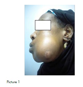
<body><![CDATA[<p>&nbsp;</p>     <p><font size="3" face="Verdana, Arial, Helvetica, sans-serif"><B> CASE REPORT</B></font></p>     <p><font size="2" face="Verdana, Arial, Helvetica, sans-serif">A 32-year-old old Libyan female patient reported to our oral and maxillofacial surgery department, her chief complaint being a painless, slowly growing, progressive swelling in the body of the left mandible of three years' duration. The medical and dental histories were both unremarkable. </font></p>     <p><font size="2" face="Verdana, Arial, Helvetica, sans-serif">The extra-oral clinical examination showed a large well-defined swelling involving the left mandible (<a href="#fig01">Figure 1</a>) measuring approximately 8x5 cm. Swelling was nontender bony hard, with a scar over its summit (old trauma to skin). Lymph nodes were not palpable, with no sensory or motor nerve deficit in the area, TMJ and jaw movements being within the normal range. On intraoral examination there was buccolingual expansion of the lesion, with normal overlying mucosa. The oral hygiene was moderate and all teeth related to the lesion were vital, tender with distolingual displacement of the second and third molars, and lingually inclined premolars with spacing. </font></p>     <p>&nbsp;</p>     <p><a name="fig01"></a></p>     <p>&nbsp;</p>     <p align="center"><img src="/img/revistas/rctbmf/v14n4/a06fig01.jpg"></p>     <p>&nbsp;</p>      <p><font size="2" face="Verdana, Arial, Helvetica, sans-serif">The following laboratory blood investigations were carried out: </font></p>     ]]></body>
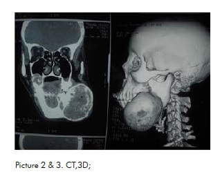
<body><![CDATA[<p><font size="2" face="Verdana, Arial, Helvetica, sans-serif">Full blood count, differential count, serum calcium, serum alkaline phosphatase, serum phosphorus, all values being within the normal ranges. </font></p>     <p><font size="2" face="Verdana, Arial, Helvetica, sans-serif">Radiographically, OPG shows a mixed lesion involving the body of the left mandible, well-defined, mixed, radiolucent and radio-opaque lesion involving the body of left mandible from canine tooth to angle of mandible distal to the lower third left molar tooth. </font></p>     <p><font size="2" face="Verdana, Arial, Helvetica, sans-serif">The CT scan shows hetero-dens, extensile lesion with well-defined borders in the body of the left mandible.(<a href="#fig02">Figure 2</a>) </font></p>     <p><font size="2" face="Verdana, Arial, Helvetica, sans-serif">The tridimensional CT scan showed the full extent of the lesion in the posterior region of the left mandible (<a href="#fig02">Figure 3</a>) </font></p>     <p><font size="2" face="Verdana, Arial, Helvetica, sans-serif">On the basis of the clinical and radiographic features, a provisional diagnosis of benign fibroosseous lesion was made, the histopathology showing bone and cementum<sup>9</sup>, in a fibrous connective tissue stroma.</font></p>     <p>&nbsp;</p>     <p><a name="fig02"></a></p>     <p>&nbsp;</p>     <p align="center"><img src="/img/revistas/rctbmf/v14n4/a06fig02.jpg"></p>     <p>&nbsp;</p>      ]]></body>
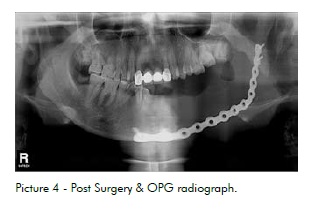
<body><![CDATA[<p><font size="3" face="Verdana, Arial, Helvetica, sans-serif"><B>TREATMENT</B></font></p>     <p><font size="2" face="Verdana, Arial, Helvetica, sans-serif">The patient was admitted to the AOA Hospital on January 16th, 2012 and placed in the care of the maxillofacial department for excision of the lesion and reconstruction under general anesthesia. </font></p>     <p><font size="2" face="Verdana, Arial, Helvetica, sans-serif">The lesion was excised completely using an extra- oral approach, and the defect was reconstructed with titanium reconstruction plates (<a href="#fig04">Figure 4</a>). </font></p>     <p><font size="2" face="Verdana, Arial, Helvetica, sans-serif">The initial postoperative period was uneventful and the patient was discharged from the hospital and followed up in the OPD maxillofacial clinic. There have been no signs of recurrence to date and bony reconstruction and dental rehabilitations are being planned.</font></p>     <p>&nbsp;</p>     <p><a name="fig04"></a></p>     <p>&nbsp;</p>     <p align="center"><img src="/img/revistas/rctbmf/v14n4/a06fig04.jpg"></p>      <p>&nbsp;</p>     <p><font size="3" face="Verdana, Arial, Helvetica, sans-serif"><b>DISCUSSION</b> </font></p>       ]]></body>
<body><![CDATA[<p><font size="2" face="Verdana, Arial, Helvetica, sans-serif">Cemento-ossifying fibroma is defined by WHO as a demarcated or rarely encapsulated neoplasm consisting of fibrous tissue containing various amounts of mineralized material (bone and/or cementum). It occurs most frequently in females (female: male=5:1) with ages ranging from 10 to 59 years. It originates in the mandible in 62 to 89% of the patients, 72% in the premolar region and 22%<sup>5</sup> involving the molar region of the maxilla, ethmoidal and orbital regions, but may also be seen exceptionally in petrous bone<sup>6</sup>. When occuring in children, it has been referred to as juvenile aggressive COF, which presents at an earlier age and is more aggressive clinically and more vascular on pathological examination<sup>6</sup>. Cemento-ossifying fibroma (COF) is dependent on the nature of the calcifications, either cementum or bone, and its behavior is either aggressive or static<sup>8</sup>. </font></p>     <p><font size="2" face="Verdana, Arial, Helvetica, sans-serif">The radiographic appearance is very important in the diagnosis of COF in order to differentiate it from other fibro-osseous lesions. In its early stages the lesion is radiolucent with an ill-defined border; as it matures it becomes more defined, with or without a sclerotic border. With increased maturity of the lesion there is an increase in calcific flecks, which may progress to form a more radio-opaque mass. The growth pattern is centrifugal (equal in all directions) and presents as a well-circumscribed mass, the soft tissue capsule making it better defined. </font></p>     <p><font size="2" face="Verdana, Arial, Helvetica, sans-serif">The radiographic differential diagnosis includes:</font></p>     <p><font size="2" face="Verdana, Arial, Helvetica, sans-serif"> 1- Fibrous dysplasia (ground glass appearance, the expanded bone resembling normal bone) </font></p>     <p><font size="2" face="Verdana, Arial, Helvetica, sans-serif">2- Cemento-osseous dysplasia (usually multifocal) </font></p>     <p><font size="2" face="Verdana, Arial, Helvetica, sans-serif">3- Condensing osteitis</font></p>     <p><font size="2" face="Verdana, Arial, Helvetica, sans-serif"> 4- Pindporg tumor (associated with impacted teeth)</font></p>     <p><font size="2" face="Verdana, Arial, Helvetica, sans-serif"> 5- Odontoma (presence of tooth-like structures)</font></p>     <p><font size="2" face="Verdana, Arial, Helvetica, sans-serif"> Treatment was complete surgical excision. As the tumor is less vascularized and well-circumscribed, it is usually easy to remove from the surrounding bone. </font></p>     <p><font size="2" face="Verdana, Arial, Helvetica, sans-serif">Prognosis is good and recurrence very rare.</font></p>     ]]></body>
<body><![CDATA[<p>&nbsp;</p>     <p><font size="3" face="Verdana, Arial, Helvetica, sans-serif"><b>CONCLUSION</b> </font></p>       <p><font size="2" face="Verdana, Arial, Helvetica, sans-serif">Establishing the correct diagnosis of cementoossifying fibroma is important because: </font></p>     <p><font size="2" face="Verdana, Arial, Helvetica, sans-serif">1- In its early stages it displays a radiolucent area and may be confused with periapical pathology.</font></p>     <p><font size="2" face="Verdana, Arial, Helvetica, sans-serif"> 2- It must not be confused with other fibro-osseous lesions as its management is different. </font></p>     <p><font size="2" face="Verdana, Arial, Helvetica, sans-serif">3- Facial asymmetry can be severe, due to its late presentation, and it is usually asymptomatic.</font></p>     <p>&nbsp;</p>     <p><font size="3" face="Verdana, Arial, Helvetica, sans-serif"><B>REFERENCES  </B></font></p>     <!-- ref --><p><font size="2" face="Verdana, Arial, Helvetica, sans-serif">1. Chia-chuan, Hsien-yen hug, Julia u fonggchang, Chuan-hangyu .Central ossifying fibroma: A Clinicopathological study of 128 cases.JFormos. Med association: 2008:107(4):208-94.    &nbsp;&nbsp;&nbsp;&nbsp;&nbsp;&nbsp;&nbsp;&nbsp;[&#160;<a href="javascript:void(0);" onclick="javascript: window.open('/scielo.php?script=sci_nlinks&ref=472711&pid=S1808-5210201400040000600001&lng=','','width=640,height=500,resizable=yes,scrollbars=1,menubar=yes,');">Links</a>&#160;]<!-- end-ref --> </font></p>     ]]></body>
<body><![CDATA[<p><font size="2" face="Verdana, Arial, Helvetica, sans-serif">2. Jayachandran S,MeenakshiR.Cemento-ossifying fibroma. Indian J.Dent Res 2004:15(1):35-39 </font></p>     <p><font size="2" face="Verdana, Arial, Helvetica, sans-serif">3. Gianfuigi Longobardi, et al, Extra osseous Cemento-ossifying Fibroma of cheek.Scolarly Research Exchange, vol.2009, Article ID: 493190.doi </font></p>     <p><font size="2" face="Verdana, Arial, Helvetica, sans-serif">4. Abdulbaset Dalghous, juma O AkhabuliCemento- ossifying fibroma occurring in Elderly patient: A case report and review of literature. Libyan journal of Medicine 2007:2:95-98 </font></p>     <p><font size="2" face="Verdana, Arial, Helvetica, sans-serif">5. Mayo Scott RF.Persistentcemento-osififying fibroma: report of a case and review of literature.J Oral Maxillofacial Surg.1988; 46:58-63 </font></p>     <p><font size="2" face="Verdana, Arial, Helvetica, sans-serif">6. Bertand B, Ely et al: juvenile aggressive cementossifying fibroma: Case report and review of litrature. Laryngoscope.1994; 103:103:1385-90 </font></p>     <p><font size="2" face="Verdana, Arial, Helvetica, sans-serif">7. A Barberi, S Cappabianca, et al. bilateral cemento-ossifying fibroma of maxillary sinus. British journal of Radiology 2003; 76:279-80 </font></p>     <p><font size="2" face="Verdana, Arial, Helvetica, sans-serif">8. Khanna maneesh et al Cemento-ossifying fibroma of Para nasal sinus presenting as orbital cellulitis. A Case report .J .Radiology 2009; 3(4):18-25 </font></p>     <p><font size="2" face="Verdana, Arial, Helvetica, sans-serif">9. Kramer IRH, Pindburgetal.Histological typing of odontogenicTumors (2nd ed).Berlin: Springer- VERLAG 1992; 27-28. </font></p>     <p><font size="2" face="Verdana, Arial, Helvetica, sans-serif">10. Robert P Lanlais, Olaf E et al; Diagnostic imaging of the jaws.Willamsand Wilkins 1995; 551-52. </font></p>      <p>&nbsp;</p>     ]]></body>
<body><![CDATA[<p><font face="Verdana, Arial, Helvetica, sans-serif" size="2"><b>Recebido para publica&ccedil;&atilde;o:</b> 13/05/2014<br/>  <b>Aceito para publica&ccedil;&atilde;o:</b> 24/06/2014</font></p>     <p>&nbsp;</p>        ]]></body>
<back>
<ref-list>
<ref id="B1">
<label>1</label><nlm-citation citation-type="journal">
<person-group person-group-type="author">
<name>
<surname><![CDATA[Chia]]></surname>
<given-names><![CDATA[chuan]]></given-names>
</name>
<name>
<surname><![CDATA[Hsien-yen]]></surname>
<given-names><![CDATA[hug]]></given-names>
</name>
<name>
<surname><![CDATA[Julia u]]></surname>
<given-names><![CDATA[fonggchang]]></given-names>
</name>
<name>
<surname><![CDATA[Chuan]]></surname>
<given-names><![CDATA[hangyu]]></given-names>
</name>
</person-group>
<article-title xml:lang="en"><![CDATA[Central ossifying fibroma: A Clinicopathological study of 128 cases.JFormos.]]></article-title>
<source><![CDATA[Med association]]></source>
<year>2008</year>
<volume>107</volume>
<numero>4</numero>
<issue>4</issue>
<page-range>208-94.</page-range></nlm-citation>
</ref>
</ref-list>
</back>
</article>