<?xml version="1.0" encoding="ISO-8859-1"?><article xmlns:mml="http://www.w3.org/1998/Math/MathML" xmlns:xlink="http://www.w3.org/1999/xlink" xmlns:xsi="http://www.w3.org/2001/XMLSchema-instance">
<front>
<journal-meta>
<journal-id>0104-5695</journal-id>
<journal-title><![CDATA[RPG. Revista de Pós-Graduação]]></journal-title>
<abbrev-journal-title><![CDATA[RPG, Rev. pós-grad.]]></abbrev-journal-title>
<issn>0104-5695</issn>
<publisher>
<publisher-name><![CDATA[Revista de Pós-Graduação da Faculdade de Odontologia da Universidade de São Paulo]]></publisher-name>
</publisher>
</journal-meta>
<article-meta>
<article-id>S0104-56952010000300008</article-id>
<title-group>
<article-title xml:lang="en"><![CDATA[Accessory mental foramen: case report]]></article-title>
<article-title xml:lang="pt"><![CDATA[Forame mentual acessório: relato de caso]]></article-title>
</title-group>
<contrib-group>
<contrib contrib-type="author">
<name>
<surname><![CDATA[Neves]]></surname>
<given-names><![CDATA[Frederico Sampaio]]></given-names>
</name>
<xref ref-type="aff" rid="A01"/>
</contrib>
<contrib contrib-type="author">
<name>
<surname><![CDATA[Oliveira]]></surname>
<given-names><![CDATA[Luciana Soares de Andrade Freitas]]></given-names>
</name>
<xref ref-type="aff" rid="A02"/>
</contrib>
<contrib contrib-type="author">
<name>
<surname><![CDATA[Torres]]></surname>
<given-names><![CDATA[Marianna Guanaes Gomes]]></given-names>
</name>
<xref ref-type="aff" rid="A02"/>
</contrib>
<contrib contrib-type="author">
<name>
<surname><![CDATA[Crusoé-Souza]]></surname>
<given-names><![CDATA[Mady]]></given-names>
</name>
<xref ref-type="aff" rid="A03"/>
</contrib>
<contrib contrib-type="author">
<name>
<surname><![CDATA[Oliveira]]></surname>
<given-names><![CDATA[Christiano]]></given-names>
</name>
<xref ref-type="aff" rid="A04"/>
</contrib>
<contrib contrib-type="author">
<name>
<surname><![CDATA[Campos]]></surname>
<given-names><![CDATA[Paulo Sérgio Flores]]></given-names>
</name>
<xref ref-type="aff" rid="A05"/>
</contrib>
<contrib contrib-type="author">
<name>
<surname><![CDATA[Crusoé-Rebello]]></surname>
<given-names><![CDATA[Ieda]]></given-names>
</name>
<xref ref-type="aff" rid="A05"/>
</contrib>
</contrib-group>
<aff id="A01">
<institution><![CDATA[,State University of Campinas Department of Oral Diagnosis ]]></institution>
<addr-line><![CDATA[Piracicaba SP]]></addr-line>
<country>Brazil</country>
</aff>
<aff id="A02">
<institution><![CDATA[,Federal University of Bahia School of Dentistry Department of Oral Radiology]]></institution>
<addr-line><![CDATA[Salvador BA]]></addr-line>
<country>Brazil</country>
</aff>
<aff id="A03">
<institution><![CDATA[,Federal University of Bahia Institute of Health Sciences Department of Morphology]]></institution>
<addr-line><![CDATA[Salvador BA]]></addr-line>
<country>Brazil</country>
</aff>
<aff id="A04">
<institution><![CDATA[,University of São Paulo Bauru School of Dentistry Department of Stomatology]]></institution>
<addr-line><![CDATA[Bauru SP]]></addr-line>
<country>Brazil</country>
</aff>
<aff id="A05">
<institution><![CDATA[,Federal University of Bahia School of Dentistry Department of Oral Radiology]]></institution>
<addr-line><![CDATA[Salvador BA]]></addr-line>
<country>Brazil</country>
</aff>
<pub-date pub-type="pub">
<day>00</day>
<month>09</month>
<year>2010</year>
</pub-date>
<pub-date pub-type="epub">
<day>00</day>
<month>09</month>
<year>2010</year>
</pub-date>
<volume>17</volume>
<numero>3</numero>
<fpage>173</fpage>
<lpage>176</lpage>
<copyright-statement/>
<copyright-year/>
<self-uri xlink:href="http://revodonto.bvsalud.org/scielo.php?script=sci_arttext&amp;pid=S0104-56952010000300008&amp;lng=en&amp;nrm=iso"></self-uri><self-uri xlink:href="http://revodonto.bvsalud.org/scielo.php?script=sci_abstract&amp;pid=S0104-56952010000300008&amp;lng=en&amp;nrm=iso"></self-uri><self-uri xlink:href="http://revodonto.bvsalud.org/scielo.php?script=sci_pdf&amp;pid=S0104-56952010000300008&amp;lng=en&amp;nrm=iso"></self-uri><abstract abstract-type="short" xml:lang="en"><p><![CDATA[Anatomical variations associated with the mental foramen are relatively uncommon. Among these variations, the presence of one or more accessory mental foramen has been reported in rare cases. The acknowledgment of their location with respect to the main mental foramen is of fundamental importance for previous evaluations for surgical and endodontic procedures. In this article, a case of accessory mental foramen was presented with computed tomography images, and the main complications related to this anatomical variation are discussed.]]></p></abstract>
<abstract abstract-type="short" xml:lang="pt"><p><![CDATA[Variações anatômicas associadas ao forame mentual são relativamente incomuns. Dentre tais variações, a presença de um ou mais forames mentuais acessórios tem sido relatada em raros casos. O conhecimento da sua localização em relação ao forame mentual principal é de fundamental importância na avaliação prévia de procedimentos cirúrgicos e endodônticos. Neste artigo, um caso de forame mentual acessório foi apresentado através de imagens por tomografia computadorizada, e as principais complicações relacionadas a essa variação anatômica são discutidas.]]></p></abstract>
<kwd-group>
<kwd lng="en"><![CDATA[Spiral computed tomography]]></kwd>
<kwd lng="en"><![CDATA[Cross-sectional anatomy]]></kwd>
<kwd lng="en"><![CDATA[Madible]]></kwd>
<kwd lng="pt"><![CDATA[Tomografia computadorizada espiral]]></kwd>
<kwd lng="pt"><![CDATA[Anatomia transversal]]></kwd>
<kwd lng="pt"><![CDATA[Mandíbula]]></kwd>
</kwd-group>
</article-meta>
</front><body><![CDATA[ <p align="right"><font size="2" face="Verdana"><b>ORIGINAL ARTICLES</b> ARTIGOS ORIGINAIS</font></p>     <p>&nbsp;</p>     <p><font size="4" face="Verdana"><b><a name="tx"></a>Accessory mental foramen: case report</b></font></p>     <p>&nbsp;</p>     <p><font size="3" face="Verdana"><b>Forame mentual acess&oacute;rio: relato de caso</b></font></p>     <p>&nbsp;</p>     <p>&nbsp;</p>     <p><font size="2" face="Verdana"><b>Frederico Sampaio Neves<sup>I</sup>; Luciana Soares de Andrade Freitas Oliveira<sup>II</sup>; Marianna Guanaes Gomes Torres<sup>II</sup>; Mady Cruso&eacute;&#45;Souza<sup>III</sup>; Christiano Oliveira<sup>IV</sup>; Paulo S&eacute;rgio Flores Campos<sup>V</sup>; Ieda Cruso&eacute;&#45;Rebello<sup>V</sup></b></font></p>     <p><font size="2" face="Verdana"><sup>I</sup>DDS, Department of Oral Diagnosis, State University of Campinas, Piracicaba Dental School – Piracicaba/SP, Brazil    <br>   <sup> II</sup>DDS, MSc, Department of Oral Radiology, Federal University of Bahia, School of Dentistry – Salvador/BA, Brazil    ]]></body>
<body><![CDATA[<br>   <sup> III</sup>DDS, MSc, Department of Morphology, Institute of Health Sciences, Federal University of Bahia – Salvador/BA, Brazil    <br>   <sup>IV</sup>DDS, MSc, Department of Stomatology, University of S&atilde;o Paulo, Bauru School of Dentistry – Bauru/SP, Brazil    <br>   <sup>V</sup>DDS, MSc, PhD, Department of Oral Radiology, Federal University of Bahia, School of Dentistry – Salvador/BA, Brazil</font></p>     <p><font size="2" face="Verdana"><a href="#nt">Corresponding address</a></font></p>     <p>&nbsp;</p>     <p>&nbsp;</p> <hr size="1" noshade>     <p><font size="2" face="Verdana"><b>ABSTRACT</b></font></p>     <p><font size="2" face="Verdana">Anatomical variations associated with the mental foramen are relatively uncommon. Among these variations, the presence of one or more accessory mental foramen has been reported in rare cases. The acknowledgment of their location with respect to the main mental foramen is of fundamental importance for previous evaluations for surgical and endodontic procedures. In this article, a case of accessory mental foramen was presented with computed tomography images, and the main complications related to this anatomical variation are discussed.</font></p>     <p><font size="2" face="Verdana"><b>Descriptors:</b> Spiral computed tomography. Cross&#45;sectional anatomy. Madible.</font></p> <hr size="1" noshade>     <p><font size="2" face="Verdana"><b>RESUMO</b></font></p>     ]]></body>
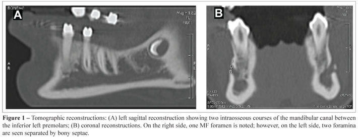
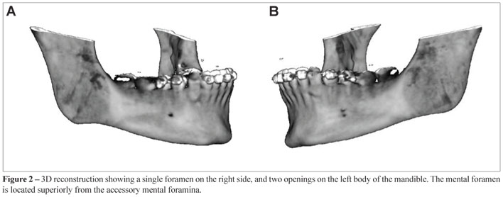
<body><![CDATA[<p><font size="2" face="Verdana">Varia&ccedil;&otilde;es anat&ocirc;micas associadas ao forame mentual s&atilde;o relativamente incomuns. Dentre tais varia&ccedil;&otilde;es, a presen&ccedil;a de um ou mais forames mentuais acess&oacute;rios tem sido relatada em raros casos. O conhecimento da sua localiza&ccedil;&atilde;o em rela&ccedil;&atilde;o ao forame mentual principal &eacute; de fundamental import&acirc;ncia na avalia&ccedil;&atilde;o pr&eacute;via de procedimentos cir&uacute;rgicos e endod&ocirc;nticos. Neste artigo, um caso de forame mentual acess&oacute;rio foi apresentado atrav&eacute;s de imagens por tomografia computadorizada, e as principais complica&ccedil;&otilde;es relacionadas a essa varia&ccedil;&atilde;o anat&ocirc;mica s&atilde;o discutidas.</font></p>     <p><font size="2" face="Verdana"><b>Descritores:</b> Tomografia computadorizada espiral. Anatomia transversal. Mand&iacute;bula.</font></p> <hr size="1" noshade>     <p>&nbsp;</p>     <p>&nbsp;</p>     <p><font size="3" face="Verdana"><b>Introduction</b></font></p>     <p><font size="2" face="Verdana">The mental foramen (MF) is located on the lateral aspect of the mandible, usually inferiorly to the interproximal region of the first and second premolars, bilaterally. The mental nerve and veins exit the bone through this foramen, which is oriented posterior&#45;superiorly in adults<SUP>11</SUP>.</font></p>     <p><font size="2" face="Verdana">The mental nerve is a somatic afferent (sensorial) nerve and corresponds to the terminal branch of the mandibular nerve, which is the third division of the trigeminal nerve. In the premolar region, the inferior alveolar nerve usually splits in two branches, the mental nerve and the incisive nerve. The incisive nerve runs intraosseously along with veins and innervates anterior mandibular teeth (incisors, canines, and premolars)<SUP>3</SUP>. The mental nerve emerges at the MF and divides into four branches: angular (innervation of the angle of the mouth region), medial and lateral inferior labial (skin of the lower lip, oral mucosa, and gingiva as far posterior as the second premolar), and mental branch (skin of the mental region)<SUP>6</SUP>.</font></p>     <p><font size="2" face="Verdana">Radiographically the MF presents as a single circular or elliptical radiolucent area in the premolar region, bilaterally<SUP>5</SUP>. The absence of MF<SUP>4</SUP> and the presence of multiple MF<SUP>9</SUP> are rarely reported. The presence of more than one MF, referred to as accessory mental foramina (AMF), has been registered by means of dissections, surgical findings, conventional radiographs, spiral computed tomography (CT), and cone beam CT.</font></p>     <p><font size="2" face="Verdana">CT has been established as a valuable imaging modality capable of providing in&#45;depth information about maxillofacial structures, allowing detailed evaluation of their topography and anatomical variations, such as AMF.</font></p>     <p><font size="2" face="Verdana">In the literature, numerous studies about the positions and anatomical variations of AMF in different races are reported. Most of the published reports are studies on dry skulls while some others have studied the panoramic radiographs of clinical cases.</font></p>     ]]></body>
<body><![CDATA[<p><font size="2" face="Verdana">The aim of this article was to report a case of AMF through multi&#45;slice CT images and to discuss relevant aspects in the literature related to this anatomical variation.</font></p>     <p>&nbsp;</p>     <p><font size="3" face="Verdana"><b>Case report</b></font></p>     <p><font size="2" face="Verdana">A 14&#45;year&#45;old asymptomatic female underwent CT exam (Multi&#45;slice LightSpeed Pro CT, GE Medical Systems) for orthodontics reasons. Axial slices of 0.5 mm slice thickness, and 0.5 mm increment were obtained from her mandible, with bone filter and standard reconstruction.</font></p>     <p><font size="2" face="Verdana">Axial slices, as well as sagittal, coronal, and 3D reconstructions showed normal mandibular foramina on both sides. However, the sagittal reconstruction (<a href="/img/revistas/rpg/v17n3/a08fig01.jpg">Figure 1A</a>) clearly showed a bifurcation on the left mandibular canal at the level of the apical third of the left inferior premolars. Distinct intraosseous courses could be observed, originating two mental foramina separated by bony septa, which could be confirmed on coronal (<a href="/img/revistas/rpg/v17n3/a08fig01.jpg">Figure 1B</a>) and 3D (<a href="/img/revistas/rpg/v17n3/a08fig02.jpg">Figure 2</a>) reconstructions.</font></p>     <p><font size="2" face="Verdana">The superior MF measured 2.3 x 1.6 mm, whilst the inferior one measured 1.3 x 1.1 mm. The contralateral MF dimensions were 2.6 x 2.1 mm (<a href="/img/revistas/rpg/v17n3/a08fig01.jpg">Figure 1B</a>).</font></p>     <p>&nbsp;</p>     <p><font size="3" face="Verdana"><b>Discussion</b></font></p>     <p><font size="2" face="Verdana">Remarkable studies about mandibular accessory foramina and canals are found in the literature. However, most of these studies are from dry mandibles<SUP>11</SUP>, dissections, and less frequently from clinical<SUP>2</SUP> and radiographic studies<SUP>8,10</SUP>. An even smaller proportion of cases present CT images, particularly from multi&#45;slice CT machines.</font></p>     <p><font size="2" face="Verdana">The presence of one or more accessory foramina are among the variations described in the literature, which are usually called mental foramina. It has been assumed that such variation results from the ramification of the mental nerve before it passes the MF<SUP>8,12</SUP>. As observed in the intraosseous bifurcation (<a href="/img/revistas/rpg/v17n3/a08fig01.jpg">Figure 1A</a>), such ramification occurs posteriorly from the MF and produces two intraosseous courses of the mental nerve and vessels, originating the MF and the AMF. It is important to differentiate the AMF from a nutritious foramen. The AMF is defined as a bony foramen originated from the mandibular canal, as observed in this case. Nutritious foramina, on the other hand, are not originated from the mandibular canal and their dimensions are significantly smaller<SUP>12</SUP>.</font></p>     ]]></body>
<body><![CDATA[<p><font size="2" face="Verdana">The dimensions of the foramina observed in the present case were 2.3 x 1.6 mm for the superior foramen, and 1.3 x 1.1 mm for the inferior one. Thus, we have considered the superior foramen as the MF and the inferior one as the AMF, since their diameters are similar to those found in other studies<SUP>8,10</SUP>. Based on the findings of Hu et al.<SUP>6</SUP>, the AMF probably carries the ramification that innervates the mental region and the medial half of inferior lip (mental and medial inferior labial branches, respectively). The angular and lateral inferior labial branches, responsible for the innervation of the angle of the mouth and the lateral portion of the inferior lip, respectively, probably emerge from the bone through the main MF.</font></p>     <p><font size="2" face="Verdana">The presence of accessory foramina and canals in the mandible is frequently undervalued in clinical procedures. It is important to highlight that these anatomical variations may only be pre&#45;surgically detected on imaging exams, and such detection may have a direct influence on therapeutic success.</font></p>     <p><font size="2" face="Verdana">The imaging exams have become extremely important in detecting such anatomical variations, being helpful for a proper treatment planning. Some cases are reported in the literature in which the presence of AMF is detected on conventional radiographs<SUP>1</SUP>. However, it has been demonstrated that two&#45;dimension radiographs may underestimate the presence of AMF<SUP>12</SUP>, particularly when their dimensions are inferior to 1 mm.</font></p>     <p><font size="2" face="Verdana">CT produces reliable high resolution images. Multi&#45;slice CT machines generate images with superior technology, with slice thickness of 0.5 mm, allowing a better visualization of structures. Additionally, 3D reconstructions may help the identification of AMFs and differentiate them from nutritious foramina. The use of 3D reconstruction in this case (<a href="/img/revistas/rpg/v17n3/a08fig02.jpg">Figure 2</a>) confirms and illustrates the presence of an AMF.</font></p>     <p><font size="2" face="Verdana">CT exams are very helpful to obtain information about the maxillofacial structures, trabecular patterns, alveolar processes, skeletal measurements, and surgical planning of jaw deformities or implant insertion. CT is the most accurate imaging modality for the identification and localization of the mandibular foramen, mandibular canal, and MF<SUP>7</SUP>. The localization of such structures, as well as their eventual anatomical variations, is of fundamental importance prior to any surgical and anesthetic procedures. Because the mental nerve supplies the skins of the chin and mucous membrane of the lower lip and gingiva, in the surgeries of the anterior region of the mandible, the surgeon should protect this anatomical structure, major when the AMF is present<SUP>13</SUP>.</font></p>     <p><font size="2" face="Verdana">The use of this imaging modality have increased in Dentistry, thus, anatomical variations that may have an influence on the diagnosis and treatment planning must be recognized. The acknowledgment of AMF may contribute to adequate anesthetic techniques, to avoid misdiagnosis of bone lesions, and eventual damages to the nerves and vessel during surgical procedures in that region.</font></p>     <p>&nbsp;</p>     <p><font size="3" face="Verdana"><b>References</b></font></p>     <!-- ref --><p><font size="2" face="Verdana"> 1. Cagirankaya LB, Kansu H. An Aaccessory mental foramen: a case report. J Contemp Dent Pract. 2008; 9:98&#45;104.    &nbsp;&nbsp;&nbsp;&nbsp;&nbsp;&nbsp;&nbsp;&nbsp;[&#160;<a href="javascript:void(0);" onclick="javascript: window.open('/scielo.php?script=sci_nlinks&ref=261583&pid=S0104-5695201000030000800001&lng=','','width=640,height=500,resizable=yes,scrollbars=1,menubar=yes,');">Links</a>&#160;]<!-- end-ref --></font></p>     ]]></body>
<body><![CDATA[<!-- ref --><p><font size="2" face="Verdana">2. Concepcion M, Rankow HJ. Accessory branch of the mental nerve. J Endod. 2000; 26(10):619&#45;20.    &nbsp;&nbsp;&nbsp;&nbsp;&nbsp;&nbsp;&nbsp;&nbsp;[&#160;<a href="javascript:void(0);" onclick="javascript: window.open('/scielo.php?script=sci_nlinks&ref=261585&pid=S0104-5695201000030000800002&lng=','','width=640,height=500,resizable=yes,scrollbars=1,menubar=yes,');">Links</a>&#160;]<!-- end-ref --></font></p>     <!-- ref --><p><font size="2" face="Verdana">3. De Andrade E, Otomo&#45;Corgel J, Pucher J, Ranganath KA, St George N Jr. The intraosseous course of the mandibular incisive nerve in the mandibular symphysis. Int J Period Restorative Dent. 2001;21(6):591&#45;7.    &nbsp;&nbsp;&nbsp;&nbsp;&nbsp;&nbsp;&nbsp;&nbsp;[&#160;<a href="javascript:void(0);" onclick="javascript: window.open('/scielo.php?script=sci_nlinks&ref=261587&pid=S0104-5695201000030000800003&lng=','','width=640,height=500,resizable=yes,scrollbars=1,menubar=yes,');">Links</a>&#160;]<!-- end-ref --></font></p>     <!-- ref --><p><font size="2" face="Verdana">4. De Freitas V, Madeira MC, Toledo Filho JL, Chagas CF. Absence of the mental foramen in dry human mandibles. Acta Anat (Basel). 1979;104(3):353&#45;5.    &nbsp;&nbsp;&nbsp;&nbsp;&nbsp;&nbsp;&nbsp;&nbsp;[&#160;<a href="javascript:void(0);" onclick="javascript: window.open('/scielo.php?script=sci_nlinks&ref=261589&pid=S0104-5695201000030000800004&lng=','','width=640,height=500,resizable=yes,scrollbars=1,menubar=yes,');">Links</a>&#160;]<!-- end-ref --></font></p>     <!-- ref --><p><font size="2" face="Verdana">5. Greenstein G, Tarnow D. The mental foramen and nerve: clinical and anatomical factors related to dental implant placement: a literature review. J Periodontol. 2006; 77(12):1933&#45;43.    &nbsp;&nbsp;&nbsp;&nbsp;&nbsp;&nbsp;&nbsp;&nbsp;[&#160;<a href="javascript:void(0);" onclick="javascript: window.open('/scielo.php?script=sci_nlinks&ref=261591&pid=S0104-5695201000030000800005&lng=','','width=640,height=500,resizable=yes,scrollbars=1,menubar=yes,');">Links</a>&#160;]<!-- end-ref --></font></p>     <!-- ref --><p><font size="2" face="Verdana">6. Hu KS, Yun HS, Hur MS, Kwon HJ, Abe S, Kim HJ. Branching patterns and intraosseous course of the mental nerve. J Oral Maxillofac Surg 2007; 65(11):2288&#45;94.    &nbsp;&nbsp;&nbsp;&nbsp;&nbsp;&nbsp;&nbsp;&nbsp;[&#160;<a href="javascript:void(0);" onclick="javascript: window.open('/scielo.php?script=sci_nlinks&ref=261593&pid=S0104-5695201000030000800006&lng=','','width=640,height=500,resizable=yes,scrollbars=1,menubar=yes,');">Links</a>&#160;]<!-- end-ref --></font></p>     ]]></body>
<body><![CDATA[<!-- ref --><p><font size="2" face="Verdana">7. Igarashi C, Kobayashi K, Yamamoto A, Morita Y, Tanaka M. Double mental foramina of the mandible on computed tomography images: a case report. Oral Radiol. 2004;20(2):68&#45;71.    &nbsp;&nbsp;&nbsp;&nbsp;&nbsp;&nbsp;&nbsp;&nbsp;[&#160;<a href="javascript:void(0);" onclick="javascript: window.open('/scielo.php?script=sci_nlinks&ref=261595&pid=S0104-5695201000030000800007&lng=','','width=640,height=500,resizable=yes,scrollbars=1,menubar=yes,');">Links</a>&#160;]<!-- end-ref --></font></p>     <!-- ref --><p><font size="2" face="Verdana">8. Katakami K, Mishima A, Shiozaki K, Shimoda S, Hamada Y, Kobayashi K. Characteristics of accessory mental foramina observed on limited cone&#45;beam computed tomography images. J Endod. 2008;34(12):1441&#45;5.    &nbsp;&nbsp;&nbsp;&nbsp;&nbsp;&nbsp;&nbsp;&nbsp;[&#160;<a href="javascript:void(0);" onclick="javascript: window.open('/scielo.php?script=sci_nlinks&ref=261597&pid=S0104-5695201000030000800008&lng=','','width=640,height=500,resizable=yes,scrollbars=1,menubar=yes,');">Links</a>&#160;]<!-- end-ref --></font></p>     <!-- ref --><p><font size="2" face="Verdana">9. Kaufman E, Serman NJ, Wang PD. Bilateral mandibular accessory foramina and canals: a case report and review of the literature. Dentomaxillofac Radiol. 2000; 29(3):170&#45;5.    &nbsp;&nbsp;&nbsp;&nbsp;&nbsp;&nbsp;&nbsp;&nbsp;[&#160;<a href="javascript:void(0);" onclick="javascript: window.open('/scielo.php?script=sci_nlinks&ref=261599&pid=S0104-5695201000030000800009&lng=','','width=640,height=500,resizable=yes,scrollbars=1,menubar=yes,');">Links</a>&#160;]<!-- end-ref --></font></p>     <!-- ref --><p><font size="2" face="Verdana">10. Naitoh M, Hiraiwa Y, Aimiya H, Gotoh K, Ariji E, Japan N. Accessory mental foramen assessment using cone&#45;beam computed tomography. Oral Surg Oral Med Oral Pathol Oral Radiol Oral Endod. 2009;107(2):289&#45;94.    &nbsp;&nbsp;&nbsp;&nbsp;&nbsp;&nbsp;&nbsp;&nbsp;[&#160;<a href="javascript:void(0);" onclick="javascript: window.open('/scielo.php?script=sci_nlinks&ref=261601&pid=S0104-5695201000030000800010&lng=','','width=640,height=500,resizable=yes,scrollbars=1,menubar=yes,');">Links</a>&#160;]<!-- end-ref --></font></p>     <!-- ref --><p><font size="2" face="Verdana">11. Sawyer DR, Kiely ML, Pyle MA. The frequency of accessory mental foramina in four ethnic groups. Arch Oral Biol. 1998; 43(5):417&#45;420.    &nbsp;&nbsp;&nbsp;&nbsp;&nbsp;&nbsp;&nbsp;&nbsp;[&#160;<a href="javascript:void(0);" onclick="javascript: window.open('/scielo.php?script=sci_nlinks&ref=261603&pid=S0104-5695201000030000800011&lng=','','width=640,height=500,resizable=yes,scrollbars=1,menubar=yes,');">Links</a>&#160;]<!-- end-ref --></font></p>     ]]></body>
<body><![CDATA[<!-- ref --><p><font size="2" face="Verdana">12. Serman NJ. Differentiation of double mental foramina from extra bony coursing of the incisive branch of the mandibular nerve&#45;&#45;an anatomic study. Refuat Hashinayim 1987;5(3):20&#45;2.    &nbsp;&nbsp;&nbsp;&nbsp;&nbsp;&nbsp;&nbsp;&nbsp;[&#160;<a href="javascript:void(0);" onclick="javascript: window.open('/scielo.php?script=sci_nlinks&ref=261605&pid=S0104-5695201000030000800012&lng=','','width=640,height=500,resizable=yes,scrollbars=1,menubar=yes,');">Links</a>&#160;]<!-- end-ref --></font></p>     <!-- ref --><p><font size="2" face="Verdana">13. Vayvada H, Demirdover C, Yilmaz M, Barutcu A. An anatomic variation of the mental nerve and foramina: a case report. Clin Anat. 2006;19(8):700&#45;1.    &nbsp;&nbsp;&nbsp;&nbsp;&nbsp;&nbsp;&nbsp;&nbsp;[&#160;<a href="javascript:void(0);" onclick="javascript: window.open('/scielo.php?script=sci_nlinks&ref=261607&pid=S0104-5695201000030000800013&lng=','','width=640,height=500,resizable=yes,scrollbars=1,menubar=yes,');">Links</a>&#160;]<!-- end-ref --></font></p>     <p>&nbsp;</p>     <p>&nbsp;</p>     <p><a name="nt"></a><a href="#tx"><img src="/img/revistas/rpg/v17n3/seta.jpg" border="0" align="baseline"></a> <font size="2" face="Verdana"><b>Corresponding address:</b>    <BR>   Frederico Sampaio Neves    <BR>   Departamento de Diagn&oacute;stico Oral    <BR>   Avenida Limeira, 901 Arei&atilde;o     ]]></body>
<body><![CDATA[<BR>   CEP: 13414&#45;018 &#150; Piracicaba/SP, Brazil     <BR>   Phone: (19) 8810&#45;8831    <BR>   E&#45;mail: <a href="mailto:fredsampaio@yahoo.com.br">fredsampaio@yahoo.com.br</a></font></p>     <p><font size="2" face="Verdana">Received: 08/03/10    <BR>   Accepted: 13/09/10</font></p>      ]]></body>
<back>
<ref-list>
<ref id="B1">
<label>1</label><nlm-citation citation-type="journal">
<person-group person-group-type="author">
<name>
<surname><![CDATA[Cagirankaya]]></surname>
<given-names><![CDATA[LB]]></given-names>
</name>
<name>
<surname><![CDATA[Kansu]]></surname>
<given-names><![CDATA[H]]></given-names>
</name>
</person-group>
<article-title xml:lang="en"><![CDATA[An Aaccessory mental foramen: a case report]]></article-title>
<source><![CDATA[J Contemp Dent Pract]]></source>
<year>2008</year>
<volume>9</volume>
<page-range>98-104</page-range></nlm-citation>
</ref>
<ref id="B2">
<label>2</label><nlm-citation citation-type="journal">
<person-group person-group-type="author">
<name>
<surname><![CDATA[Concepcion]]></surname>
<given-names><![CDATA[M]]></given-names>
</name>
<name>
<surname><![CDATA[Rankow]]></surname>
<given-names><![CDATA[HJ]]></given-names>
</name>
</person-group>
<article-title xml:lang="en"><![CDATA[Accessory branch of the mental nerve]]></article-title>
<source><![CDATA[J Endod]]></source>
<year>2000</year>
<volume>26</volume>
<numero>10</numero>
<issue>10</issue>
<page-range>619-20</page-range></nlm-citation>
</ref>
<ref id="B3">
<label>3</label><nlm-citation citation-type="journal">
<person-group person-group-type="author">
<name>
<surname><![CDATA[De Andrade]]></surname>
<given-names><![CDATA[E]]></given-names>
</name>
<name>
<surname><![CDATA[Otomo-Corgel]]></surname>
<given-names><![CDATA[J]]></given-names>
</name>
<name>
<surname><![CDATA[Pucher]]></surname>
<given-names><![CDATA[J]]></given-names>
</name>
<name>
<surname><![CDATA[Ranganath]]></surname>
<given-names><![CDATA[KA]]></given-names>
</name>
<name>
<surname><![CDATA[St George]]></surname>
<given-names><![CDATA[N Jr]]></given-names>
</name>
</person-group>
<article-title xml:lang="en"><![CDATA[The intraosseous course of the mandibular incisive nerve in the mandibular symphysis]]></article-title>
<source><![CDATA[Int J Period Restorative Dent]]></source>
<year>2001</year>
<volume>21</volume>
<numero>6</numero>
<issue>6</issue>
<page-range>591-7</page-range></nlm-citation>
</ref>
<ref id="B4">
<label>4</label><nlm-citation citation-type="journal">
<person-group person-group-type="author">
<name>
<surname><![CDATA[De Freitas]]></surname>
<given-names><![CDATA[V]]></given-names>
</name>
<name>
<surname><![CDATA[Madeira]]></surname>
<given-names><![CDATA[MC]]></given-names>
</name>
<name>
<surname><![CDATA[Toledo Filho]]></surname>
<given-names><![CDATA[JL]]></given-names>
</name>
<name>
<surname><![CDATA[Chagas]]></surname>
<given-names><![CDATA[CF]]></given-names>
</name>
</person-group>
<article-title xml:lang="en"><![CDATA[Absence of the mental foramen in dry human mandibles]]></article-title>
<source><![CDATA[Acta Anat (Basel)]]></source>
<year>1979</year>
<volume>104</volume>
<numero>3</numero>
<issue>3</issue>
<page-range>353-5</page-range></nlm-citation>
</ref>
<ref id="B5">
<label>5</label><nlm-citation citation-type="journal">
<person-group person-group-type="author">
<name>
<surname><![CDATA[Greenstein]]></surname>
<given-names><![CDATA[G]]></given-names>
</name>
<name>
<surname><![CDATA[Tarnow]]></surname>
<given-names><![CDATA[D]]></given-names>
</name>
</person-group>
<article-title xml:lang="en"><![CDATA[The mental foramen and nerve: clinical and anatomical factors related to dental implant placement: a literature review]]></article-title>
<source><![CDATA[J Periodontol]]></source>
<year>2006</year>
<volume>77</volume>
<numero>12</numero>
<issue>12</issue>
<page-range>1933-43</page-range></nlm-citation>
</ref>
<ref id="B6">
<label>6</label><nlm-citation citation-type="journal">
<person-group person-group-type="author">
<name>
<surname><![CDATA[Hu]]></surname>
<given-names><![CDATA[KS]]></given-names>
</name>
<name>
<surname><![CDATA[Yun]]></surname>
<given-names><![CDATA[HS]]></given-names>
</name>
<name>
<surname><![CDATA[Hur]]></surname>
<given-names><![CDATA[MS]]></given-names>
</name>
<name>
<surname><![CDATA[Kwon]]></surname>
<given-names><![CDATA[HJ]]></given-names>
</name>
<name>
<surname><![CDATA[Abe]]></surname>
<given-names><![CDATA[S]]></given-names>
</name>
<name>
<surname><![CDATA[Kim]]></surname>
<given-names><![CDATA[HJ]]></given-names>
</name>
</person-group>
<article-title xml:lang="en"><![CDATA[Branching patterns and intraosseous course of the mental nerve]]></article-title>
<source><![CDATA[J Oral Maxillofac Surg]]></source>
<year>2007</year>
<volume>65</volume>
<numero>11</numero>
<issue>11</issue>
<page-range>2288-94</page-range></nlm-citation>
</ref>
<ref id="B7">
<label>7</label><nlm-citation citation-type="journal">
<person-group person-group-type="author">
<name>
<surname><![CDATA[Igarashi]]></surname>
<given-names><![CDATA[C]]></given-names>
</name>
<name>
<surname><![CDATA[Kobayashi]]></surname>
<given-names><![CDATA[K]]></given-names>
</name>
<name>
<surname><![CDATA[Yamamoto]]></surname>
<given-names><![CDATA[A]]></given-names>
</name>
<name>
<surname><![CDATA[Morita]]></surname>
<given-names><![CDATA[Y]]></given-names>
</name>
<name>
<surname><![CDATA[Tanaka]]></surname>
<given-names><![CDATA[M]]></given-names>
</name>
</person-group>
<article-title xml:lang="en"><![CDATA[Double mental foramina of the mandible on computed tomography images: a case report]]></article-title>
<source><![CDATA[Oral Radiol]]></source>
<year>2004</year>
<volume>20</volume>
<numero>2</numero>
<issue>2</issue>
<page-range>68-71</page-range></nlm-citation>
</ref>
<ref id="B8">
<label>8</label><nlm-citation citation-type="journal">
<person-group person-group-type="author">
<name>
<surname><![CDATA[Katakami]]></surname>
<given-names><![CDATA[K]]></given-names>
</name>
<name>
<surname><![CDATA[Mishima]]></surname>
<given-names><![CDATA[A]]></given-names>
</name>
<name>
<surname><![CDATA[Shiozaki]]></surname>
<given-names><![CDATA[K]]></given-names>
</name>
<name>
<surname><![CDATA[Shimoda]]></surname>
<given-names><![CDATA[S]]></given-names>
</name>
<name>
<surname><![CDATA[Hamada]]></surname>
<given-names><![CDATA[Y]]></given-names>
</name>
<name>
<surname><![CDATA[Kobayashi]]></surname>
<given-names><![CDATA[K]]></given-names>
</name>
</person-group>
<article-title xml:lang="en"><![CDATA[Characteristics of accessory mental foramina observed on limited cone-beam computed tomography images]]></article-title>
<source><![CDATA[J Endod]]></source>
<year>2008</year>
<volume>34</volume>
<numero>12</numero>
<issue>12</issue>
<page-range>1441-5</page-range></nlm-citation>
</ref>
<ref id="B9">
<label>9</label><nlm-citation citation-type="journal">
<person-group person-group-type="author">
<name>
<surname><![CDATA[Kaufman]]></surname>
<given-names><![CDATA[E]]></given-names>
</name>
<name>
<surname><![CDATA[Serman]]></surname>
<given-names><![CDATA[NJ]]></given-names>
</name>
<name>
<surname><![CDATA[Wang]]></surname>
<given-names><![CDATA[PD]]></given-names>
</name>
</person-group>
<article-title xml:lang="en"><![CDATA[Bilateral mandibular accessory foramina and canals: a case report and review of the literature]]></article-title>
<source><![CDATA[Dentomaxillofac Radiol]]></source>
<year>2000</year>
<volume>29</volume>
<numero>3</numero>
<issue>3</issue>
<page-range>170-5</page-range></nlm-citation>
</ref>
<ref id="B10">
<label>10</label><nlm-citation citation-type="journal">
<person-group person-group-type="author">
<name>
<surname><![CDATA[Naitoh]]></surname>
<given-names><![CDATA[M]]></given-names>
</name>
<name>
<surname><![CDATA[Hiraiwa]]></surname>
<given-names><![CDATA[Y]]></given-names>
</name>
<name>
<surname><![CDATA[Aimiya]]></surname>
<given-names><![CDATA[H]]></given-names>
</name>
<name>
<surname><![CDATA[Gotoh]]></surname>
<given-names><![CDATA[K]]></given-names>
</name>
<name>
<surname><![CDATA[Ariji]]></surname>
<given-names><![CDATA[E]]></given-names>
</name>
<name>
<surname><![CDATA[Japan]]></surname>
<given-names><![CDATA[N]]></given-names>
</name>
</person-group>
<article-title xml:lang="en"><![CDATA[Accessory mental foramen assessment using cone-beam computed tomography]]></article-title>
<source><![CDATA[Oral Surg Oral Med Oral Pathol Oral Radiol Oral Endod]]></source>
<year>2009</year>
<volume>107</volume>
<numero>2</numero>
<issue>2</issue>
<page-range>289-94</page-range></nlm-citation>
</ref>
<ref id="B11">
<label>11</label><nlm-citation citation-type="journal">
<person-group person-group-type="author">
<name>
<surname><![CDATA[Sawyer]]></surname>
<given-names><![CDATA[DR]]></given-names>
</name>
<name>
<surname><![CDATA[Kiely]]></surname>
<given-names><![CDATA[ML]]></given-names>
</name>
<name>
<surname><![CDATA[Pyle]]></surname>
<given-names><![CDATA[MA]]></given-names>
</name>
</person-group>
<article-title xml:lang="en"><![CDATA[The frequency of accessory mental foramina in four ethnic groups]]></article-title>
<source><![CDATA[Arch Oral Biol]]></source>
<year>1998</year>
<volume>43</volume>
<numero>5</numero>
<issue>5</issue>
<page-range>417-420</page-range></nlm-citation>
</ref>
<ref id="B12">
<label>12</label><nlm-citation citation-type="journal">
<person-group person-group-type="author">
<name>
<surname><![CDATA[Serman]]></surname>
<given-names><![CDATA[NJ]]></given-names>
</name>
</person-group>
<article-title xml:lang="en"><![CDATA[Differentiation of double mental foramina from extra bony coursing of the incisive branch of the mandibular nerve--an anatomic study]]></article-title>
<source><![CDATA[Refuat Hashinayim]]></source>
<year>1987</year>
<volume>5</volume>
<numero>3</numero>
<issue>3</issue>
<page-range>20-2</page-range></nlm-citation>
</ref>
<ref id="B13">
<label>13</label><nlm-citation citation-type="journal">
<person-group person-group-type="author">
<name>
<surname><![CDATA[Vayvada]]></surname>
<given-names><![CDATA[H]]></given-names>
</name>
<name>
<surname><![CDATA[Demirdover]]></surname>
<given-names><![CDATA[C]]></given-names>
</name>
<name>
<surname><![CDATA[Yilmaz]]></surname>
<given-names><![CDATA[M]]></given-names>
</name>
<name>
<surname><![CDATA[Barutcu]]></surname>
<given-names><![CDATA[A]]></given-names>
</name>
</person-group>
<article-title xml:lang="en"><![CDATA[An anatomic variation of the mental nerve and foramina: a case report]]></article-title>
<source><![CDATA[Clin Anat]]></source>
<year>2006</year>
<volume>19</volume>
<numero>8</numero>
<issue>8</issue>
<page-range>700-1</page-range></nlm-citation>
</ref>
</ref-list>
</back>
</article>
