<?xml version="1.0" encoding="ISO-8859-1"?><article xmlns:mml="http://www.w3.org/1998/Math/MathML" xmlns:xlink="http://www.w3.org/1999/xlink" xmlns:xsi="http://www.w3.org/2001/XMLSchema-instance">
<front>
<journal-meta>
<journal-id>1413-4012</journal-id>
<journal-title><![CDATA[RFO UPF]]></journal-title>
<abbrev-journal-title><![CDATA[RFO UPF]]></abbrev-journal-title>
<issn>1413-4012</issn>
<publisher>
<publisher-name><![CDATA[Faculdade de Odontologia da UPF]]></publisher-name>
</publisher>
</journal-meta>
<article-meta>
<article-id>S1413-40122012000100014</article-id>
<title-group>
<article-title xml:lang="en"><![CDATA[Osteoinduction of human bone marrow cells: an in vitro study]]></article-title>
<article-title xml:lang="pt"><![CDATA[Introdução osteogênica de células da medula óssea humana: um estudo in vitro]]></article-title>
</title-group>
<contrib-group>
<contrib contrib-type="author">
<name>
<surname><![CDATA[Loro]]></surname>
<given-names><![CDATA[Raphael Carlos Drumond]]></given-names>
</name>
<xref ref-type="aff" rid="A01"/>
</contrib>
<contrib contrib-type="author">
<name>
<surname><![CDATA[Machado]]></surname>
<given-names><![CDATA[Denise Cantarelli]]></given-names>
</name>
<xref ref-type="aff" rid="A02"/>
</contrib>
<contrib contrib-type="author">
<name>
<surname><![CDATA[Oliveira]]></surname>
<given-names><![CDATA[Marília Gerhardt de]]></given-names>
</name>
<xref ref-type="aff" rid="A03"/>
</contrib>
<contrib contrib-type="author">
<name>
<surname><![CDATA[Fregapani]]></surname>
<given-names><![CDATA[Patrícia Wehmeyer]]></given-names>
</name>
<xref ref-type="aff" rid="A04"/>
</contrib>
<contrib contrib-type="author">
<name>
<surname><![CDATA[Viezzer]]></surname>
<given-names><![CDATA[Christian]]></given-names>
</name>
<xref ref-type="aff" rid="A05"/>
</contrib>
<contrib contrib-type="author">
<name>
<surname><![CDATA[Beltrão]]></surname>
<given-names><![CDATA[Gilson Correia]]></given-names>
</name>
<xref ref-type="aff" rid="A06"/>
</contrib>
</contrib-group>
<aff id="A01">
<institution><![CDATA[,Universidade Católica do Rio Grande do Sul Oral and Maxillofacial Surgery Department ]]></institution>
<addr-line><![CDATA[ ]]></addr-line>
</aff>
<aff id="A02">
<institution><![CDATA[,Universidade Católica do Rio Grande do Sul Biomedical Research Institute School of Medicine]]></institution>
<addr-line><![CDATA[ ]]></addr-line>
</aff>
<aff id="A03">
<institution><![CDATA[,Universidade Católica do Rio Grande do Sul Oral and Maxillofacial Surgery Department ]]></institution>
<addr-line><![CDATA[ ]]></addr-line>
</aff>
<aff id="A04">
<institution><![CDATA[,Universidade Luterana do Brasil Oral and Maxillofacial Surgery Program ]]></institution>
<addr-line><![CDATA[ ]]></addr-line>
</aff>
<aff id="A05">
<institution><![CDATA[,Universidade Federal do Rio Grande do Sul Materials Engineering ]]></institution>
<addr-line><![CDATA[ ]]></addr-line>
</aff>
<aff id="A06">
<institution><![CDATA[,Hospital Municipal de Pronto Socorro Oral and Maxillofacial Department ]]></institution>
<addr-line><![CDATA[ ]]></addr-line>
</aff>
<pub-date pub-type="pub">
<day>00</day>
<month>04</month>
<year>2012</year>
</pub-date>
<pub-date pub-type="epub">
<day>00</day>
<month>04</month>
<year>2012</year>
</pub-date>
<volume>17</volume>
<numero>1</numero>
<fpage>72</fpage>
<lpage>77</lpage>
<copyright-statement/>
<copyright-year/>
<self-uri xlink:href="http://revodonto.bvsalud.org/scielo.php?script=sci_arttext&amp;pid=S1413-40122012000100014&amp;lng=en&amp;nrm=iso"></self-uri><self-uri xlink:href="http://revodonto.bvsalud.org/scielo.php?script=sci_abstract&amp;pid=S1413-40122012000100014&amp;lng=en&amp;nrm=iso"></self-uri><self-uri xlink:href="http://revodonto.bvsalud.org/scielo.php?script=sci_pdf&amp;pid=S1413-40122012000100014&amp;lng=en&amp;nrm=iso"></self-uri><abstract abstract-type="short" xml:lang="en"><p><![CDATA[Objective: This study evaluated the osteogenic induction of human bone marrow cells by human recombinant bone morphogenetic protein-4 (rhBMP-4) and proteins released by Saos-2 (human osteosarcoma cell line). Study design: Osteoinduction in the presence or absence of Saos-2 and/or rhBMP-4 was evaluated in cultured human bone marrow cells. Morphological aspects and bone protein markers (osteonectin, osteopontin, and osteocalcin) were analyzed on days 1, 2, 5, 8, 11 and 14. Osteonectin expression was evaluated using immunohistochemistry with anti-secreted protein acidic and rich in cystein (anti-SPARC) antibody. mRNA transcripts for osteopontin were determined using RT- -PCR with specific primers. Results: Bone marrow cells were adherent since the first day of culture and were positive for osteonectin. mRNA transcripts were detected in all culture conditions since the first day of culture. As human osteosarcoma cells are a source of additional growth they did not affect osteoinduction. rhBMP-4 up regulates osteoinduction during the first days of culture only. Osteoblasts were obtained from human bone marrow cells even in the absence of growth factors and showed a typical morphology. Cells derived from bone marrow can undergo osteoinduction in vitro in the absence of osteoinductive factors such as bone morphogenetic proteins. Conclusions: This study showed that an osteoblastic cell lineage may be obtained from human bone marrow derived from adherent cells, and that the presence of the rhBMP-4 seems to have an effect during the first stages of differentiation only.]]></p></abstract>
<abstract abstract-type="short" xml:lang="pt"><p><![CDATA[Objetivo: este estudo avaliou a indução osteogênica de células da medula óssea humana por proteína-4 morfogenética óssea recombinante humana (rhBMP-4) e proteínas liberadas pela Saos-2 (linha de células do osteossarcoma humano). Metodologia: a osteoindução, na presença ou na ausência de Saos-2 e/ou rhBMP-4, foi avaliada em células cultivadas de medula óssea humana. Aspectos morfológicos e marcadores de proteínas ósseas (osteonectina, osteopontina e osteocalcina) foram analisados nos dias 1, 2, 5, 8, 11 e 14. A expressão da osteonectina foi avaliada usando imuno-histoquímica com proteína ácida antissecretada e rica em anticorpo cisteína (anti-SPARC). Transcrições de mRNA para osteopontina foram determinadas através de RTPCR, com primers específicos. Resultados: as células de medula óssea aderiram desde o primeiro dia da cultura e se mostraram positivas para osteonectina. Transcrições de mRNA foram detectadas em todas as condições de cultura, desde o primeiro dia. O fato de que células do osteossarcoma humano são fonte adicional do fator de crescimento não afetou a osteoindução. RhBMP-4 regulou a osteoindução apenas durante os primeiros dias da cultura. Osteoblastos foram obtidos a partir de células de medula óssea humana, mesmo na ausência de fatores de crescimento e apresentaram morfologia característica. Células derivadas de medula óssea podem sofrer osteoindução in vitro na ausência de fatores de osteocondução, tais como proteínas morfogenéticas ósseas. Conclusões: este estudo revelou que uma linhagem celular osteoblástica pode ser obtida a partir de células aderentes derivadas de medula óssea humana e que a presença de rhBMP-4 parece ter efeito apenas durante os primeiros estágios da diferenciação.]]></p></abstract>
<kwd-group>
<kwd lng="en"><![CDATA[Stem cells]]></kwd>
<kwd lng="en"><![CDATA[Immunohistochemistry]]></kwd>
<kwd lng="en"><![CDATA[Osteoblasts]]></kwd>
<kwd lng="en"><![CDATA[Stem cells]]></kwd>
<kwd lng="pt"><![CDATA[Células-tronco]]></kwd>
<kwd lng="pt"><![CDATA[Imunoistoquímica]]></kwd>
<kwd lng="pt"><![CDATA[Osteoblastos]]></kwd>
</kwd-group>
</article-meta>
</front><body><![CDATA[ <p>&nbsp;</p>     <p>&nbsp;</p>     <p><font size="4" face="Verdana"><a name="tx"></a><B>Osteoinduction of human bone marrow cells: an in vitro study</B></font></p>     <p>&nbsp;</p>     <p><font size="3" face="Verdana"><B>Introdu&ccedil;&atilde;o osteog&ecirc;nica de c&eacute;lulas da medula &oacute;ssea humana: um estudo in vitro</B></font></p>     <p>&nbsp;</p>     <p>&nbsp;</p>     <p><font size="2" face="Verdana"><b>Raphael Carlos Drumond Loro<sup>I</sup>; Denise Cantarelli Machado<sup>II</sup>; Mar&iacute;lia Gerhardt de Oliveira<sup>III</sup>; Patr&iacute;cia Wehmeyer Fregapani<sup>IV</sup>; Christian Viezzer<sup>V</sup>; Gilson Correia Beltr&atilde;o<sup>VI</sup></b></font></p>     <p><font size="2" face="Verdana"><sup>I</sup>DDS, MSc, PhD, Associate Professor, Oral and Maxillofacial Surgery Department, Pontif&iacute;cia Universidade Cat&oacute;lica do Rio Grande do Sul, Porto Alegre, RS,   Brazil.    <br>   <sup>II</sup>MSc, PhD, Associate Professor, School of Medicine, Biomedical Research Institute, Pontif&iacute;cia Universidade Cat&oacute;lica do Rio Grande do Sul, Porto Alegre, RS,   Brazil.    ]]></body>
<body><![CDATA[<br> <sup>III</sup>DDS, MSc, PhD, Associate Professor, Oral and Maxillofacial Surgery Department, Pontif&iacute;cia Universidade Cat&oacute;lica do Rio Grande do Sul, Porto Alegre, RS, Brazil.    <br> <sup>IV</sup>DDS, PhD Student, Oral and Maxillofacial Surgery Program, Universidade Luterana do Brasil, Canoas, RS, Brazil.    <br>     <sup>V</sup>PhD student, Mining, Steel, and Materials Engineering, Universidade Federal do Rio Grande do Sul, Porto Alegre, Brazil.    <br>   <sup>VI</sup>DDS, MSc, PhD, Surgeon, Oral and Maxillofacial Department; Hospital Municipal de Pronto Socorro, Porto Alegre, RS, Brazil. </font></p>     <p><font size="2" face="Verdana"><a href="#back">Correspondence</a></font></p>     <p>&nbsp;</p>     <p>&nbsp;</p> <hr size="1" noshade>     <p>&nbsp;</p>     <p><font size="2" face="Verdana"><b>ABSTRACT</b> </font></p>     <p><font size="2" face="Verdana">Objective: This study evaluated the osteogenic induction   of human bone marrow cells by human recombinant   bone morphogenetic protein-4 (rhBMP-4) and   proteins released by Saos-2 (human osteosarcoma cell   line). Study design: Osteoinduction in the presence or   absence of Saos-2 and/or rhBMP-4 was evaluated in   cultured human bone marrow cells. Morphological   aspects and bone protein markers (osteonectin, osteopontin,   and osteocalcin) were analyzed on days 1, 2,   5, 8, 11 and 14. Osteonectin expression was evaluated   using immunohistochemistry with anti-secreted protein   acidic and rich in cystein (anti-SPARC) antibody. mRNA   transcripts for osteopontin were determined using RT-   -PCR with specific primers. Results: Bone marrow cells   were adherent since the first day of culture and were   positive for osteonectin. mRNA transcripts were detected   in all culture conditions since the first day of culture.   As human osteosarcoma cells are a source of additional   growth they did not affect osteoinduction. rhBMP-4 up   regulates osteoinduction during the first days of culture   only. Osteoblasts were obtained from human bone   marrow cells even in the absence of growth factors and   showed a typical morphology. Cells derived from bone   marrow can undergo osteoinduction in vitro in the absence   of osteoinductive factors such as bone morphogenetic   proteins. Conclusions: This study showed that an   osteoblastic cell lineage may be obtained from human   bone marrow derived from adherent cells, and that the   presence of the rhBMP-4 seems to have an effect during the first stages of differentiation only.</font></p>     ]]></body>
<body><![CDATA[<p><font size="2" face="Verdana"><b>Keywords</b>: </B>Stem cells. Immunohistochemistry. Osteoblasts. Stem cells.</font></p> <hr size="1" noshade>     <p>&nbsp;</p>     <p><font size="2" face="Verdana"><B>RESUMO</B> </font></p>     <p><font size="2" face="Verdana">Objetivo: este estudo avaliou a indu&ccedil;&atilde;o osteog&ecirc;nica   de c&eacute;lulas da medula &oacute;ssea humana por prote&iacute;na-4   morfogen&eacute;tica &oacute;ssea recombinante humana (rhBMP-4)   e prote&iacute;nas liberadas pela Saos-2 (linha de c&eacute;lulas do   osteossarcoma humano). Metodologia: a osteoindu&ccedil;&atilde;o,   na presen&ccedil;a ou na aus&ecirc;ncia de Saos-2 e/ou rhBMP-4,   foi avaliada em c&eacute;lulas cultivadas de medula &oacute;ssea humana.   Aspectos morfol&oacute;gicos e marcadores de prote&iacute;nas  &oacute;sseas (osteonectina, osteopontina e osteocalcina)   foram analisados nos dias 1, 2, 5, 8, 11 e 14. A express&atilde;o   da osteonectina foi avaliada usando imuno-histoqu&iacute;mica   com prote&iacute;na &aacute;cida antissecretada e rica em   anticorpo ciste&iacute;na (anti-SPARC). Transcri&ccedil;&otilde;es de mRNA   para osteopontina foram determinadas atrav&eacute;s de RTPCR,   com primers espec&iacute;ficos. Resultados: as c&eacute;lulas de   medula &oacute;ssea aderiram desde o primeiro dia da cultura   e se mostraram positivas para osteonectina. Transcri&ccedil;&otilde;es   de mRNA foram detectadas em todas as condi&ccedil;&otilde;es   de cultura, desde o primeiro dia. O fato de que c&eacute;lulas   do osteossarcoma humano s&atilde;o fonte adicional do fator   de crescimento n&atilde;o afetou a osteoindu&ccedil;&atilde;o. RhBMP-4   regulou a osteoindu&ccedil;&atilde;o apenas durante os primeiros   dias da cultura. Osteoblastos foram obtidos a partir de   c&eacute;lulas de medula &oacute;ssea humana, mesmo na aus&ecirc;ncia   de fatores de crescimento e apresentaram morfologia   caracter&iacute;stica. C&eacute;lulas derivadas de medula &oacute;ssea podem   sofrer osteoindu&ccedil;&atilde;o in vitro na aus&ecirc;ncia de fatores   de osteocondu&ccedil;&atilde;o, tais como prote&iacute;nas morfogen&eacute;ticas  &oacute;sseas. Conclus&otilde;es: este estudo revelou que uma linhagem   celular osteobl&aacute;stica pode ser obtida a partir de   c&eacute;lulas aderentes derivadas de medula &oacute;ssea humana   e que a presen&ccedil;a de rhBMP-4 parece ter efeito apenas durante os primeiros est&aacute;gios da diferencia&ccedil;&atilde;o.</font></p>     <p><font size="2" face="Verdana"><B>Palavras-chave: </B>C&eacute;lulas-tronco. Imunoistoqu&iacute;mica. Osteoblastos.</font> </p> <hr noshade size="1">     <p>&nbsp;</p>     <p>&nbsp;</p>     <p><font size="3" face="Verdana"><B> Introduction</B></font></p>     <p><font size="2" face="Verdana">Undifferentiated mesenchymal stem cells are   pluripotent cells that can differentiate into various   phenotypes and are a source of osteogenic Cells<sup>1,2</sup>.   The osteogenic process involves maturation and   proliferation of precursor primitive cells into functional   osteoblasts. The bone cell lineages originated   from undifferentiated mesenchymal cells give rise   to osteoprogenitor cells, preosteoblasts, osteoblasts   and osteocytes. The development of osteoblastic   cells from stem cells occurs throughout a series of   transitional events that can be characterized by   several morphological, biochemical and molecular criteria.</font></p>     <p><font size="2" face="Verdana">Alternative strategies, created by tissue engineering,   will allow the development of new tools for   bone regeneration<sup>3</sup>. In recent years, the osteogenic   potential of demineralized bovine bone matrix implants   has been investigated in studies based on the   hypothesis that mesenchymal cells can differentiate   into osteoblasts and chondroblasts for new bone<sup>4</sup>.   Isolation and expansion of stem or osteoprogenitor   cells and appropriated osteoinduction factors that   mimic a proper environment are essential components   for bone reconstitution. Osteoblasts, chondrocytes,   myocytes and adipocytes are derived fromundifferentiated mesenchymal cells or mesenchymal   stem cells. During the differentiation process,   progenitor cells acquire specific phenotypes under   the control of regulatory factors<sup>5</sup>.</font></p>     ]]></body>
<body><![CDATA[<p><font size="2" face="Verdana">Molecules, known as bone morphogenetic proteins   (BMPs), induce bone formation, as well as   some growth and differentiation factors. BMPs   have a major role in the differentiation of mesenchymal   cells into osteoblasts<sup>6</sup>. These are osteoinductive   cytokines, although some are not secreted   molecules<sup>7</sup>. Bone formation is dependent on a number   of microenvironmental signals, such as cytokines   and growth factors, molecules from the extracellular   matrix and cell-to-cell interactions that act   in concert.</font></p>     <p><font size="2" face="Verdana">Ectopic bone formations have being described   as a result of BMP inductions. Osteoblasts are believed   to be the main source of BMP secretion and   deposition into the extracellular matrix. Moreover,   several studies are under way to examine the molecular   mechanism of ectopic bone formation and   the biological effects of recombinant BMPs on osteoblast   differentiation through primary or lineage   cell cultures<sup>6,8</sup>.</font></p>     <p><font size="2" face="Verdana">Human osteosarcoma cell lineages can synthesize,   store and secrete several BMPs and induce   bone formation<sup>9</sup>. Several studies are under way to   identify the mechanisms of osteoinduction by those   molecules<sup>7,10,11</sup>. The Saos-2 can mainly synthesize   BMP-1, BMP-2, BMP-3, BMP-4, BMP-6 and TGF-&beta;.</font></p>     <p><font size="2" face="Verdana">Osteopontin, osteonectin and osteocalcin are   bone markers that can help to determine the several   stages of osteogenic differentiation during the   osteoinductive process and have been used for the   differential diagnosis of osteosarcoma<sup>12</sup>. Osteonectin,   also known as SPARC, is synthesized by osteoblasts   in vitro, in the early phases of osteoblastic differentiation<sup>13</sup>.   Moreover, this protein is found in several   tissues undergoing remodeling and repair<sup>13,14</sup>.   Osteopontin expression occurs in the early stages of   differentiation during bone precursor proliferation   and is produced in large amounts in osteoblasts<sup>15,16</sup>.</font></p>     <p><font size="2" face="Verdana">Once the mechanisms that involve differentiation   of precursor cells into several cell types are   known, surgical procedures to treat a variety of pathologies   related to tissue neoformation and remodeling   would be favored by a shortened period of healing,   which may result in more successful surgical   interventions. Moreover, several reports have described   the isolation and expansion of bone-marrow-derived mesenchymal stem cells (MSCs). Some of   them focused on scaffolds and their putative application   to bone repair<sup>17</sup>, tooth bioengeneering<sup>18</sup> and   more effective osseointegration<sup>19</sup>.</font></p>     <p><font size="2" face="Verdana">Bone-marrow-derived cells seem to have the   largest capability to differentiate into diverse cell   types, including endothelium and myoblasts, and to   became part of the neural system, the liver and the   heart<sup>20</sup>.</font></p>     <p><font size="2" face="Verdana">This study evaluated the osteogenic induction of   human bone marrow cells by human recombinant   bone morphogenetic protein-4 (rhBMP-4) and proteins   released by Saos-2 (human osteosarcoma cell   line).</font></p>     <p>&nbsp;</p>     <p><font size="3" face="Verdana"><b>Material and methods</b> </font></p>     <p><font size="2" face="Verdana"><b>Material </b> </font></p>     ]]></body>
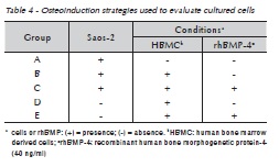
<body><![CDATA[<p><font size="2" face="Verdana">Dulbeco's Modified Eagle's Medium (DMEM),   fetal bovine serum (FBS) for cell culture,   Histopaque&reg;-1077 and DPBS were purchased from   GIBCO&reg; (Grand Island, NY, USA). Human recombinant   bone morphogenetic protein-4 (rhBMP-4) was   purchased from R&amp;D Systems Inc. (Minneapolis,   MN, USA). All other routine reagents were analytical grade.</font></p>     <p><font size="2" face="Verdana"><b>Human bone marrow harvesting and preparation of mesenchymal cells </b> </font></p>     <p><font size="2" face="Verdana">Human bone marrow cells were obtained after   written informed consent from a patient undergoing   maxillary reconstruction surgery. This study   was approved by the Ethics Committee of Hospital   S&atilde;o Lucas, Pontif&iacute;cia Universidade Cat&oacute;lica do   Rio Grande do Sul, Brazil, protocol no 06/03481.   Human bone marrow cells were aspirated with a   syringe from the iliac crest of a 28-year-old woman.   The cells were separated by centrifugation over a   Histopaque&reg;-1077 (GIBCO&reg;, Grand Island, NY,   USA) gradient at 400 g for 30 minutes. The mononuclear   cell layer was aspirated, washed with 10   ml of DPBS (GIBCO&reg;, Grand Island, NY, USA) by   centrifugation at 500 g during 5 minutes and resuspended at 0.5 x 10⁵ cell/ml with DMEM.</font></p>     <p><font size="2" face="Verdana"><b>Saos-2 culture </b> </font></p>     <p><font size="2" face="Verdana">Human osteosarcoma cell line (Saos-2) was purchased   from the American Type Cell Culture Collection   (HTB-85) (Manassas, VA, USA) and grown   in DMEM culture medium supplemented with 10%    <br> FBS.</font></p>     <p><font size="2" face="Verdana"><b>Co-culture of human bone marrow derived cells with Saos-2 </b> </font></p>     <p><font size="2" face="Verdana">Osteoinduction of human bone marrow derived   cells (HBMC) was performed using various conditions,   as described in <a href="#tab01">Table 1</a>. Briefly, 2.5 ml of   Saos-2 at a density of 0.5 x 10⁵ cells/ml of DMEM   was plated into 6 well plates to evaluate morphological   and proliferative features. Immunohistochemistry   was performed in cell cultures in 96 well plates   seeded with 100 &mu;l of cells at 0.5 x 10⁵ cells/ml. Because Saos-2 is an adherent cell line, Saos-2 wasplated 24 hours before addition of mesenchymal cells when cells were co-cultured. Cell morphology and proliferation were observed at 1, 2, 5, 8, 11 and 14 days of culture.</font></p>     <p><font size="2" face="Verdana"><b>Osteonectin detection by Immunohistochemistry </b> </font></p>     <p><font size="2" face="Verdana">Anti-SPARC antibody (R&amp;D Systems Inc., Minneapolis,   MN, USA) at a concentration of 15 &mu;g/ml was used to detect intracellular expression of   osteonectin by cultured cells at 1, 2, 5, 8, 11 and    ]]></body>
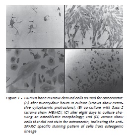
<body><![CDATA[<br>   14 culture days. Briefly, the cells were washed with   DPBS and fixed with 3.7% formaldehyde in DPBS   for 10 min, incubated for 10 min with H2O2 to inhibit   endogenous peroxidase, followed by methanol   incuba tion for 6 min at &ndash;20 oC. After two washes   with DPBS, cells were incubated with anti-goat immunoglobulin-biotin conjugated (Sigma, St. Louis,   MO, USA) antibody (15 &mu;g/ml) for 40 min at 37 oC.   Cells were incubated with one drop of streptavidinperoxidase   conjugate (Dako, Carpinteria, CA, USA)   for 10 min at room temperature. After two washes   with DPBS, the cells were incubated with 50 &mu;l/well   of a solution containing diaminobenzidine (Dako,   Carpinteria, CA, USA). Finally, after a wash with   DPBS, cells were analyzed under an inverted light   microscope (Axiovert 25, Carl Zeiss AG, Oberkochen, Germany)</font><font size="2" face="Verdana">.</font></p>     <p><font size="2" face="Verdana"><b>Reverse transcription polymerase   chain reaction (RT-PCR) to detect osteopontin expression </b> </font></p>     <p><font size="2" face="Verdana">Expression of osteopontin mRNA by HBMC, co-cultured or not with Saos-2 in the presence or absence   of rhBMP-4 on days 1, 2, 5, 8, 11 and 14 were   evaluated by RT-PCR. Total RNA was isolated from   cultured cells using Trizol LS reagent (Invitrogen   Inc., Carlsbad, CA, USA). First strand cDNA syntheses   were carried out using oligo-dT12-18 primers   (Invitrogen Inc., Carlsbad, CA, USA). After DNAse   treatment of the samples, reverse transcription   was carried out using Superscript II reverse transcriptase   (Invitrogen Inc, Carlsbad, CA, USA). PCR   reactions (50 &mu;l) were performed with 5 &mu;l of cDNA,   using 2 U of Easy Taq-DNA polymerase (LabTrade   do Brasil Ltda, S&atilde;o Paulo, SP, Brazil) and 25 pmol   of osteopontin specific primers (forward: 5'CAT   CTC AGA AGC AGA ATC TCC 3'; reverse: 5'CCA   TAA ACC ACA CTA TCA CCT C 3') and run on a   thermocycler (Peltier Thermal Cycler-200, MJ Research   Inc., Waltham, MA, USA). The 35 cycles of   PCR were performed as follows: denaturation step   at 94 oC for 30 sec, annealing at 55 oC for 45 sec and extension at 72 oC for 1 min.</font></p>     <p>&nbsp;</p>     <p><font size="3" face="Verdana"><B>Results</B> </font></p>     <p><font size="2" face="Verdana"><B>Morphological and proliferative   features of human bone marrow derived cells</B> </font></p>     <p><font size="2" face="Verdana">Human bone marrow derived cells adhered after   24 hours in culture. However, they show proliferation   and colony formation in the presence of   rhBMP-4. These characteristics were not detected   when they were co-cultured with Saos-2. Cell colonies   in intense proliferation were observed in all   conditions between the 5th and 8th culture days,   and HBMC alone showed extensive communicating   branches. Moreover, HBMC had multiple layers of interconnected cells after 14 culture days.</font></p>     <p><font size="2" face="Verdana"><B>Osteonectin detection by Immunohistochemistry</B> </font></p>     <p><font size="2" face="Verdana">Human bone marrow cells stained for osteonectin   after 24 hours in culture as detected by immunohistochemistry.   Co-cultures of HBMC and Saos-2 were both positive for anti-SPARC antibody, but   HBMC always had a more intense staining. Cell differentiation   and proliferation were confirmed with   anti-SPARC antibody after five days. Cells with a   distinct morphology were seen, as well as cells that   were apparently growing in culture but that did not stain for osteonectin (<a href="#fig01">Figure 1 A-D</a>).</font></p>     <p>&nbsp;</p>     ]]></body>
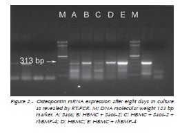
<body><![CDATA[<p><a name="fig01"></a></p>     <p>&nbsp;</p>     <p align="center"><img src="/img/revistas/rfo/v17n1/a14fig01.jpg"></p>     <p>&nbsp;</p>     <p><font size="2" face="Verdana"><B>Osteopontin mRNA expression</B> </font></p>     <p><font size="2" face="Verdana">Messenger RNA expression was revealed by   RT-PCR using total RNA from culture cells as described.   As expected, Saos-2 expressed mRNA for   osteopontin from day 1. After 8 days(<a href="#fig02">Figure 2</a>), cultured   human bone marrow derived cells expressed   osteopontin mRNA. However, cultures supplemented   with rhBMP-4 seemed to produce higher levels   of osteopontin mRNA. Similarly, high expression   levels of osteopontin were synthesized by cultured   cells (HBMC and Saos-2) in the presence or not of rhBMP-4 on days 11 and 14 (<a href="#fig03">Figure 3</a>).</font></p>     <p>&nbsp;</p>     <p><a name="fig02"></a></p>     <p>&nbsp;</p>     <p align="center"><img src="/img/revistas/rfo/v17n1/a14fig02.jpg"></p>     ]]></body>
<body><![CDATA[<p>&nbsp;</p>     <p><a name="fig03"></a></p>     <p>&nbsp;</p>     <p align="center"><img src="/img/revistas/rfo/v17n1/a14fig03.jpg"></p>     <p>&nbsp;</p>     <p><font size="3" face="Verdana"><B>Discussion</B> </font></p>     <p><font size="2" face="Verdana">Bone marrow derived cells can growth adherent   or in suspension<sup>21,22</sup>. Cells that adhere can differentiate   into mesenchymal cells, while cells that grow   in suspension will give rise to hematopoietic cells.   Several studies have confirmed the adhesion property   of mesenchymal cells<sup>23-25</sup> and we found that   HBMCs are already attached to the culture substrate after 24 hours in culture.</font></p>     <p><font size="2" face="Verdana">Petite et al.<sup>25</sup> (2000) have demonstrated that,   after a few days, adherent bone marrow cells or   mesenchymal cells in culture have three distinct   morphological cell types, that is, compact spindleshaped,   slender cells with long cytoplasmic extensions,   and large and amorphous cells with many   cytoplasmic extensions, as described in this study.</font></p>     <p><font size="2" face="Verdana">Bone morphogenetic proteins are molecules   that can induce osteogenesis, and rhBMP-4 has the   property to induce cell aggregates and colony formation,   mainly in the first five days in culture<sup>26-28</sup>.   The effect of rhBMP-4 on HBMC co-cultured with   Saos-2 seems to be intensified, probably due to the   fact that the osteosarcoma cell line Saos-2 also expresses   and secretes BMP-4 and other BMPs9. In   contrast, non-adherent cells decreased in number   after five days in culture, probably because they   are hematopoietic precursor cells that require other   growth factors to undergo differentiation in culture,   as already described in the literature<sup>23,25</sup>. Cocultures   of HBMC with Saos-2 were not affected by   rhBMP-4 after a longer period of culture, which is   in agreement with observations made by Virdi et   al.<sup>28</sup> (1998), who showed an inhibition of mature osteoblast   proliferation in the later phases of osteogenesis.   Therefore, the differentiated cells obtained   in this study may be classified as osteoblasts. In   fact, mesenchymal adherent cells reached a stage   at which they did not undergo further proliferation.   Similar results<sup>10,15,16</sup> have shown that cultured osteoprogenitor   cells have a limited self-renewing capability.</font></p>     <p><font size="2" face="Verdana">Osteonectin, also known as SPARC, is characteristic   of bone lineage cells, whose expression occurs   from the preosteoblastic to the osteoblastic phase.   The osteosarcoma and adherent mesenchymal cells   studied here expressed osteonectin, as demonstrated   by immunohistochemistry, which additionally   confirms their osteoblastic origin, although adherent   HBMC seems to produce higher levels of osteonectin.   Our findings are confirmed by studies   that used immunohistochemistry or RT-PCR techniques<sup>29</sup>.</font></p>     ]]></body>
<body><![CDATA[<p>&nbsp;</p>     <p><a name="tab01"></a></p>     <p>&nbsp;</p>     <p align="center"><img src="/img/revistas/rfo/v17n1/a14tab01.jpg"></p>     <p>&nbsp;</p>     <p><font size="2" face="Verdana">Moreover, adherent mesenchymal cells produced   mRNAs for osteopontin in all conditions tested.   Some studies have described a detectable expression   of osteopontin in the early stages of osteogenesis   with higher levels from the preosteoblastic to   the osteoblastic stage<sup>15,16</sup>. This finding suggested   that, after eight days in culture, adherent mesenchymal   cells reached the preosteoblastic or osteoblastic phase of osteogenesis.</font></p>     <p><font size="2" face="Verdana">In the near future, autologous bone marrow cells   will be applied to bone reconstitution, and the knowledge   of their physiology and adequate manipulation,   together with appropriate scaffolding techniques,   will be necessary to restore bone structures   in vitro and to replace damaged bone tissues, which   will reduce the morbidity of this type of treatment.</font></p>     <p>&nbsp;</p>     <p><font size="3" face="Verdana"><B>Conclusions</B> </font></p>     <p><font size="2" face="Verdana">This preliminary study results suggests that an   osteoblastic cell lineage can be obtained from human   bone marrow derived adherent cells even in   the absence of osteoinductive factors such as BMPs,   and that rhBMP-4 seems to have an effect only during   the early differentiation stages. Human bone   marrow stromal cells have an osteogenic potential   and are prone to undergoing osteogenesis in short term cultures.</font></p>     ]]></body>
<body><![CDATA[<p>&nbsp;</p>     <p><font size="3" face="Verdana"><B>Acknowledgements</B> </font></p>     <p><font size="2" face="Verdana">The authors thank Dr. Raphael Ott for providing   the human bone marrow cells and Vinicius   Schenk Michaelsen for the help with cell cultures.   This study was funded by Coordena&ccedil;&atilde;o de Aperfei&ccedil;oamento   de Pessoal de N&iacute;vel Superior (CAPES - Brazil) and 3i Implants, Brazil.</font></p>     <p>&nbsp;</p>     <p><font size="3" face="Verdana"><B>References</B></font></p>     <!-- ref --><p><font size="2" face="Verdana">1. Conrad C, Huss R. Adult stem cell lines in regenerative   medicine and reconstructive surgery. J Surg Res. 2005; 124:201-8.    &nbsp;&nbsp;&nbsp;&nbsp;&nbsp;&nbsp;&nbsp;&nbsp;[&#160;<a href="javascript:void(0);" onclick="javascript: window.open('/scielo.php?script=sci_nlinks&ref=142080&pid=S1413-4012201200010001400001&lng=','','width=640,height=500,resizable=yes,scrollbars=1,menubar=yes,');">Links</a>&#160;]<!-- end-ref --></font></p>     <!-- ref --><p><font size="2" face="Verdana">2. Schliephake H, Knebel JW, Aufderheide M, Tauscher M.   Use of cultivated osteoprogenitor cells to increase bone   formation in segmental mandibular defects: an experimental   pilot study in sheep. Int J Oral Maxillofac Surg. 2001;   30:531-7.    &nbsp;&nbsp;&nbsp;&nbsp;&nbsp;&nbsp;&nbsp;&nbsp;[&#160;<a href="javascript:void(0);" onclick="javascript: window.open('/scielo.php?script=sci_nlinks&ref=142082&pid=S1413-4012201200010001400002&lng=','','width=640,height=500,resizable=yes,scrollbars=1,menubar=yes,');">Links</a>&#160;]<!-- end-ref --></font></p>     <!-- ref --><p><font size="2" face="Verdana">3. Sonoyama W, Coppe C, Gronthos S, Shi S. Skeletal stem   cells in regenerative medicine. Curr Top Dev Biol. 2005;   67:305-23.    &nbsp;&nbsp;&nbsp;&nbsp;&nbsp;&nbsp;&nbsp;&nbsp;[&#160;<a href="javascript:void(0);" onclick="javascript: window.open('/scielo.php?script=sci_nlinks&ref=142084&pid=S1413-4012201200010001400003&lng=','','width=640,height=500,resizable=yes,scrollbars=1,menubar=yes,');">Links</a>&#160;]<!-- end-ref --></font></p>     <!-- ref --><p><font size="2" face="Verdana">4. Leite FR, Ramalho LT. Bone regeneration after demineralized   bone matrix and castor oil (Ricinus communis) polyurethane   implantation. J Appl Oral Sci. 2008; 16:122-6.    &nbsp;&nbsp;&nbsp;&nbsp;&nbsp;&nbsp;&nbsp;&nbsp;[&#160;<a href="javascript:void(0);" onclick="javascript: window.open('/scielo.php?script=sci_nlinks&ref=142086&pid=S1413-4012201200010001400004&lng=','','width=640,height=500,resizable=yes,scrollbars=1,menubar=yes,');">Links</a>&#160;]<!-- end-ref --></font></p>     <!-- ref --><p><font size="2" face="Verdana">5. Partridge K, Yang X, Clarke NM, Okubo Y, Bessho K, Sebald   W, et al. Adenoviral BMP-2 gene transfer in mesenchymal   stem cells: in vitro and in vivo bone formation on biodegradable   polymer scaffolds. Biochem Biophys Res Commun.   2002; 292:144-52.    &nbsp;&nbsp;&nbsp;&nbsp;&nbsp;&nbsp;&nbsp;&nbsp;[&#160;<a href="javascript:void(0);" onclick="javascript: window.open('/scielo.php?script=sci_nlinks&ref=142088&pid=S1413-4012201200010001400005&lng=','','width=640,height=500,resizable=yes,scrollbars=1,menubar=yes,');">Links</a>&#160;]<!-- end-ref --></font></p>     <!-- ref --><p><font size="2" face="Verdana">6. Katagiri T, Takahashi N. Regulatory mechanisms of osteoblast   and osteoclast differentiation. Oral Dis. 2002; 8:147-59.    &nbsp;&nbsp;&nbsp;&nbsp;&nbsp;&nbsp;&nbsp;&nbsp;[&#160;<a href="javascript:void(0);" onclick="javascript: window.open('/scielo.php?script=sci_nlinks&ref=142090&pid=S1413-4012201200010001400006&lng=','','width=640,height=500,resizable=yes,scrollbars=1,menubar=yes,');">Links</a>&#160;]<!-- end-ref --></font></p>     <!-- ref --><p><font size="2" face="Verdana">7. Yoon ST, Boden SD. Osteoinductive molecules in orthopaedics:   basic science and preclinical studies. Clin Orthop Relat   Res. 2002; (395):33-43.    &nbsp;&nbsp;&nbsp;&nbsp;&nbsp;&nbsp;&nbsp;&nbsp;[&#160;<a href="javascript:void(0);" onclick="javascript: window.open('/scielo.php?script=sci_nlinks&ref=142092&pid=S1413-4012201200010001400007&lng=','','width=640,height=500,resizable=yes,scrollbars=1,menubar=yes,');">Links</a>&#160;]<!-- end-ref --></font></p>     <!-- ref --><p><font size="2" face="Verdana">8. Gamradt SC, Lieberman JR. Genetic modification of stem   cells to enhance bone repair. Ann Biomed Eng. 2004; 32:136-47.    &nbsp;&nbsp;&nbsp;&nbsp;&nbsp;&nbsp;&nbsp;&nbsp;[&#160;<a href="javascript:void(0);" onclick="javascript: window.open('/scielo.php?script=sci_nlinks&ref=142094&pid=S1413-4012201200010001400008&lng=','','width=640,height=500,resizable=yes,scrollbars=1,menubar=yes,');">Links</a>&#160;]<!-- end-ref --></font></p>     <!-- ref --><p><font size="2" face="Verdana">9. Raval P, Hsu HH, Anderson HC. Osteoinductive ability of   confluent Saos-2 cell correlates with enhanced expression of   bone morphogenetic proteins. J Orthop Res. 1996; 14:605-10.    &nbsp;&nbsp;&nbsp;&nbsp;&nbsp;&nbsp;&nbsp;&nbsp;[&#160;<a href="javascript:void(0);" onclick="javascript: window.open('/scielo.php?script=sci_nlinks&ref=142096&pid=S1413-4012201200010001400009&lng=','','width=640,height=500,resizable=yes,scrollbars=1,menubar=yes,');">Links</a>&#160;]<!-- end-ref --></font></p>     <!-- ref --><p><font size="2" face="Verdana">10. Muschler GF, Midura RJ. Connective tissue progenitors:   practical concepts for clinical applications. Clin Orthop Relat   Res. 2002; (395):66-80.    &nbsp;&nbsp;&nbsp;&nbsp;&nbsp;&nbsp;&nbsp;&nbsp;[&#160;<a href="javascript:void(0);" onclick="javascript: window.open('/scielo.php?script=sci_nlinks&ref=142098&pid=S1413-4012201200010001400010&lng=','','width=640,height=500,resizable=yes,scrollbars=1,menubar=yes,');">Links</a>&#160;]<!-- end-ref --></font></p>     <!-- ref --><p><font size="2" face="Verdana">11. Strayhorn CL, Garrett JS, Dunn RL, Benedict JJ, Somerman   MJ. Growth factors regulate expression of osteoblastassociated   genes. J Periodontol. 1999; 70:1345-54.    &nbsp;&nbsp;&nbsp;&nbsp;&nbsp;&nbsp;&nbsp;&nbsp;[&#160;<a href="javascript:void(0);" onclick="javascript: window.open('/scielo.php?script=sci_nlinks&ref=142100&pid=S1413-4012201200010001400011&lng=','','width=640,height=500,resizable=yes,scrollbars=1,menubar=yes,');">Links</a>&#160;]<!-- end-ref --></font></p>     <!-- ref --><p><font size="2" face="Verdana">12. Fanburg-Smith JC, Bratthauer GL, Miettinen M. Osteocalcin   and osteonectin immunoreactivity in extraskeletal osteosarcoma:   a study of 28 cases. Hum Pathol. 1999; 30:32-8.    &nbsp;&nbsp;&nbsp;&nbsp;&nbsp;&nbsp;&nbsp;&nbsp;[&#160;<a href="javascript:void(0);" onclick="javascript: window.open('/scielo.php?script=sci_nlinks&ref=142102&pid=S1413-4012201200010001400012&lng=','','width=640,height=500,resizable=yes,scrollbars=1,menubar=yes,');">Links</a>&#160;]<!-- end-ref --></font></p>     <!-- ref --><p><font size="2" face="Verdana">13. Motamed K. SPARC (osteonectin/BM-40). Int J Biochem   Cell Biol. 1999; 31:1363-6.    &nbsp;&nbsp;&nbsp;&nbsp;&nbsp;&nbsp;&nbsp;&nbsp;[&#160;<a href="javascript:void(0);" onclick="javascript: window.open('/scielo.php?script=sci_nlinks&ref=142104&pid=S1413-4012201200010001400013&lng=','','width=640,height=500,resizable=yes,scrollbars=1,menubar=yes,');">Links</a>&#160;]<!-- end-ref --></font></p>     <!-- ref --><p><font size="2" face="Verdana">14. Meyer U, Meyer T, Vosshans J, Joos U. Decreased expression   of osteocalcin and osteonectin in relation to high strains   and decreased mineralization in mandibular distraction osteogenesis. J Craniomaxillofac Surg. 1999; 27:222-7.    &nbsp;&nbsp;&nbsp;&nbsp;&nbsp;&nbsp;&nbsp;&nbsp;[&#160;<a href="javascript:void(0);" onclick="javascript: window.open('/scielo.php?script=sci_nlinks&ref=142106&pid=S1413-4012201200010001400014&lng=','','width=640,height=500,resizable=yes,scrollbars=1,menubar=yes,');">Links</a>&#160;]<!-- end-ref --></font></p>     <!-- ref --><p><font size="2" face="Verdana">15. Aubin JE. Advances in the osteoblast lineage. Biochem Cell   Biol. 1998; 76:899-910.    &nbsp;&nbsp;&nbsp;&nbsp;&nbsp;&nbsp;&nbsp;&nbsp;[&#160;<a href="javascript:void(0);" onclick="javascript: window.open('/scielo.php?script=sci_nlinks&ref=142108&pid=S1413-4012201200010001400015&lng=','','width=640,height=500,resizable=yes,scrollbars=1,menubar=yes,');">Links</a>&#160;]<!-- end-ref --></font></p>     <!-- ref --><p><font size="2" face="Verdana">16. Aubin JE. Bone stem cells. J Cell Biochem Suppl. 1998; 30-31:73-82.    &nbsp;&nbsp;&nbsp;&nbsp;&nbsp;&nbsp;&nbsp;&nbsp;[&#160;<a href="javascript:void(0);" onclick="javascript: window.open('/scielo.php?script=sci_nlinks&ref=142110&pid=S1413-4012201200010001400016&lng=','','width=640,height=500,resizable=yes,scrollbars=1,menubar=yes,');">Links</a>&#160;]<!-- end-ref --></font></p>     <!-- ref --><p><font size="2" face="Verdana">17. De Kok IJ, Drapeau SJ, Young R, Cooper LF. Evaluation   of mesenchymal stem cells following implantation in alveolar   sockets: a canine safety study. Int J Oral Maxillofac Implants.   2005; 20:511-8.    &nbsp;&nbsp;&nbsp;&nbsp;&nbsp;&nbsp;&nbsp;&nbsp;[&#160;<a href="javascript:void(0);" onclick="javascript: window.open('/scielo.php?script=sci_nlinks&ref=142112&pid=S1413-4012201200010001400017&lng=','','width=640,height=500,resizable=yes,scrollbars=1,menubar=yes,');">Links</a>&#160;]<!-- end-ref --></font></p>     <!-- ref --><p><font size="2" face="Verdana">18. Young CS, Abukawa H, Asrican R, Ravens M, Troulis MJ,   Kaban LB, et al. Tissue-engineered hybrid tooth and bone.   Tissue Eng. 2005; 11:1599-610.    &nbsp;&nbsp;&nbsp;&nbsp;&nbsp;&nbsp;&nbsp;&nbsp;[&#160;<a href="javascript:void(0);" onclick="javascript: window.open('/scielo.php?script=sci_nlinks&ref=142114&pid=S1413-4012201200010001400018&lng=','','width=640,height=500,resizable=yes,scrollbars=1,menubar=yes,');">Links</a>&#160;]<!-- end-ref --></font></p>     <!-- ref --><p><font size="2" face="Verdana">19. Yamada Y, Ueda M, Naiki T, Nagasaka T. Tissue-engineered   injectable bone regeneration for osseointegrated dental implants.   Clin Oral Implants Res. 2004; 15:589-97.    &nbsp;&nbsp;&nbsp;&nbsp;&nbsp;&nbsp;&nbsp;&nbsp;[&#160;<a href="javascript:void(0);" onclick="javascript: window.open('/scielo.php?script=sci_nlinks&ref=142116&pid=S1413-4012201200010001400019&lng=','','width=640,height=500,resizable=yes,scrollbars=1,menubar=yes,');">Links</a>&#160;]<!-- end-ref --></font></p>     <!-- ref --><p><font size="2" face="Verdana">20. Yen AH, Sharpe PT. Stem cells and tooth tissue engineering.   Cell Tissue Res. 2008; 331:359-72.    &nbsp;&nbsp;&nbsp;&nbsp;&nbsp;&nbsp;&nbsp;&nbsp;[&#160;<a href="javascript:void(0);" onclick="javascript: window.open('/scielo.php?script=sci_nlinks&ref=142118&pid=S1413-4012201200010001400020&lng=','','width=640,height=500,resizable=yes,scrollbars=1,menubar=yes,');">Links</a>&#160;]<!-- end-ref --></font></p>     <!-- ref --><p><font size="2" face="Verdana">21. Bertram H, Mayer H, Schliephake H. Effect of donor characteristics,   technique of harvesting and in vitro processing   on culturing of human marrow stroma cells for tissue   engineered growth of bone. Clin Oral Implants Res. 2005;   16:524-31.    &nbsp;&nbsp;&nbsp;&nbsp;&nbsp;&nbsp;&nbsp;&nbsp;[&#160;<a href="javascript:void(0);" onclick="javascript: window.open('/scielo.php?script=sci_nlinks&ref=142120&pid=S1413-4012201200010001400021&lng=','','width=640,height=500,resizable=yes,scrollbars=1,menubar=yes,');">Links</a>&#160;]<!-- end-ref --></font></p>     <!-- ref --><p><font size="2" face="Verdana">22. Chen J, Sotome S, Wang J, Orii H, Uemura T, Shinomiya K.   Correlation of in vivo bone formation capability and in vitro   differentiation of human bone marrow stromal cells. J Med   Dent Sci. 2005; 52:27-34.    &nbsp;&nbsp;&nbsp;&nbsp;&nbsp;&nbsp;&nbsp;&nbsp;[&#160;<a href="javascript:void(0);" onclick="javascript: window.open('/scielo.php?script=sci_nlinks&ref=142122&pid=S1413-4012201200010001400022&lng=','','width=640,height=500,resizable=yes,scrollbars=1,menubar=yes,');">Links</a>&#160;]<!-- end-ref --></font></p>     <!-- ref --><p><font size="2" face="Verdana">23. Aubin JE. Osteoprogenitor cell frequency in rat bone marrow   stromal populations: role for heterotypic cell-cell interactions   in osteoblast differentiation. J Cell Biochem. 1999;   72:396-410.    &nbsp;&nbsp;&nbsp;&nbsp;&nbsp;&nbsp;&nbsp;&nbsp;[&#160;<a href="javascript:void(0);" onclick="javascript: window.open('/scielo.php?script=sci_nlinks&ref=142124&pid=S1413-4012201200010001400023&lng=','','width=640,height=500,resizable=yes,scrollbars=1,menubar=yes,');">Links</a>&#160;]<!-- end-ref --></font></p>     <!-- ref --><p><font size="2" face="Verdana">24. Krebsbach PH, Kuznetsov SA, Bianco P, Robey PG. Bone   marrow stromal cells: characterization and clinical application.   Crit Rev Oral Biol Med. 1999; 10:165-81.    &nbsp;&nbsp;&nbsp;&nbsp;&nbsp;&nbsp;&nbsp;&nbsp;[&#160;<a href="javascript:void(0);" onclick="javascript: window.open('/scielo.php?script=sci_nlinks&ref=142126&pid=S1413-4012201200010001400024&lng=','','width=640,height=500,resizable=yes,scrollbars=1,menubar=yes,');">Links</a>&#160;]<!-- end-ref --></font></p>     <!-- ref --><p><font size="2" face="Verdana">25. Petite H, Viateau V, Bensaid W, Meunier A, de Pollak C,   Bourguignon M, et al. Tissue-engineered bone regeneration.   Nat Biotechnol. 2000; 18:959-63.    &nbsp;&nbsp;&nbsp;&nbsp;&nbsp;&nbsp;&nbsp;&nbsp;[&#160;<a href="javascript:void(0);" onclick="javascript: window.open('/scielo.php?script=sci_nlinks&ref=142128&pid=S1413-4012201200010001400025&lng=','','width=640,height=500,resizable=yes,scrollbars=1,menubar=yes,');">Links</a>&#160;]<!-- end-ref --></font></p>     <!-- ref --><p><font size="2" face="Verdana">26. Kessler S, Kastler S, Mayr-Wohlfart U, Puhl W, Gunther   KP. Stimulation of primary osteoblast cultures with rh-TGF-beta, rh-bFGF, rh-BMP 2 and rx-BMP 4 in an in vitro   model. Orthopade. 2000; 29:107-11.    &nbsp;&nbsp;&nbsp;&nbsp;&nbsp;&nbsp;&nbsp;&nbsp;[&#160;<a href="javascript:void(0);" onclick="javascript: window.open('/scielo.php?script=sci_nlinks&ref=142130&pid=S1413-4012201200010001400026&lng=','','width=640,height=500,resizable=yes,scrollbars=1,menubar=yes,');">Links</a>&#160;]<!-- end-ref --></font></p>     <!-- ref --><p><font size="2" face="Verdana">27. Li G, Berven S, Simpson H, Triffitt JT. Expression of BMP-4   mRNA during distraction osteogenesis in rabbits. Acta Orthop   Scand. 1998; 69:420-5.    &nbsp;&nbsp;&nbsp;&nbsp;&nbsp;&nbsp;&nbsp;&nbsp;[&#160;<a href="javascript:void(0);" onclick="javascript: window.open('/scielo.php?script=sci_nlinks&ref=142132&pid=S1413-4012201200010001400027&lng=','','width=640,height=500,resizable=yes,scrollbars=1,menubar=yes,');">Links</a>&#160;]<!-- end-ref --></font></p>     <!-- ref --><p><font size="2" face="Verdana">28. Virdi AS, Cook LJ, Oreffo RO, Triffitt JT. Modulation of   bone morphogenetic protein-2 and bone morphogenetic protein-4 gene expression in osteoblastic cell lines. Cell Mol   Biol (Noisy-le-grand). 1998; 44:1237-46.    &nbsp;&nbsp;&nbsp;&nbsp;&nbsp;&nbsp;&nbsp;&nbsp;[&#160;<a href="javascript:void(0);" onclick="javascript: window.open('/scielo.php?script=sci_nlinks&ref=142134&pid=S1413-4012201200010001400028&lng=','','width=640,height=500,resizable=yes,scrollbars=1,menubar=yes,');">Links</a>&#160;]<!-- end-ref --></font></p>     <!-- ref --><p><font size="2" face="Verdana">29. Malaval L, Modrowski D, Gupta AK, Aubin JE. Cellular expression   of bone-related proteins during in vitro osteogenesis   in rat bone marrow stromal cell cultures. J Cell Physiol.   1994; 158:555-72.    &nbsp;&nbsp;&nbsp;&nbsp;&nbsp;&nbsp;&nbsp;&nbsp;[&#160;<a href="javascript:void(0);" onclick="javascript: window.open('/scielo.php?script=sci_nlinks&ref=142136&pid=S1413-4012201200010001400029&lng=','','width=640,height=500,resizable=yes,scrollbars=1,menubar=yes,');">Links</a>&#160;]<!-- end-ref --></font></p>     <p>&nbsp;</p>     <p>&nbsp;</p>     <p><font size="2" face="Verdana"><a name="back"/></a><a href="#top"><img src="/img/revistas/rfo/v17n1/seta.jpg" border="0" align="absmiddle"/></a><b>Address for Correspondence:</b>    <br>   </font><font size="2" face="Verdana">Mar&iacute;lia Gerhardt de Oliveira    <br>   Av. Coronel Lucas de Oliveira, 1841/203,    <br>   Bairro Petr&oacute;polis    <br>   90460.001 Porto Alegre - RS</font><font size="2" face="Verdana">    ]]></body>
<body><![CDATA[<br> e-mail: </font><font size="2" face="Verdana"><a href="mailto:gerhardtoliveira@gmail.com" target="_blank">gerhardtoliveira@gmail.com</a></font></p>     <p><font size="2" face="Verdana">Recebido: 24/10/2011    <br> Aceito: 19/03/2012</b></font></p>     <p>&nbsp;</p>       ]]></body>
<back>
<ref-list>
<ref id="B1">
<label>1</label><nlm-citation citation-type="journal">
<person-group person-group-type="author">
<name>
<surname><![CDATA[Conrad]]></surname>
<given-names><![CDATA[C]]></given-names>
</name>
<name>
<surname><![CDATA[Huss]]></surname>
<given-names><![CDATA[R]]></given-names>
</name>
</person-group>
<article-title xml:lang="en"><![CDATA[Adult stem cell lines in regenerative medicine and reconstructive surgery]]></article-title>
<source><![CDATA[J Surg Res]]></source>
<year>2005</year>
<volume>124</volume>
<page-range>201-8</page-range></nlm-citation>
</ref>
<ref id="B2">
<label>2</label><nlm-citation citation-type="journal">
<person-group person-group-type="author">
<name>
<surname><![CDATA[Schliephake]]></surname>
<given-names><![CDATA[H]]></given-names>
</name>
<name>
<surname><![CDATA[Knebel]]></surname>
<given-names><![CDATA[JW]]></given-names>
</name>
<name>
<surname><![CDATA[Aufderheide]]></surname>
<given-names><![CDATA[M]]></given-names>
</name>
<name>
<surname><![CDATA[Tauscher]]></surname>
<given-names><![CDATA[M]]></given-names>
</name>
</person-group>
<article-title xml:lang="en"><![CDATA[Use of cultivated osteoprogenitor cells to increase bone formation in segmental mandibular defects: an experimental pilot study in sheep]]></article-title>
<source><![CDATA[Int J Oral Maxillofac Surg]]></source>
<year>2001</year>
<volume>30</volume>
<page-range>531-7</page-range></nlm-citation>
</ref>
<ref id="B3">
<label>3</label><nlm-citation citation-type="journal">
<person-group person-group-type="author">
<name>
<surname><![CDATA[Sonoyama]]></surname>
<given-names><![CDATA[W]]></given-names>
</name>
<name>
<surname><![CDATA[Coppe]]></surname>
<given-names><![CDATA[C]]></given-names>
</name>
<name>
<surname><![CDATA[Gronthos]]></surname>
<given-names><![CDATA[S]]></given-names>
</name>
<name>
<surname><![CDATA[Shi]]></surname>
<given-names><![CDATA[S]]></given-names>
</name>
</person-group>
<article-title xml:lang="en"><![CDATA[Skeletal stem cells in regenerative medicine]]></article-title>
<source><![CDATA[Curr Top Dev Biol]]></source>
<year>2005</year>
<volume>67</volume>
<page-range>305-23</page-range></nlm-citation>
</ref>
<ref id="B4">
<label>4</label><nlm-citation citation-type="journal">
<person-group person-group-type="author">
<name>
<surname><![CDATA[Leite]]></surname>
<given-names><![CDATA[FR]]></given-names>
</name>
<name>
<surname><![CDATA[Ramalho]]></surname>
<given-names><![CDATA[LT]]></given-names>
</name>
</person-group>
<article-title xml:lang="en"><![CDATA[Bone regeneration after demineralized bone matrix and castor oil (Ricinus communis) polyurethane implantation]]></article-title>
<source><![CDATA[J Appl Oral Sci]]></source>
<year>2008</year>
<volume>16</volume>
<page-range>122-6</page-range></nlm-citation>
</ref>
<ref id="B5">
<label>5</label><nlm-citation citation-type="journal">
<person-group person-group-type="author">
<name>
<surname><![CDATA[Partridge]]></surname>
<given-names><![CDATA[K]]></given-names>
</name>
<name>
<surname><![CDATA[Yang]]></surname>
<given-names><![CDATA[X]]></given-names>
</name>
<name>
<surname><![CDATA[Clarke]]></surname>
<given-names><![CDATA[NM]]></given-names>
</name>
<name>
<surname><![CDATA[Okubo]]></surname>
<given-names><![CDATA[Y]]></given-names>
</name>
<name>
<surname><![CDATA[Bessho]]></surname>
<given-names><![CDATA[K]]></given-names>
</name>
<name>
<surname><![CDATA[Sebald]]></surname>
<given-names><![CDATA[W]]></given-names>
</name>
</person-group>
<article-title xml:lang="en"><![CDATA[Adenoviral BMP-2 gene transfer in mesenchymal stem cells: in vitro and in vivo bone formation on biodegradable polymer scaffolds]]></article-title>
<source><![CDATA[Biochem Biophys Res Commun]]></source>
<year>2002</year>
<volume>292</volume>
<page-range>144-52</page-range></nlm-citation>
</ref>
<ref id="B6">
<label>6</label><nlm-citation citation-type="journal">
<person-group person-group-type="author">
<name>
<surname><![CDATA[Katagiri]]></surname>
<given-names><![CDATA[T]]></given-names>
</name>
<name>
<surname><![CDATA[Takahashi]]></surname>
<given-names><![CDATA[N]]></given-names>
</name>
</person-group>
<article-title xml:lang="en"><![CDATA[Regulatory mechanisms of osteoblast and osteoclast differentiation]]></article-title>
<source><![CDATA[Oral Dis]]></source>
<year>2002</year>
<volume>8</volume>
<page-range>147-59</page-range></nlm-citation>
</ref>
<ref id="B7">
<label>7</label><nlm-citation citation-type="journal">
<person-group person-group-type="author">
<name>
<surname><![CDATA[Yoon]]></surname>
<given-names><![CDATA[ST]]></given-names>
</name>
<name>
<surname><![CDATA[Boden]]></surname>
<given-names><![CDATA[SD]]></given-names>
</name>
</person-group>
<article-title xml:lang="en"><![CDATA[Osteoinductive molecules in orthopaedics: basic science and preclinical studies]]></article-title>
<source><![CDATA[Clin Orthop Relat Res]]></source>
<year>2002</year>
<numero>395</numero>
<issue>395</issue>
<page-range>33-43</page-range></nlm-citation>
</ref>
<ref id="B8">
<label>8</label><nlm-citation citation-type="journal">
<person-group person-group-type="author">
<name>
<surname><![CDATA[Gamradt]]></surname>
<given-names><![CDATA[SC]]></given-names>
</name>
<name>
<surname><![CDATA[Lieberman]]></surname>
<given-names><![CDATA[JR]]></given-names>
</name>
</person-group>
<article-title xml:lang="en"><![CDATA[Genetic modification of stem cells to enhance bone repair]]></article-title>
<source><![CDATA[Ann Biomed Eng]]></source>
<year>2004</year>
<volume>32</volume>
<page-range>136-47</page-range></nlm-citation>
</ref>
<ref id="B9">
<label>9</label><nlm-citation citation-type="journal">
<person-group person-group-type="author">
<name>
<surname><![CDATA[Raval]]></surname>
<given-names><![CDATA[P]]></given-names>
</name>
<name>
<surname><![CDATA[Hsu]]></surname>
<given-names><![CDATA[HH]]></given-names>
</name>
<name>
<surname><![CDATA[Anderson]]></surname>
<given-names><![CDATA[HC]]></given-names>
</name>
</person-group>
<article-title xml:lang="en"><![CDATA[Osteoinductive ability of confluent Saos-2 cell correlates with enhanced expression of bone morphogenetic proteins]]></article-title>
<source><![CDATA[J Orthop Res]]></source>
<year>1996</year>
<volume>14</volume>
<page-range>605-10</page-range></nlm-citation>
</ref>
<ref id="B10">
<label>10</label><nlm-citation citation-type="journal">
<person-group person-group-type="author">
<name>
<surname><![CDATA[Muschler]]></surname>
<given-names><![CDATA[GF]]></given-names>
</name>
<name>
<surname><![CDATA[Midura]]></surname>
<given-names><![CDATA[RJ]]></given-names>
</name>
</person-group>
<article-title xml:lang="en"><![CDATA[Connective tissue progenitors: practical concepts for clinical applications]]></article-title>
<source><![CDATA[Clin Orthop Relat Res]]></source>
<year>2002</year>
<numero>395</numero>
<issue>395</issue>
<page-range>66-80</page-range></nlm-citation>
</ref>
<ref id="B11">
<label>11</label><nlm-citation citation-type="journal">
<person-group person-group-type="author">
<name>
<surname><![CDATA[Strayhorn]]></surname>
<given-names><![CDATA[CL]]></given-names>
</name>
<name>
<surname><![CDATA[Garrett]]></surname>
<given-names><![CDATA[JS]]></given-names>
</name>
<name>
<surname><![CDATA[Dunn]]></surname>
<given-names><![CDATA[RL]]></given-names>
</name>
<name>
<surname><![CDATA[Benedict]]></surname>
<given-names><![CDATA[JJ]]></given-names>
</name>
<name>
<surname><![CDATA[Somerman]]></surname>
<given-names><![CDATA[MJ]]></given-names>
</name>
</person-group>
<article-title xml:lang="en"><![CDATA[Growth factors regulate expression of osteoblastassociated genes]]></article-title>
<source><![CDATA[J Periodontol]]></source>
<year>1999</year>
<volume>70</volume>
<page-range>1345-54</page-range></nlm-citation>
</ref>
<ref id="B12">
<label>12</label><nlm-citation citation-type="journal">
<person-group person-group-type="author">
<name>
<surname><![CDATA[Fanburg-Smith]]></surname>
<given-names><![CDATA[JC]]></given-names>
</name>
<name>
<surname><![CDATA[Bratthauer]]></surname>
<given-names><![CDATA[GL]]></given-names>
</name>
<name>
<surname><![CDATA[Miettinen]]></surname>
<given-names><![CDATA[M]]></given-names>
</name>
</person-group>
<article-title xml:lang="en"><![CDATA[Osteocalcin and osteonectin immunoreactivity in extraskeletal osteosarcoma: a study of 28 cases]]></article-title>
<source><![CDATA[Hum Pathol]]></source>
<year>1999</year>
<volume>30</volume>
<page-range>32-8</page-range></nlm-citation>
</ref>
<ref id="B13">
<label>13</label><nlm-citation citation-type="journal">
<person-group person-group-type="author">
<name>
<surname><![CDATA[Motamed]]></surname>
<given-names><![CDATA[K]]></given-names>
</name>
</person-group>
<article-title xml:lang="en"><![CDATA[SPARC (osteonectin/BM-40)]]></article-title>
<source><![CDATA[Int J Biochem Cell Biol]]></source>
<year>1999</year>
<volume>31</volume>
<page-range>1363-6</page-range></nlm-citation>
</ref>
<ref id="B14">
<label>14</label><nlm-citation citation-type="journal">
<person-group person-group-type="author">
<name>
<surname><![CDATA[Meyer]]></surname>
<given-names><![CDATA[U]]></given-names>
</name>
<name>
<surname><![CDATA[Meyer]]></surname>
<given-names><![CDATA[T]]></given-names>
</name>
<name>
<surname><![CDATA[Vosshans]]></surname>
<given-names><![CDATA[J]]></given-names>
</name>
<name>
<surname><![CDATA[Joos]]></surname>
<given-names><![CDATA[U]]></given-names>
</name>
</person-group>
<article-title xml:lang="en"><![CDATA[Decreased expression of osteocalcin and osteonectin in relation to high strains and decreased mineralization in mandibular distraction osteogenesis]]></article-title>
<source><![CDATA[J Craniomaxillofac Surg]]></source>
<year>1999</year>
<volume>27</volume>
<page-range>222-7</page-range></nlm-citation>
</ref>
<ref id="B15">
<label>15</label><nlm-citation citation-type="journal">
<person-group person-group-type="author">
<name>
<surname><![CDATA[Aubin]]></surname>
<given-names><![CDATA[JE]]></given-names>
</name>
</person-group>
<article-title xml:lang="en"><![CDATA[Advances in the osteoblast lineage]]></article-title>
<source><![CDATA[Biochem Cell Biol]]></source>
<year>1998</year>
<volume>76</volume>
<page-range>899-910</page-range></nlm-citation>
</ref>
<ref id="B16">
<label>16</label><nlm-citation citation-type="journal">
<person-group person-group-type="author">
<name>
<surname><![CDATA[Aubin]]></surname>
<given-names><![CDATA[JE]]></given-names>
</name>
</person-group>
<article-title xml:lang="en"><![CDATA[Bone stem cells]]></article-title>
<source><![CDATA[Cell Biochem Suppl]]></source>
<year>1998</year>
<volume>30-31</volume>
<page-range>73-82</page-range></nlm-citation>
</ref>
<ref id="B17">
<label>17</label><nlm-citation citation-type="journal">
<person-group person-group-type="author">
<name>
<surname><![CDATA[De Kok]]></surname>
<given-names><![CDATA[IJ]]></given-names>
</name>
<name>
<surname><![CDATA[Drapeau]]></surname>
<given-names><![CDATA[SJ]]></given-names>
</name>
<name>
<surname><![CDATA[Young]]></surname>
<given-names><![CDATA[R]]></given-names>
</name>
<name>
<surname><![CDATA[Cooper]]></surname>
<given-names><![CDATA[LF]]></given-names>
</name>
</person-group>
<article-title xml:lang="en"><![CDATA[Evaluation of mesenchymal stem cells following implantation in alveolar sockets: a canine safety study]]></article-title>
<source><![CDATA[Int J Oral Maxillofac Implants]]></source>
<year>2005</year>
<volume>20</volume>
<page-range>511-8</page-range></nlm-citation>
</ref>
<ref id="B18">
<label>18</label><nlm-citation citation-type="journal">
<person-group person-group-type="author">
<name>
<surname><![CDATA[Young]]></surname>
<given-names><![CDATA[CS]]></given-names>
</name>
<name>
<surname><![CDATA[Abukawa]]></surname>
<given-names><![CDATA[H]]></given-names>
</name>
<name>
<surname><![CDATA[Asrican]]></surname>
<given-names><![CDATA[R]]></given-names>
</name>
<name>
<surname><![CDATA[Ravens]]></surname>
<given-names><![CDATA[M]]></given-names>
</name>
<name>
<surname><![CDATA[Troulis]]></surname>
<given-names><![CDATA[MJ]]></given-names>
</name>
<name>
<surname><![CDATA[Kaban]]></surname>
<given-names><![CDATA[LB]]></given-names>
</name>
</person-group>
<article-title xml:lang="en"><![CDATA[Tissue-engineered hybrid tooth and bone]]></article-title>
<source><![CDATA[Tissue Eng]]></source>
<year>2005</year>
<volume>11</volume>
<page-range>1599-610</page-range></nlm-citation>
</ref>
<ref id="B19">
<label>19</label><nlm-citation citation-type="journal">
<person-group person-group-type="author">
<name>
<surname><![CDATA[Yamada]]></surname>
<given-names><![CDATA[Y]]></given-names>
</name>
<name>
<surname><![CDATA[Ueda]]></surname>
<given-names><![CDATA[M]]></given-names>
</name>
<name>
<surname><![CDATA[Naiki]]></surname>
<given-names><![CDATA[T]]></given-names>
</name>
<name>
<surname><![CDATA[Nagasaka]]></surname>
<given-names><![CDATA[T]]></given-names>
</name>
</person-group>
<article-title xml:lang="en"><![CDATA[Tissue-engineered injectable bone regeneration for osseointegrated dental implants]]></article-title>
<source><![CDATA[Clin Oral Implants Res]]></source>
<year>2004</year>
<volume>15</volume>
<page-range>589-97</page-range></nlm-citation>
</ref>
<ref id="B20">
<label>20</label><nlm-citation citation-type="journal">
<person-group person-group-type="author">
<name>
<surname><![CDATA[Yen]]></surname>
<given-names><![CDATA[AH]]></given-names>
</name>
<name>
<surname><![CDATA[Sharpe]]></surname>
<given-names><![CDATA[PT]]></given-names>
</name>
</person-group>
<article-title xml:lang="en"><![CDATA[Stem cells and tooth tissue engineering]]></article-title>
<source><![CDATA[Cell Tissue Res]]></source>
<year>2008</year>
<volume>331</volume>
<page-range>359-72</page-range></nlm-citation>
</ref>
<ref id="B21">
<label>21</label><nlm-citation citation-type="journal">
<person-group person-group-type="author">
<name>
<surname><![CDATA[Bertram]]></surname>
<given-names><![CDATA[H]]></given-names>
</name>
<name>
<surname><![CDATA[Mayer]]></surname>
<given-names><![CDATA[H]]></given-names>
</name>
<name>
<surname><![CDATA[Schliephake]]></surname>
<given-names><![CDATA[H]]></given-names>
</name>
</person-group>
<article-title xml:lang="en"><![CDATA[Effect of donor characteristics, technique of harvesting and in vitro processing on culturing of human marrow stroma cells for tissue engineered growth of bone]]></article-title>
<source><![CDATA[Clin Oral Implants Res]]></source>
<year>2005</year>
<volume>16</volume>
<page-range>524-31</page-range></nlm-citation>
</ref>
<ref id="B22">
<label>22</label><nlm-citation citation-type="journal">
<person-group person-group-type="author">
<name>
<surname><![CDATA[Chen]]></surname>
<given-names><![CDATA[J]]></given-names>
</name>
<name>
<surname><![CDATA[Sotome]]></surname>
<given-names><![CDATA[S]]></given-names>
</name>
<name>
<surname><![CDATA[Wang]]></surname>
<given-names><![CDATA[J]]></given-names>
</name>
<name>
<surname><![CDATA[Orii]]></surname>
<given-names><![CDATA[H]]></given-names>
</name>
<name>
<surname><![CDATA[Uemura]]></surname>
<given-names><![CDATA[T]]></given-names>
</name>
<name>
<surname><![CDATA[Shinomiya]]></surname>
<given-names><![CDATA[K]]></given-names>
</name>
</person-group>
<article-title xml:lang="en"><![CDATA[Correlation of in vivo bone formation capability and in vitro differentiation of human bone marrow stromal cells]]></article-title>
<source><![CDATA[J Med Dent Sci]]></source>
<year>2005</year>
<volume>52</volume>
<page-range>27-34</page-range></nlm-citation>
</ref>
<ref id="B23">
<label>23</label><nlm-citation citation-type="journal">
<person-group person-group-type="author">
<name>
<surname><![CDATA[Aubin]]></surname>
<given-names><![CDATA[JE]]></given-names>
</name>
</person-group>
<article-title xml:lang="en"><![CDATA[Osteoprogenitor cell frequency in rat bone marrow stromal populations: role for heterotypic cell-cell interactions in osteoblast differentiation]]></article-title>
<source><![CDATA[J Cell Biochem]]></source>
<year>1999</year>
<volume>72</volume>
<page-range>396-410</page-range></nlm-citation>
</ref>
<ref id="B24">
<label>24</label><nlm-citation citation-type="journal">
<person-group person-group-type="author">
<name>
<surname><![CDATA[Krebsbach]]></surname>
<given-names><![CDATA[PH]]></given-names>
</name>
<name>
<surname><![CDATA[Kuznetsov]]></surname>
<given-names><![CDATA[SA]]></given-names>
</name>
<name>
<surname><![CDATA[Bianco]]></surname>
<given-names><![CDATA[P]]></given-names>
</name>
<name>
<surname><![CDATA[Robey]]></surname>
<given-names><![CDATA[PG]]></given-names>
</name>
</person-group>
<article-title xml:lang="en"><![CDATA[Bone marrow stromal cells: characterization and clinical application]]></article-title>
<source><![CDATA[Crit Rev Oral Biol Med]]></source>
<year>1999</year>
<volume>10</volume>
<page-range>165-81</page-range></nlm-citation>
</ref>
<ref id="B25">
<label>25</label><nlm-citation citation-type="journal">
<person-group person-group-type="author">
<name>
<surname><![CDATA[Petite]]></surname>
<given-names><![CDATA[H]]></given-names>
</name>
<name>
<surname><![CDATA[Viateau]]></surname>
<given-names><![CDATA[V]]></given-names>
</name>
<name>
<surname><![CDATA[Bensaid]]></surname>
<given-names><![CDATA[W]]></given-names>
</name>
<name>
<surname><![CDATA[Meunier]]></surname>
<given-names><![CDATA[A]]></given-names>
</name>
<name>
<surname><![CDATA[de Pollak]]></surname>
<given-names><![CDATA[C]]></given-names>
</name>
<name>
<surname><![CDATA[Bourguignon]]></surname>
<given-names><![CDATA[M]]></given-names>
</name>
</person-group>
<article-title xml:lang="en"><![CDATA[Tissue-engineered bone regeneration]]></article-title>
<source><![CDATA[Nat Biotechnol]]></source>
<year>2000</year>
<volume>18</volume>
<page-range>959-63</page-range></nlm-citation>
</ref>
<ref id="B26">
<label>26</label><nlm-citation citation-type="journal">
<person-group person-group-type="author">
<name>
<surname><![CDATA[Kessler]]></surname>
<given-names><![CDATA[S]]></given-names>
</name>
<name>
<surname><![CDATA[Kastler]]></surname>
<given-names><![CDATA[S]]></given-names>
</name>
<name>
<surname><![CDATA[Mayr-Wohlfart]]></surname>
<given-names><![CDATA[U]]></given-names>
</name>
<name>
<surname><![CDATA[Puhl]]></surname>
<given-names><![CDATA[W]]></given-names>
</name>
<name>
<surname><![CDATA[Gunther]]></surname>
<given-names><![CDATA[KP]]></given-names>
</name>
</person-group>
<article-title xml:lang="en"><![CDATA[Stimulation of primary osteoblast cultures with rh-TGF-beta, rh-bFGF, rh-BMP 2 and rx-BMP 4 in an in vitro model]]></article-title>
<source><![CDATA[Orthopade]]></source>
<year>2000</year>
<volume>29</volume>
<page-range>107-11</page-range></nlm-citation>
</ref>
<ref id="B27">
<label>27</label><nlm-citation citation-type="journal">
<person-group person-group-type="author">
<name>
<surname><![CDATA[Li]]></surname>
<given-names><![CDATA[G]]></given-names>
</name>
<name>
<surname><![CDATA[Berven]]></surname>
<given-names><![CDATA[S]]></given-names>
</name>
<name>
<surname><![CDATA[Simpson]]></surname>
<given-names><![CDATA[H]]></given-names>
</name>
<name>
<surname><![CDATA[Triffitt]]></surname>
<given-names><![CDATA[JT]]></given-names>
</name>
</person-group>
<article-title xml:lang="en"><![CDATA[Expression of BMP-4 mRNA during distraction osteogenesis in rabbits]]></article-title>
<source><![CDATA[Acta Orthop Scand]]></source>
<year>1998</year>
<volume>69</volume>
<page-range>420-5</page-range></nlm-citation>
</ref>
<ref id="B28">
<label>28</label><nlm-citation citation-type="journal">
<person-group person-group-type="author">
<name>
<surname><![CDATA[Virdi]]></surname>
<given-names><![CDATA[AS]]></given-names>
</name>
<name>
<surname><![CDATA[Cook]]></surname>
<given-names><![CDATA[LJ]]></given-names>
</name>
<name>
<surname><![CDATA[Oreffo]]></surname>
<given-names><![CDATA[RO]]></given-names>
</name>
<name>
<surname><![CDATA[Triffitt]]></surname>
<given-names><![CDATA[JT]]></given-names>
</name>
</person-group>
<article-title xml:lang="en"><![CDATA[Modulation of bone morphogenetic protein-2 and bone morphogenetic protein-4 gene expression in osteoblastic cell lines]]></article-title>
<source><![CDATA[Cell Mol Biol (Noisy-le-grand)]]></source>
<year>1998</year>
<volume>44</volume>
<page-range>1237-46</page-range></nlm-citation>
</ref>
<ref id="B29">
<label>29</label><nlm-citation citation-type="journal">
<person-group person-group-type="author">
<name>
<surname><![CDATA[Malaval]]></surname>
<given-names><![CDATA[L]]></given-names>
</name>
<name>
<surname><![CDATA[Modrowski]]></surname>
<given-names><![CDATA[D]]></given-names>
</name>
<name>
<surname><![CDATA[Gupta]]></surname>
<given-names><![CDATA[AK]]></given-names>
</name>
<name>
<surname><![CDATA[Aubin]]></surname>
<given-names><![CDATA[JE]]></given-names>
</name>
</person-group>
<article-title xml:lang="en"><![CDATA[Cellular expression of bone-related proteins during in vitro osteogenesis in rat bone marrow stromal cell cultures]]></article-title>
<source><![CDATA[J Cell Physiol]]></source>
<year>1994</year>
<volume>158</volume>
<page-range>555-72</page-range></nlm-citation>
</ref>
</ref-list>
</back>
</article>