Effects of low-level laser therapy on distraction osteogenesis: a histological analysis
Efeito do laser de baixa intensidade na distração osteogênica: análise histológica
Luciano Mayer I; Angelo Luis Freddo II; Diego Segatto Blaya III; Marília Gerhardt de Oliveira IV; Ferdinando De Conto V
I Researcher, Pontifícia Universidade Católica do Rio Grande do Sul, Porto Alegre, RS, Brazil
II PhD, Oral surgeon. School of Dentistry, Universidade Federal do Rio Grande do Sul, Porto Alegre, RS, Brazil
III PhD, Oral surgeon. School of Dentistry, Centro Universitário Franciscano, Santa Maria, Brazil ]]>
IV PhD, Researcher, National Council for Scientific and Technological Development (CNPq), Brazil
V PhD, Oral surgeon. School of Dentistry, Universidade de Passo Fundo, Passo Fundo, RS, Brazil
ABSTRACT
Objective: The aim of this study was to assess the effect of low-level laser therapy on tissue repair in the rabbit mandible after osteotomy and distraction osteogenesis, through histological analysis of the area of bone neoformation and measurement of the amount of neoformed bone. Methods: Twenty-four male New Zealand white rabbits were randomly allocated into one of two groups: experiment (laser applied directly over the site of fracture and distraction osteogenesis) or control (non-irradiated animals). Distraction osteogenesis was performed according to the following protocol: 3 days of latency, 7 days of activation (0.8 mm/day), and 10 days of consolidation. In the experiment group, irradiation was performed with an infrared laser (AlGaAs; wavelength 830 nm, CW, time 101 seconds, 40mW), at a dose of 10 J/cm2 per session, every 48 hours during the activation period. Twenty days after surgery, all rabbits were sacrificed. Results: The amount of neoformed bone was significantly higher in the laser-treated group (62.68%) than in the control group (43.09%) (p < 0.001). Conclusion: The application of low-level laser therapy following the irradiation protocol used in this study had a positive effect on the tissue repair process in a rabbit model of mandibular fracture and distraction osteogenesis, as shown by histological analysis.
Keywords: Osteogenesis. Distraction. Lasers. Histology.
]]> RESUMO
Objetivo: Avaliar o efeito do laser de baixa intensidade no tecido de reparo em mandíbula de coelhos, após ter sido provocada osteotomia e distração osteogênica local, poar meio de análise histológica da área de neoformação óssea e mensuração do percentual de osso neoformado. Métodos: Vinte e quatro coelhos brancos neozelandeses foram randomizados e distribuídos aleatoriamente em dois grupos: experimental (laser aplicado diretamente sobre o local da fratura e distração osteogênica) ou de controle (animais não irradiados). A distração osteogênica foi realizada seguindo o seguinte protocolo: três dias de latência, sete dias de ativação (0.8 mm/dia) e dez dias de consolidação. No grupo experimental a irradiação foi feita com laser infravermelho (AlGaAs; comprimento de onda de 830 nm, CW, tempo de 101 segundos, 40 m W), com uma dose de 10 J/cm2 por sessão, a cada 48h durante o período de ativação. Resultados: A quantidade de neoformação óssea foi significativamente mais alta no grupo que recebeu o tratamento com laser (62,68%) em ralação ao grupo de controle (43,09%) (p < 0.001). Conclusão: A aplicação do laser de baixa intensidade seguindo o protocolo de irradiação apresentado teve um efeito positivo no processo de reparo tecidual da mandíbula de coelhos que foram submetidos à fratura e distração osteogênica, como mostra a análise histológica.
Palavras-chave: Osteogênese por distração. Lasers. Histologia.
Introdução
The concept of distraction osteogenesis (DO) was introduced by G. A. Ilizarov in the 1950s and knowledge of this method has added countless benefits to the treatment of a variety of musculoskeletal disorders, enabling bone lengthening, correction of angular deformities of the limbs, and correction of pseudarthrosis, as well as a technique for alveolar ridge augmentation1.
In the practice of dentistry and oral and maxillofacial surgery, treatment of facial deformities still poses a challenge, as it frequently involves multiple, high-cost procedures that carry a high morbidity and often produce unsatisfactory outcomes. Autogenous tissue is the ideal substrate for bone augmentation, but harvesting procedures increase operative time and cost and require a second surgical field, which may increase postoperative discomfort2. Furthermore, the process of obtaining graft tissue in sufficient quantity for bone reconstruction is fraught with difficulty, and is also often associated with unsatisfactory results3.
In this context, DO constitutes a promising and increasingly established alternative for bone reconstruction that may be used in the treatment of congenital deformity, trauma, or after oncologic surgery. DO is based on the use of distraction devices, which are designed to enable growth of bone at a deformed site4. Nevertheless, the long-term stability of the outcomes of DO has yet to be well documented; instability and recurrence of deformity have been reported4,5.
Faced with this scenario, some researchers have sought ways to speed the bone maturation process and enhance the physical properties of tissue at the elongated bone site6,7. As any process involving tissue repair, DO entails a series of metabolic activities. This process of neoformation of bone is amenable to biomodulation through use of low-level laser therapy (LLLT), which has been shown to benefit the tissue repair process in soft tissue and bone and has the potential to reduce treatment time8-10. Laser radiation has been used for therapeutic purposes since the 1960s, in view of its low power intensity and wavelengths capable of tissue penetration.
]]> Laser therapy exerts a major biomodulatory effect on the tissue repair process, and is widely used in a variety of healthcare fields. Dentistry is one of the health sciences to use it most broadly, for a wide range of clinical procedures; however, information on the combined use of laser therapy and DO is still lacking.In light of the various potential applications of DO, particularly in the field of dentistry, there is a need for more precise characterization of the histological features of bone tissue generated by means of this method, especially when subjected to LLLT.
Materials and methods
This study was approved by the Pontifícia Universidade Católica do Rio Grande do Sul (PUCRS) Research Ethics Committee (CEP 0219/07 – CEUA 075/08) and was conducted in accordance with the Ethical Principles for Experimental Research set forth by the Brazilian Society for Laboratory Animal Science (SBCAL).
Animals
The study sample consisted of 24 male New Zealand white rabbits (order Lagomorpha, genus Oryctolagos, species O. cuniculus) of known adult age and weight 3.5-4.5 kg. The animals were randomly allocated into two groups of 12: control group "C" (nonirradiated) and experiment group "L" (laser- irradiated). Animals were weighed, placed into individual cages, identified with cage cards, and provided a commercially available solid diet (Purina ®, Nestlé Purina Petcare, St. Louis, MO) and water ad lib. The vivarium was climate-controlled so as to ensure normal lighting, relative humidity, and temperature conditions. After one week of adaptation and observation, animals were examined by Universidade Federal de Pelotas (UFPel) vivarium staff veterinarians and cleared for the study.
Operative technique
All stages of anesthesia were performed by trained veterinarians, who were also responsible for observing all animals in the pre-, intra-, and postoperative periods of the study.
Animals were anesthetized with 2% xylazine hydrochloride (Anasedan®, Ceva Santé Animale, Brazil) and tiletamine/zolazepam (Zoletil®, Virbac, Brazil) IM. Xylazine is a muscle relaxant and analgesic, whereas tiletamine/zolazepam is a combination dissociative anesthetic that exerts direct action on the cerebral cortex to induce analgesia and loss of consciousness. The combined action of these drugs provides effective anesthesia for surgical procedures involving moderate stimuli.
]]> After induction of anesthesia, the left submandibular region was shaved with an electric razor. Animals were then taken to the operating theater, placed in the right lateral recumbent position, and tied to the operating table with restraints. The surgical field was disinfected with an iodophor-in-alcohol solution (0.5% available iodine).Sterile surgical drapes were used to isolate the field. The planned incision site was further anesthetized with local infiltration of 1% lidocaine/epinephrine 1:100000 into the subcutaneous space (approximately 2 mL per specimen) to achieve adequate vasoconstriction and enhance visibility during surgery. Antibiotic prophylaxis was provided with a single dose of enrofloxacin (Flotril®, Schering Plough, Brasil) IM.
A single, approximately 3 cm-long linear incision was made on the skin overlying the inferior border of the mandible with a #15 disposable scalpel. Metzenbaum scissors were used to dissect down to the periosteum, which, along with its muscle insertions, was carefully elevated with Molt and Seldin periosteal elevators at the lateral and inferior surfaces of the mandible.
Stay sutures (4-0 nylon monofilament) were placed through the skin and deep muscle planes to provide traction during placement of the DO device, respecting the natural position of these tissues to ensure correct repositioning at the conclusion of the procedure.
After exposure of the body of the mandible, the inferior alveolar nerve was identified at its emergence from the mental foramen and dissected. A vertical monocortical osteotomy was performed on the vestibular surface of the mandible between the premolar and mental foramen, using a 1.0-mm straight-shank carbide bur, at low rotation, with constant normal saline irrigation. Throughout the procedure, skin and muscle tissue were retracted using the stay sutures, whereas the lingual plate and adjacent periosteum were kept intact.
The distraction device (PROMM®, Brazil) was installed with 5-mm monocortical screws, placed into three holes drilled through a miniplate at each side of the osteotomy, below the mental foramen and tooth roots. All six screws were placed equidistant to the osteotomy. The distraction screw was then placed and activated for two complete turns or until frank resistance to activation was felt. The osteotomy was then completed with application of chisels in a lever motion. The wound bed was irrigated copiously and layered closure of the incision was performed with 4-0 nylon monofilament using simple interrupted stitches.
Postoperative period
After the procedure, animals were returned to the UFPel vivarium and remained under the care of the investigators and staff veterinarians. Animals recovered from anesthesia in a heated room to avoid hypothermia. Postoperative diet consisted of green leaves and feed ground mixed with water. During the first three postoperative days, metamizole (Novalgina®) and enrofloxacin (50 mg/day) were administered for analgesia and antibiotic coverage respectively.
Distraction osteogenesis
Latency period – 3 days (postoperative days 1 through 3): During the first three postoperative days, the distractor was not activated, but merely inspected and disinfected with a 1% iodophor-in-alcohol solution. Each animal were placed in an Elizabethan collar to prevent displacement of the distraction device. Diet consisted of commercially available feed (Purina®, Nestlé Purina Petcare, St. Louis, MO, USA), ground and mixed with water to a semisolid consistency.
]]> Activation period – 7 days (postoperative days 4 through 10): After the third postoperative day, the distraction device was activated in daily 0.8-mm increments over a total of 7 days (approximately 5.6 mm total).Maturation period – 10 days (postoperative days 11 through 20): After the latency and activation periods, the distraction device remained in place for a further 10 days, serving as an external fixator so that bone maturation could be achieved.
Laser irradiation
Infrared laser irradiation was performed with an aluminum gallium arsenide diode laser (AlGa- As). This procedure is painless, and therefore required no sedation or anesthesia.
In the experiment group, spot laser was applied to three points overlying the site of DO, at an energy density of 4 J/cm2 per point, with the irradiation tip placed perpendicularly over the area, for a total dose of 12 J/cm2 per session. An AlGaAs infrared laser (Thera Lase®, DMC Equipamentos, São Carlos, SP, Brazil) was used at a wavelength of 830 nm, with 40 mW output power, and continuous wave irradiation for a spot application time of 00:01:41 (101 seconds, automatically controlled by the laser device as determined by other parameters). The irradiation protocol was instituted immediately after activation of the distractor and repeated every 48 hours thereafter, for a total of four sessions having a total dose of 48 J/cm2. Nonirradiated animals (control group) underwent sham irradiation: the same procedure described above was performed, but with the laser unit switched off.
Euthanasia (postoperative day 20)
Twenty days after surgery, rabbits were anesthetized and sacrificed by a veterinarian using a thiopental injection at 200 mg/kg, according to the guidelines of the Brazilian Resolution no. 714, issued on June 20 2002 by the Brazilian Federal Council of Veterinary Medicine, regarding euthanasia procedures and methods for laboratory animals.
A specimen of the mandible corresponding to the site of osteotomy and DO was obtained from each animal and sent to the UFPel Oral Pathology Lab for processing and histomorphometric analysis.
Histological and histomorphometric analysis
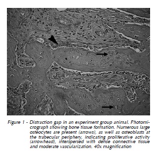
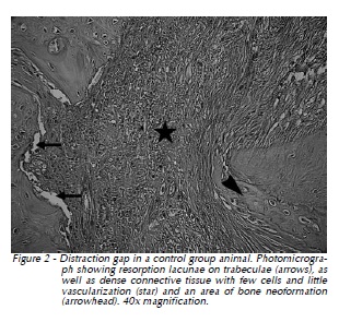
Samples were fixated in 10% neutral buffered formalin solution for no less than 24 and no more than 72 hours. Shortly thereafter, samples were decalcified in 20% formic acid solution under agitation at room temperature, rinsed in running water for a few hours, dehydrated in a graded ethanol series (80-100%), embedded in paraffin blocks, labeled, and cut into 10 μm slices, which were then mounted and stained with hematoxylin and eosin (H&E). Slides were examined under a light microscope for histological and histomorphometric analysis Figures (1 and 2).
]]>
Statistical analysis
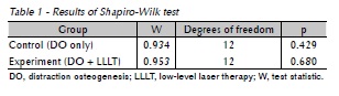
]]> Statistical analyses were performed in the SPSS® 17.0 (Statistical Package for Social Science) software environment (SPSS Inc, Chicago, IL, USA). The Student t test was used for between-group comparison of the amount of bone neoformation in the distraction gap. The significance level was set at 5% (α = 0.05).The mean percentage of bone formation was significantly higher in the experiment than in the control group (Table 1). Table 1 shows results of the Shapiro-Wilk test for normality, proving that the variable of interest followed a normal distribution.
Results
Histomorphometric analysis of the distracted area in experimental group animals showed more abundant bone neoformation in the laser therapy groups. Laser had a demonstrably positive effect on bone repair, as shown by the greater number of active osteoblasts, secreting collagen matrix more intensely, as well as the smaller areas of hemorrhage and inflammatory infiltration (Figure 1).
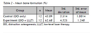
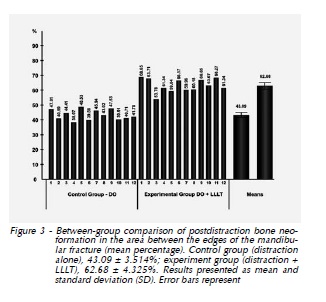
The mean percentage of bone neoformation in the distraction gap (the area between the edges of the mandibular fracture) was significantly higher in the experiment group – that is, animals subjected to LLLT performed during the activation period in addition to DO – than in the control group of animals subjected to DO alone (p < 0.001) (Table 2, Figure 3).
]]>
Discussion
]]> In this experiment, to create an initial clinical condition for laser therapy, rabbits underwent osteotomy of the body of the mandible between the premolar and mental foramen with subsequent DO.LLLT has proved to be an effective and beneficial adjunct to a wide range of oral and dental treatments. Due to the wide use of laser therapy by healthcare providers, the effects of LLLT on different anatomical structures and for a variety of clinical applications have been the object of extensive research10.
Promotion of faster and improved bone tissue repair stands out among the countless indications for LLLT reported in the literature1,2,10,11.
The objective of combining laser therapy and DO was to ascertain whether LLLT has a positive biomodulatory effect on – that is, whether it encourages – maturation of the bone wound. DO can cause exquisite patient discomfort, particularly when performed with the use of external distraction devices, which can be associated with superficial infection, paresthesia, and keloid formation, in addition to negative social impacts (as DO hinders participation in social activities) and instability and recurrence after the distraction process4,5,12,13. Therefore, we believe that the development of therapeutic strategies capable of shortening the consolidation stage of DO is a very important research objective.
We chose to perform LLLT with infrared laser to speed tissue regeneration, as studies have shown that wavelengths in the infrared range provide the best outcomes in terms of positive biomodulation of bone tissue healing14-17.
An AlGaAs diode (λ = 830 nm) was used in this study due to its improved tissue penetration profile as compared to that of helium-neon (HeNe) lasers, which operate at a wavelength of 632.8 nm. Infrared lasers penetrate better into subcutaneous tissue due to poor absorption of their energy by water and skin pigments18.
There is still no defined protocol as to the optimal timing of LLLT as an adjuvant to distraction osteogenesis. However, the biomodulatory effect of laser therapy during the activation phase led to higher bone neoformation rates in the distraction zone as compared with laser irradiation during the consolidation phase13,19.
Several investigators have used histological analysis to evaluate the process of bone neoformation3,20-22. Although the events that occur during bone lengthening by DO are well-established and have been described in detail, we believe the present study was justified as a means of proving the occurrence of bone neoformation, describing its characteristics and, above all, assessing the percentage difference in neoformation when the site of distraction is subjected to low-level laser irradiation23.
Descriptive analysis confirmed that bone neoformation occurred in both study groups, following similar patterns of formation of trabecular bone interspersed with fibrovascular tissue. In both groups, trabecular bone was aligned with the direction of distraction and regeneration of bone was sparser in the central area, indicating that bone formation began at the edges of the preexisting fractured bone and became thinner toward the center of the gap.
Histological analysis also revealed bone formation in both groups (control and experiment). Bone formation is directly associated with activation time and the length of each increment of distraction. The device was activated in daily increments of 0.8 mm, as, according to the literature, activation rates of 0.5 to 1 mm/day lead to bone formation, whereas faster distraction (with larger increments) would lead to fibrous union20,24.
]]> The consolidation period was defined as only 10 days, as we sought to assess the initial stages of bone formation in the same animal model (rabbit) used in other studies on the theme20,21,25, which employed fixation periods of 8 to 6 weeks.In this study, LLLT had a positive biomodulatory effect when applied during the activation period, increasing the amount of bone neoformation in comparison to the control group. These findings are promising and similar to those reported in the literature for studies in which laser therapy was applied during the maturation period, where authors also found increased bone neoformation in irradiated animals3,20,26.
Statistical testing revealed significantly greater neoformation of bone in the laser therapy group (62.68%) as compared to the group of animals that underwent DO alone (43.09%).
Ao observar os maiores escores obtidos (Tab. 2), pode-se perceber que algumas
Conclusion
]]> According to the irradiation protocol used in this study, LLLT performed during the activation period has a positive effect on the tissue repair process in a rabbit model of DO of the mandible, as shown by histological analysis.Although the results of this study are encouraging and reveal laser therapy as a viable adjunct to DO, further studies using different sample sizes, doses, power settings, and treatment periods are warranted, in order to provide a more in-depth understanding of the effects of LLLT on the bone neoformation process.
Acknowledgments
Prof. Dr. Adriana Etges - Pathology/UFPel and Prof. Dr. João Feliz Duarte de Moraes - Mathematics/ UFRGS.
Author disclosure statement
No competing financial interests exist.
Referências
1. Kucerova H, Dostalova T, Himmlova L, Bartova J, Mazanek J. Low-level laser therapy after molar extraction. J Clin Laser Med Surg 2000; 18(6): 309-15. [ Links ]
2. Valiati R, Paes JV, Moraes AN, Gava A, Agostini M, Masiero AV et al. Effect of Low-Level Laser Therapy on Incorporation of Block Allografts. Int. J. Med. Sci. 2010; 9(10): 853-861.
3. Cerqueira A, Silveira RL, Oliveira MG, Sant'ana Filho M, Heitz C. Bone tissue microscopic findings related to the use of diode laser (830 nm) in ovine mandible submitted to distraction osteogenesis. Acta Cir Bras 2007; 22(2): 92-7.
4. Mofid MM, Manson PN, Robertson BC, Tufaro AP, Elias JJ, Vander Kolk CA. Craniofacial distraction osteogenesis: a review of 3278 cases. Plast Reconstr Surg 2001; 108(5): 1103- 14; discussion 15-7.
5. Marquez IM, Fish LC, Stella JP. Two-year follow-up of distraction osteogenesis: its effect on mandibular ramus height in hemifacial microsomia. Am J Orthod Dentofacial Orthop 2000; 117(2): 130-9.
6. Saito S, Shimizu N. Stimulatory effects of low-power laser irradiation on bone regeneration in midpalatal suture during expansion in the rat. Am J Orthod Dentofacial Orthop 1997; 111(5): 525-32.
7. Shimazaki A, Inui K, Azuma Y, Nishimura N, Yamano Y. Low-intensity pulsed ultrasound accelerates bone maturation in distraction osteogenesis in rabbits. J Bone Joint Surg Br 2000; 82(7): 1077-82.
8. Mester E, Spiry T, Szende B, Tota JG. Effect of laser rays on wound healing. Am J Surg 1971; 122(4): 532-5.
9. Weber JB, Pinheiro AL, de Oliveira MG, Oliveira FA, Ramalho LM. Laser therapy improves healing of bone defects submitted to autologous bone graft. Photomed Laser Surg 2006; 24(1): 38-44.
10. Fronza B, Somacal T, Mayer L, de Moraes JF, de Oliveira MG, Weber JB. Assessment of the systemic effects of low-level laser therapy (LLLT) on thyroid hormone function in a rabbit model. Int. J. Oral Maxillofac. Surg. 2013; 42: 26-30.
]]> 11. Takeda Y. Irradiation effect of low-energy laser on alveolar bone after tooth extraction. Experimental study in rats. Int J Oral Maxillofac Surg. 1988; 17(6): 388-91.12. Diner PA, Kollar E, Martinez H, Vazquez MP. Submerged intraoral device for mandibular lengthening. J Craniomaxillofac Surg 1997; 25(3): 116-23.
13. Freddo AL, Hübler R, de Castro-Beck CA, Heitz C, de Oliveira MG. A preliminary study of hardness and modulus of elasticity in sheep mandibles submitted to distraction osteogenesis and low-level laser therapy. Med Oral Patol Oral Cir Bucal. 2012; 17(1): e102-107.
14. Blaya DS, Guimaraes MB, Pozza DH, Weber JB, de Oliveira MG. Histologic study of the effect of laser therapy on bone repair. J Contemp Dent Pract 2008; 9(6): 41-8.
15. Kazem Shakouri S, Soleimanpour J, Salekzamani Y, Oskuie MR. Effect of low-level laser therapy on the fracture healing process. Lasers Med Sci 2010; 25(1): 73-7.
16. Pretel H, Lizarelli RF, Ramalho LT. Effect of low-level laser therapy on bone repair: histological study in rats. Lasers Surg Med 2007; 39(10): 788-96.
17. Nascimento AGLE, Sant'anna EF, Ruellas CA, Nojima IL, Filho ACG. Laser Versus Ultrasound on Bone Density Recuperation After Distraction Osteogenesis - A Cone-Beam Computer Tomographic Analysis. J of Oral and Maxillof Surg. 2013 Disponível em URL: http://www.ncbi.nlm.nih. gov/pubmed/23351480.
18. Kolarova H, Ditrichova D, Wagner J. Penetration of the laser light into the skin in vitro. Lasers Surg Med 1999; 24(3): 231- 5.
19. Kreisner PE, Blaya DS, Gaiao L, Maciel-Santos ME, Etges A, Santana-Filho M, et al. Histological evaluation of the effect of low-level laser on distraction osteogenesis in rabbit mandibles. Med Oral Patol Oral Cir Bucal 2010; 15(4): e616- 8.
20. Djasim UM, Mathot BJ, Wolvius EB, van Neck JW, van der Wal KG. Histomorphometric comparison between continuous and discontinuous distraction osteogenesis. J Craniomaxillofac Surg 2009; 37(7): 398-404.
]]> 21. Miloro M, Miller JJ, Stoner JA. Low-level laser effect on mandibular distraction osteogenesis. J Oral Maxillofac Surg 2007; 65(2): 168-76.22. Zimmermann CE, Thurmuller P, Troulis MJ, Perrott DH, Rahn B, Kaban LB. Histology of the porcine mandibular distraction wound. Int J Oral Maxillofac Surg 2005; 34(4): 411- 9.
23. Kocyigit ID, Coskunses FM, Pala E, Tugcu F, Onder E, Mocan A. A Comparison of the Low-Level Laser Versus Low Intensity Pulsed Ultrasound on New Bone Formed Through Distraction Osteogenesis. Photomed. Laser. Surg. 2012; 30(8): 438-443.
24. Heiple KG, Herndon CH. The pathologic physiology of nonunion. Clin Orthop Relat Res 1965; 43: 11-21.
25. Zheng LW, Ma L, Cheung LK. Changes in blood perfusion and bone healing induced by nicotine during distraction osteogenesis. Bone 2008; 43(2): 355-61.
26. Hubler R, Blando E, Gaiao L, Kreisner PE, Post LK, Xavier CB, et al. Effects of low-level laser therapy on bone formed after distraction osteogenesis. Lasers Med Sci 2010; 25(2): 213-9.
 Endereço para correspondência:
Marília Gerhardt de Oliveira ]]>
Rua Visconde do Herval, 725, apt 404.B
Bairro Menino Deus, Porto Alegre, RS, Brazil 0460–001
Zipcode: 90.130-151
Porto Alegre, RS, Brazil
e-mail: gerhardtoliveira@gmail.com
]]>