<?xml version="1.0" encoding="ISO-8859-1"?><article xmlns:mml="http://www.w3.org/1998/Math/MathML" xmlns:xlink="http://www.w3.org/1999/xlink" xmlns:xsi="http://www.w3.org/2001/XMLSchema-instance">
<front>
<journal-meta>
<journal-id>1677-3888</journal-id>
<journal-title><![CDATA[Odontologia Clínico-Científica (Online)]]></journal-title>
<abbrev-journal-title><![CDATA[Odontol. Clín.-Cient. (Online)]]></abbrev-journal-title>
<issn>1677-3888</issn>
<publisher>
<publisher-name><![CDATA[Conselho Regional de Odontologia de Pernambuco]]></publisher-name>
</publisher>
</journal-meta>
<article-meta>
<article-id>S1677-38882011000100007</article-id>
<title-group>
<article-title xml:lang="pt"><![CDATA[Citologia esfoliativa convencional versus citologia em meio líquido para prevenção e diagnóstico precoce do carcinoma escamo celular (CEC) oral.]]></article-title>
<article-title xml:lang="en"><![CDATA[Conventional Cytology versus liquid-based cytology for prevention and early diagnosis of oral squamous cell carcioma (OSCC)]]></article-title>
</title-group>
<contrib-group>
<contrib contrib-type="author">
<name>
<surname><![CDATA[Vidal]]></surname>
<given-names><![CDATA[Aurora Karla de Lacerda]]></given-names>
</name>
<xref ref-type="aff" rid="A01"/>
</contrib>
<contrib contrib-type="author">
<name>
<surname><![CDATA[Caldas Júnior]]></surname>
<given-names><![CDATA[Arnaldo de França]]></given-names>
</name>
<xref ref-type="aff" rid="A02"/>
</contrib>
<contrib contrib-type="author">
<name>
<surname><![CDATA[Mello]]></surname>
<given-names><![CDATA[Roberto José Vieira de]]></given-names>
</name>
<xref ref-type="aff" rid="A03"/>
</contrib>
<contrib contrib-type="author">
<name>
<surname><![CDATA[Brandão]]></surname>
<given-names><![CDATA[Vírginia Ribes Amorim]]></given-names>
</name>
<xref ref-type="aff" rid="A04"/>
</contrib>
<contrib contrib-type="author">
<name>
<surname><![CDATA[Lima]]></surname>
<given-names><![CDATA[Paula Abreu e]]></given-names>
</name>
<xref ref-type="aff" rid="A05"/>
</contrib>
<contrib contrib-type="author">
<name>
<surname><![CDATA[Figueiroa]]></surname>
<given-names><![CDATA[José Natal]]></given-names>
</name>
<xref ref-type="aff" rid="A06"/>
</contrib>
</contrib-group>
<aff id="A01">
<institution><![CDATA[,University of Pernambuco Biological Institute ]]></institution>
<addr-line><![CDATA[ ]]></addr-line>
<country>Brazil</country>
</aff>
<aff id="A02">
<institution><![CDATA[,University of Pernambuco School of Dentistry ]]></institution>
<addr-line><![CDATA[ ]]></addr-line>
<country>Brazil</country>
</aff>
<aff id="A03">
<institution><![CDATA[,Federal University of Pernambuco Department of Pathology ]]></institution>
<addr-line><![CDATA[ ]]></addr-line>
<country>Brazil</country>
</aff>
<aff id="A04">
<institution><![CDATA[,University of Pernambuco School of Medicine Department of Anatomic Pathology]]></institution>
<addr-line><![CDATA[ ]]></addr-line>
<country>Brazil</country>
</aff>
<aff id="A05">
<institution><![CDATA[,Cancer Hospital of Pernambuco  ]]></institution>
<addr-line><![CDATA[ ]]></addr-line>
<country>Brazil</country>
</aff>
<aff id="A06">
<institution><![CDATA[,Infant Maternal Institute of Pernambuco  ]]></institution>
<addr-line><![CDATA[ ]]></addr-line>
<country>Brazil</country>
</aff>
<pub-date pub-type="pub">
<day>00</day>
<month>03</month>
<year>2011</year>
</pub-date>
<pub-date pub-type="epub">
<day>00</day>
<month>03</month>
<year>2011</year>
</pub-date>
<volume>10</volume>
<numero>1</numero>
<fpage>31</fpage>
<lpage>36</lpage>
<copyright-statement/>
<copyright-year/>
<self-uri xlink:href="http://revodonto.bvsalud.org/scielo.php?script=sci_arttext&amp;pid=S1677-38882011000100007&amp;lng=en&amp;nrm=iso"></self-uri><self-uri xlink:href="http://revodonto.bvsalud.org/scielo.php?script=sci_abstract&amp;pid=S1677-38882011000100007&amp;lng=en&amp;nrm=iso"></self-uri><self-uri xlink:href="http://revodonto.bvsalud.org/scielo.php?script=sci_pdf&amp;pid=S1677-38882011000100007&amp;lng=en&amp;nrm=iso"></self-uri><abstract abstract-type="short" xml:lang="pt"><p><![CDATA[Objetivando avaliar a concordância, sensibilidade e especificidade diagnóstica dos métodos citológicos: convencional e em meio líquido, foi realizado um estudo de caso-controle, no Ambulatório de Cabeça e Pescoço do Hospital de Câncer de Pernambuco, sendo selecionados 182 indivíduos doentes e 179 sãos, no período de setembro de 2002 a janeiro de 2004, os quais, permitiram através de assinatura de termo de consentimento livre e esclarecido a realização de exames (clínico/ citológico esfoliativo - cytobrush) na área carcinomatosa e em área sã, constituindo, respectivamente, o grupo de casos e controles. Os dados obtidos foram analisados através dos softwares estatísticos SPSS (Statistical Package for Social Science) e Epi-Info versão 6.4. Os resultados evidenciaram que os métodos diagnósticos citológicos apresentaram entre os casos: sensibilidade= 96,9% (IC= 95%: 92,5% a 98,8%); especificidade= 75,0% (IC= 95%: 21,9% a 98,7%) e acurácia= 96,3% (IC=95%: 92,5% a 98,5%); e entre os controles: sensibilidade= 91,0% (IC=95%: 84,1% a 95,2%); especificidade = 57,5% (IC=95%: 41,0% a 72,6%) e acurácia= 82,7% (IC=95%: 76,3% a 88,0%). A citologia convencional e em base-líquida apresentaram concordância com o diagnóstico histopatológico em mais de 90%. Conclui-se, portanto, que, se bem indicado e executado, o exame citológico pode ser utilizado rotineiramente como exame complementar, pois ambos apresentaram alta sensibilidade e razoável especificidade, cabendo à citologia convencional maior especificidade para o diagnóstico das lesões orais.]]></p></abstract>
<abstract abstract-type="short" xml:lang="en"><p><![CDATA[The purpose of this study was to evaluate the sensitivity, specificity, and concordance between conventional cytology and liquid-based cytology. A case-control study was developed at the Head and Neck ambulatory service of Hospital do Cancer de Pernambuco. A hundred and eighty-two patients with primary Oral Squamous Cellular Carcinoma (OSCC) (case group) and 179 individuals with normal buccal mucosa (control group) were selected, from September 2002 to January 2004. They agreed to be submitted to clinical examination and exfoliative cytology of the oral cavity, by signing a document of consent. The data were analysed by Statistical Package for Social Science (SPSS) and Epi-Info 6.4 program. Among the cases, the cytologic methods demontrated a sensitivity= 96.9% (IC= 95%: 92.5% to 98.8%), specificity= 75.0% (IC=95%: 21.9% to 98.7%), and accuracy= 96.3% (IC=95%: 92.5% to 98.5%). Among the controls the results were: sensitivity= 91.0% (IC=95%: 84.1% to 95.2%);, specificity= 57.5% (IC=95%: 41.0% to 72.6%), and accuracy= 82.7% (IC=95%: 76.3% to 88.0%). Conventional cytology and liquid-based cytology demonstrated a diagnostic concordance with histopathology of more than 90%. In conclusion if properly indicated and executed, they can be routinely used as complementary diagnostic methods. When compared to each other, conventional cytology and liquid-based cytology showed a high sensitivity and reasonable specificity, conventional cytology having higher specificity.]]></p></abstract>
<kwd-group>
<kwd lng="pt"><![CDATA[Carcinoma Escamo Celular (CEC)]]></kwd>
<kwd lng="pt"><![CDATA[Prevenção]]></kwd>
<kwd lng="pt"><![CDATA[Métodos Diagnósticos]]></kwd>
<kwd lng="pt"><![CDATA[Citologia Convencional]]></kwd>
<kwd lng="pt"><![CDATA[Citologia em meio líquido]]></kwd>
<kwd lng="en"><![CDATA[Oral Squamous Cell Carcinoma (OSCC)]]></kwd>
<kwd lng="en"><![CDATA[Prevention]]></kwd>
<kwd lng="en"><![CDATA[Diagnostic Methods]]></kwd>
<kwd lng="en"><![CDATA[Conventional Cytology]]></kwd>
<kwd lng="en"><![CDATA[Liquid-based Cytology]]></kwd>
</kwd-group>
</article-meta>
</front><body><![CDATA[ <p align="right"><font size="2" face="Verdana"> <b>ARTIGO ORIGINAL</b>  ORIGINAL ARTICE</font></p>     <p>&nbsp;</p>     <p><font size="4" face="verdana"><B><a name="tx"></a>Citologia esfoliativa convencional <I>versus</I> citologia em meio l&iacute;quido para preven&ccedil;&atilde;o e diagn&oacute;stico precoce do carcinoma escamo celular (CEC) oral.</B></font></p>     <p>&nbsp;</p>     <p><font size="3" face="Verdana"><b>Conventional Cytology <i>versus</i> liquid&#45;based cytology for prevention and early diagnosis of oral squamous cell carcioma (OSCC)</b></font></p>     <p>&nbsp;</p>     <p>&nbsp;</p>     <p><font size="2" face="Verdana"><b>Aurora Karla de Lacerda Vidal<sup>I</sup>; Arnaldo de Fran&ccedil;a Caldas J&uacute;nior<sup>II</sup>; Roberto Jos&eacute; Vieira de Mello<sup>III</sup>; V&iacute;rginia Ribes Amorim Brand&atilde;o<sup>IV</sup>; Paula Abreu e Lima<sup>V</sup>; Jos&eacute; Natal Figueiroa<sup>VI</sup></b></font></p>     <p><font size="2" face="Verdana"><sup>I</sup>Department of Pathology, Biological Institute of University of Pernambuco &#45; Brazil    <br> <sup>II</sup>Department of Preventive and Social Dentistry, School of Dentistry of University of Pernambuco &#45; Brazil    ]]></body>
<body><![CDATA[<br> <sup>III</sup>Department of Pathology, Cancer Hospital of Pernambuco and the Federal University of Pernambuco &#45; Brazil    <br> <sup>IV</sup>Department of Anatomic Pathology, School of Medicine of University of Pernambuco &#45; Brazil    <br> <sup>V</sup>Department of Pathology, Cancer Hospital of Pernambuco &#45; Brazil    <br> <sup>VI</sup>Infant Maternal Institute of Pernambuco &#45; Brazil</font></p>     <p><font size="2" face="Verdana"><a href="#nt">Endere&ccedil;o para correspond&ecirc;ncia</a></font></p>     <p>&nbsp;</p>     <p>&nbsp;</p> <hr size="1" noshade>     <p><font size="2" face="Verdana"><B>RESUMO</B></font></p>     <p><font size="2" face="Verdana">Objetivando avaliar a concord&acirc;ncia, sensibilidade e especificidade diagn&oacute;stica dos m&eacute;todos citol&oacute;gicos: convencional e em meio l&iacute;quido, foi realizado um estudo de caso&#45;controle, no Ambulat&oacute;rio de Cabe&ccedil;a e Pesco&ccedil;o do Hospital de C&acirc;ncer de Pernambuco, sendo selecionados 182 indiv&iacute;duos doentes e 179 s&atilde;os, no per&iacute;odo de setembro de 2002 a janeiro de 2004, os quais, permitiram atrav&eacute;s de assinatura de termo de consentimento livre e esclarecido a realiza&ccedil;&atilde;o de exames (cl&iacute;nico/ citol&oacute;gico esfoliativo &#45; cytobrush) na &aacute;rea carcinomatosa e em &aacute;rea s&atilde;, constituindo, respectivamente, o grupo de casos e controles. Os dados obtidos foram analisados atrav&eacute;s dos softwares estat&iacute;sticos SPSS (Statistical Package for Social Science) e Epi&#45;Info vers&atilde;o 6.4. Os resultados evidenciaram que os m&eacute;todos diagn&oacute;sticos citol&oacute;gicos apresentaram entre os casos: sensibilidade= 96,9% (IC= 95%: 92,5% a 98,8%); especificidade= 75,0% (IC= 95%: 21,9% a 98,7%) e acur&aacute;cia= 96,3% (IC=95%: 92,5% a 98,5%); e entre os controles: sensibilidade= 91,0% (IC=95%: 84,1% a 95,2%); especificidade = 57,5% (IC=95%: 41,0% a 72,6%) e acur&aacute;cia= 82,7% (IC=95%: 76,3% a 88,0%). A citologia convencional e em base&#45;l&iacute;quida apresentaram concord&acirc;ncia com o diagn&oacute;stico histopatol&oacute;gico em mais de 90%. Conclui&#45;se, portanto, que, se bem indicado e executado, o exame citol&oacute;gico pode ser utilizado rotineiramente como exame complementar, pois ambos apresentaram alta sensibilidade e razo&aacute;vel especificidade, cabendo &agrave; citologia convencional maior especificidade para o diagn&oacute;stico das les&otilde;es orais.</font></p>     <p><font size="2" face="Verdana"><B>Descritores:</B> Carcinoma Escamo Celular (CEC), Preven&ccedil;&atilde;o, M&eacute;todos Diagn&oacute;sticos, Citologia Convencional, Citologia em meio l&iacute;quido</font></p> <hr size="1" noshade>     ]]></body>
<body><![CDATA[<p><font size="2" face="Verdana"><B>ABSTRACT</B></font></p>     <p><font size="2" face="Verdana">The purpose of this study was to evaluate the sensitivity, specificity, and concordance between conventional cytology and liquid&#45;based cytology. A case&#45;control study was developed at the Head and Neck ambulatory service of Hospital do Cancer de Pernambuco. A hundred and eighty&#45;two patients with primary Oral Squamous Cellular Carcinoma (OSCC) (case group) and 179 individuals with normal buccal mucosa (control group) were selected, from September 2002 to January 2004. They agreed to be submitted to clinical examination and exfoliative cytology of the oral cavity, by signing a document of consent. The data were analysed by Statistical Package for Social Science (SPSS) and Epi&#45;Info 6.4 program. Among the cases, the cytologic methods demontrated a sensitivity= 96.9% (IC= 95%: 92.5% to 98.8%), specificity= 75.0% (IC=95%: 21.9% to 98.7%), and accuracy= 96.3% (IC=95%: 92.5% to 98.5%). Among the controls the results were: sensitivity= 91.0% (IC=95%: 84.1% to 95.2%);, specificity= 57.5% (IC=95%: 41.0% to 72.6%), and accuracy= 82.7% (IC=95%: 76.3% to 88.0%). Conventional cytology and liquid&#45;based cytology demonstrated a diagnostic concordance with histopathology of more than 90%. In conclusion if properly indicated and executed, they can be routinely used as complementary diagnostic methods. When compared to each other, conventional cytology and liquid&#45;based cytology showed a high sensitivity and reasonable specificity, conventional cytology having higher specificity.</font></p>     <p><font size="2" face="Verdana"><B>Keywords:</B> Oral Squamous Cell Carcinoma (OSCC), Prevention, Diagnostic Methods, Conventional Cytology, Liquid&#45;based Cytology.</font></p> <hr size="1" noshade>     <p>&nbsp;</p>     <p>&nbsp;</p>     <p><font size="3" face="Verdana"><B>INTRODUCTION</B></font></p>     <p><font size="2" face="Verdana">Approximately 5% of all human cancers are localized in the mouth. More than 90% of them are squamous cell carcinomas that could be identified without the need for special techniques, since it is easily accessible by direct examination of the oral cavity. They are responsible for 99% of the deaths from oral cancer<sup>1,2,3,4,5</sup> .</font></p>     <p><font size="2" face="Verdana">The need to increase the number of cases diagnosed at early stages has to be recognized as essential in order to provide real benefit for the patients. In this manner, the attention should be directed toward asymptomatic patients, so that an early diagnosis can be made<sup>6,7,8</sup>.</font></p>     <p><font size="2" face="Verdana">Conventional cytology, whose criteria were established by Papanicolaou9, is still widely employed. Many authors <sup>7, 8, 10,11,12,13,14,15,16,17,18,19,20,21,22,23,24,25</sup> defend it as a very useful diagnostic method in stomatology and in the study of material found on the suface of lesions. The latter helps clarify lesions found in pemphigus, herpes simplex, paracocciodiomycosis, actinomycosis, and candidiasis.</font></p>     <p><font size="2" face="Verdana">Exfoliative cytology is a simple method that could be added to the routine of all dental surgeons. This idea was defended by Christian<sup>26</sup>, who developed a preventive program with participation of dental surgeons and dental hygiene technicians. Vidal et al<sup>8</sup> also included dentist assistants and health agents in programs aimed at making early diagnosis, identifying risk factors, and teaching how to perform self&#45;examination.</font></p>     ]]></body>
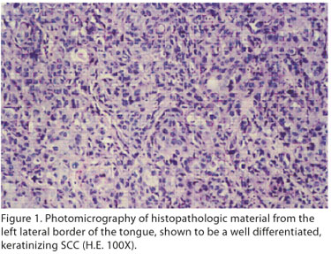
<body><![CDATA[<p><font size="2" face="Verdana">Diagnostic cytology has contributed to tracking precursor as well as malignant lesions, with the identification of potentially fatal cases and the decrease in the impact of potentially irreversible lesions. It has shown to be efficient in detecting atypia also in ulcerated and non&#45;ketatinizing lesions.</font></p>     <p><font size="2" face="Verdana">In countries (like Brazil) where preventive programs based on the Papanicolaou test have been largely implemented and continuously administered, the prevalence of cervical cancer decreased dramatically. As it has proved to be successful in the control of cervical cancer, we believe it can be employed in oral cancer for the same purpose.</font></p>     <p><font size="2" face="Verdana">Cytology in liquid medium (DNA&#45;CITOLIQ), also referred to as "monolayer cytology", "thin&#45;layer cytology", or preferentially "liquid sample cytology" or "liquid&#45;based cytology", was idealized, developed and released in March 2002 by Digene do Brasil. The tests were conducted at UNIFESP and in eight Brazilian private laboratories. This method represents an improvement of Papanicolaou, and began a new era in cytology, connecting morphologic and biomolecular pathology. The studies performed on cervical samples demonstrated that the DNA&#45;CITOLIQ System is sensitive for detecting epithelial lesions as well as microorganisms, with excellent results <sup>27</sup>.</font></p>     <p><font size="2" face="Verdana">The purpose of this study was to evaluate and compare the sensitivity and specificity of conventional cytology and liquid&#45;based cytology, as diagnostic tools employed in buccal malignant lesions and clinically normal mucosa.</font></p>     <p>&nbsp;</p>     <p><font size="3" face="Verdana"><B>MATERIALS AND METHODS</B></font></p>     <p><font size="2" face="Verdana">A case&#45;control study was performed at the head and neck ambulatory service of Hospital do C&acirc;ncer de Pernambuco, which is a reference center for oral cancer in the state of Pernambuco, serving rural, urban areas, and neighboring states.</font></p>     <p><font size="2" face="Verdana">The case group included 182 patients with squamous cell carcinoma in the oral cavity (CID&#45;O 140&#45;145)<sup>28,29</sup>. They were diagnosed according to the WHO classification of tumors, which describes it as a malignant epithelial tumor with squamous cell differentiation, presenting microscopically with cells that resemble keratinocytes, intercellular bridges and/or keratinization <sup>30,31</sup> (<a href="#fig01">Figure 1</a>). None of the selected patients had started treatment and none presented with other neoplasia elsewhere. The patients spontaneously agreed to participate in this study and signed a document of consent.</font></p>     <p><a name="fig01"></a></p>     <p>&nbsp;</p>     ]]></body>
<body><![CDATA[<p align="center"><img src="/img/revistas/occ/v10n1/a07fig01.jpg"></p>     <p>&nbsp;</p>     <p><font size="2" face="Verdana">The control group included 179 clinically healthy individuals that were either relatives or neighbors of the case group. They spontaneously agreed to participate in this study and signed a document of consent.</font></p>     <p><font size="2" face="Verdana"><B>Clinical and laboratorial procedures performed on both groups</B></font></p>     <p><font size="2" face="Verdana">History and physical examination were performed. The latter included careful examination of the oral cavity and was followed by exfoliative cytology (cytobrush) of the areas previously diagnosed as squamous cell carcinoma, in the case group, and of the correspondent clinically normal areas, in the control group. The smears were prepared on properly identified slides and immersed in absolute alcohol. The processing (Papanicolaou staining, preparation of slides) and conventional microscopic interpretation took place at Centro Integrado de Anatomia Patol&oacute;gica (CIAP) &#150; HUOC/UPE. Right after submitting the first sample for conventional cytology evaluation, a second sample was collected for liquid&#45;based<sup>&#174;</sup> cytology and accomodated in the specific Digene kit (with a solution that conserves DNA), known as Universal Collection Medium &#45; UCM<sup>&#174;</sup>. The second samples were sent to Laborat&oacute;rio Digene do Brasil, S&atilde;o Paulo, for processing.</font></p>     <p><font size="2" face="Verdana">The material was collected in loco by a technician who was previously evaluated by the Kappa test, described in Andrade and Zicker32. She demonstated an excellent intra (Kappa= 0.854) and inter&#45;examiner (Kappa= 0.869) reliability. The conventional cytology and liquid&#45;based<sup>&#174;</sup> cytology interpretations were carried out by a cytopathologist (examiner 1) who received training in DNA&#45;Citoliq at Centro de Treinamento Digene (CTD). She was also evaluated by the Kappa test for intra and inter&#45;examiner (with 2 other cytopathologists) reliability in conventional cytology. A specific cytogram was used.</font></p>     <p><font size="2" face="Verdana">Each interpretation, both in conventional and liquid&#45;based cytology, were performed independently, with no sharing of results.</font></p>     <p><font size="2" face="Verdana">In conventional cytology, the intra&#45;examiner (examiner 1: gold&#45;standard) concordance was perfect (100%) in the case group, and excellent in the control group (Kappa= 0.908). In liquid&#45;based<sup>&#174;</sup> cytology, the concordance was excellent both in the case (kappa= 0,869) and control group (Kappa= 0.901).</font></p>     <p><font size="2" face="Verdana">In the case group, the conventional cytology inter&#45;examiners agreement between examiners 1 and 2 was excellent (kappa= 0,805); good between 1 and 3 (Kappa= 0.726), and excellent between 2 and 3 (Kappa= 0.909). In the control group, examiner 1 showed good concordance with examiners 2 and 3 (Kappa= 0.713 for both); the concordance between examiners 2 and 3 was also good (Kappa= 0.608).</font></p>     <p><font size="2" face="Verdana"><B>Laboratorial methods</B></font></p>     ]]></body>
<body><![CDATA[<p><font size="2" face="Verdana">Liquid&#45;based cytology has not been proposed as a substitute for conventional cytology, but rather as an improvement of the Papanicolaou test. It is a product that aggregates the basic principles of liquid&#45;based<sup>&#174;</sup> cytology, with a reduced operational cost and a fast and reproducible protocol. Similarly to the cytology applied in the uterine cervix, the goal is to detect precursor lesions and cancer in the oral cavity.</font></p>     <p><font size="2" face="Verdana">The technician and cytopathologist (examiner 1) who participated in this study were trained at Centro de Treinamento da Digene (CTD) do Brasil, in S&atilde;o Paulo. The training included every step of processing, from using the pipette and preparing slides, to the final diagnostic evaluation. The criteria utilized are internationally recognized and are the ones applied in conventional cytology (The Bethesda System &#150;TBS) <sup>33</sup>.</font></p>     <p><font size="2" face="Verdana"><B>Cytologic interpretation</B></font></p>     <p><font size="2" face="Verdana">1. Type of sample:    <br> 1.1. Conventional Cytology    <br> 1.2. Liquid medium<sup>&#174;</sup> cytology</font></p>     <p><font size="2" face="Verdana">2. Pre&#45;analytical evaluation    <br> Causes of rejection of the sample:    <br> 2.1. Lack of identification or misidentification of slides and/or slide recipients    <br> 2.2. Damaged or absent slides    ]]></body>
<body><![CDATA[<br> 2.3. Causes unrelated to the laboratory (specify)    <br> 2.4. Other causes (specify)</font></p>     <p><font size="2" face="Verdana">3. Adequacy of the sample:    <br> 3.1. Satisfactory    <br> 3.2. Unsatisfactory (specify)</font></p>     <p><font size="2" face="Verdana">4. Descriptive diagnosis    <br> 4.1. Within normal limits    <br> 4.2. Benign cellular alterations    <br> 4.2.1. Inflammation    <br> 4.2.2. Repair    ]]></body>
<body><![CDATA[<br> 4.2.3. Immature squamous metaplasia    <br> 4.2.4. Atrophy due to inflammation    <br> 4.2.5. Radiation    <br> 4.2.6. Others (specify)    <br> 4.3. Atypia    <br> 4.3.1.1 Of squamous cells    <br> 4.3.1.2. ASCUS: characterized by morphologic alterations of undetermined significance (Bethesda 2001)    <br> 4.3.1.3. LSIL: Low grade squamous intraepithelial lesion (HPV and NIC I)    <br> 4.3.1.4. HSIL: High grade squamous intraepithelial lesion (NIC II and III)    <br> 4.3.1.5. High grade squamous intraepithelial lesion, unable to exclude invasion (in situ)    ]]></body>
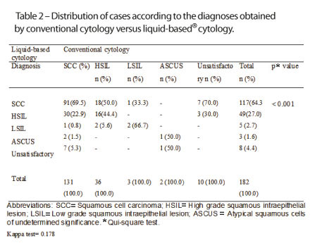
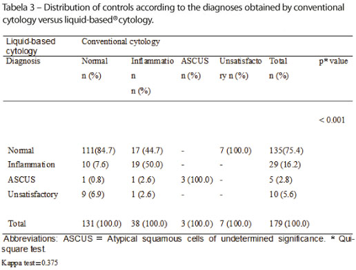
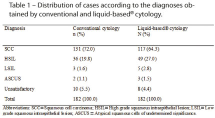
<body><![CDATA[<br> 4.3.1.6. Invasive squamous cell carcinoma</font></p>     <p><font size="2" face="Verdana"><B>Results and discussion</B></font></p>     <p><font size="2" face="Verdana">Among the 182 cases, ten (5.5%) and eight (4.4%) samples were considered unsatisfactory on conventional cytology and liquid&#45;based cytology, respectively. Out of the 179 controls, seven (3.9%) and ten (5.6%) samples were considered unsatisfactory on conventional cytology and liquid&#45;based cytology, respectively. In this manner, in conventional cytology 17 (5.2%) samples were disregarded due to excessive hemorrhage, scarcity of cells, or inappropriate staining. In liquid&#45;based<sup>&#174;</sup> cytology, 18 (5.0%) samples were rejected for presenting scarcity of cells &#45; perhaps caused by delay in pipettage, or conditions related to the patient himself or collect &#150; and inappropriate staining (see <a href="#tab01">tables 1</a>, <a href="#tab02">2</a> and <a href="#tab03">3</a>). However, this was not shown to be a satistically significant loss.</font></p>     <p><a name="tab01"></a></p>     <p>&nbsp;</p>     <p align="center"><img src="/img/revistas/occ/v10n1/a07tab01.jpg"></p>     <p>&nbsp;</p>     <p><a name="tab02"></a></p>     <p>&nbsp;</p>     <p align="center"><img src="/img/revistas/occ/v10n1/a07tab02.jpg"></p>     ]]></body>
<body><![CDATA[<p>&nbsp;</p>     <p><a name="tab03"></a></p>     <p>&nbsp;</p>     <p align="center"><img src="/img/revistas/occ/v10n1/a07tab03.jpg"></p>     <p>&nbsp;</p>     <p><font size="2" face="Verdana">In the case group, conventional cytology and liquid&#45;based cytology showed diagnostic agreement with histopathology in 131 (76.2%), and 117 (67.3%) SCC cases, respectively. However, if HSIL diagnoses &#45; which corresponds to carcinoma in situ &#45; were considered positive for disease, those numbers would incresase to 167 (97.1%) and 166 (955%), respectively. Thus, in conventional cytology 5 cases did not correspond to the histopathologic result; in liquid&#45;based<sup>&#174;</sup> cytology 8 cases did not agree with the histopathologic diagnoses (<a href="#tab01">Table 1</a>).</font></p>     <p><font size="2" face="Verdana">Among the cases, the agreement between the two cytologic methods was weak (Kappa= 0.178), with sensitivity= 96.9% (IC= 95%: 92.5% to 98.8%); specificity= 75.0% (IC= 95%: 21.9% to 98.7%); positive predictive value= 99.4%; negative predictive value= 37.5%, and accuracy= 96.3% (IC=95%: 92.5% to 98.5%). The results classified as SCC and HSIL were considered positive; the remainder were considered negative, and the unsatisfactory were excluded (<a href="#tab02">Table 2</a>).</font></p>     <p><font size="2" face="Verdana">In the control group, the agreement between the two cytologic methods was very weak (Kappa= 0.375), with sensitivity= 91.0% (IC= 95%: 84.1% to 95.2%); specificity= 57.5% (IC= 95%: 41.0% to 72.6%); positive predictive value= 86.7% (IC= 95%: 79.3% to 91.8%); negative predictive value= 67.6% (IC= 95%: 49.4% to 82.0%) and accuracy= 82.7% (IC= 95%: 76.3% to 88.0%). The results classified as normal were considered positive; the remainder were considered negative, and the unsatisfactory were excluded (<a href="#tab03">Table 3</a>).</font></p>     <p><font size="2" face="Verdana">Conventional cytology and liquid&#45;based cytology proved to be able to identify and classify cellular alterations characteristic of malignancy. Therefore, they are valuable as auxiliary diagnostic methods. When compared to each other, they showed a high sensitivity and reasonable specificity, conventional cytology being more reliable. The latter was defended and largely used by Mao<sup>34</sup>, and according to Vidal and Silva<sup>7</sup>, if used appropriately, it has an excellent sensitivity and specificity when compared to histolopathologic diagnoses.</font></p>     <p><font size="2" face="Verdana">Some authors<sup>7,8,26</sup>, defend the use of cytology as a screening method for prevention and early diagnosis of oral cancer. Sciubba5 further<sup>35</sup> mentions the computer&#45;assisted exfoliative cytology as an accurate method for detecting premalignant and malignant buccal lesions.</font></p>     ]]></body>
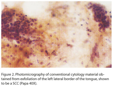
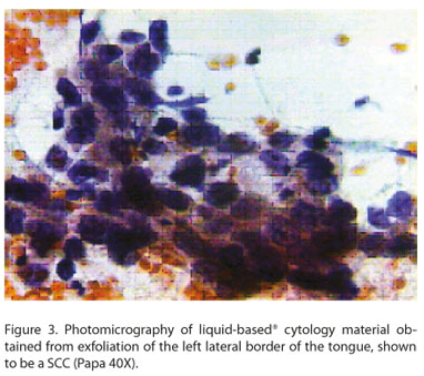
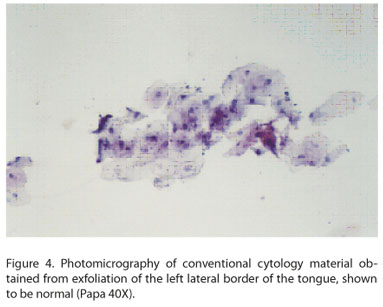
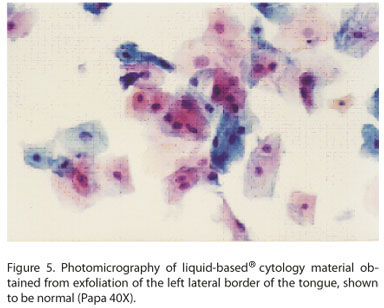
<body><![CDATA[<p><font size="2" face="Verdana">Gynther, Rozell and Heimdahl<sup>36</sup> cite direct buccal microscopy as a method for selecting areas to be submitted to cytology and biopsy. Moreover, Sciubba<sup>35</sup> mentions the need to carry out studies to evaluate the sensitivity and specificity of oral scan laboratories inc. (Oral CDX). This is a computer&#45;assisted method for the analysis of exfoliative cytology material, which adds to clinical examination in the differentiation of premalignant and malignant alterations found in "benign" lesions, the purpose being to find carcinomas of innocuous appearance, at the most curable and early stages.</font></p>     <p><font size="2" face="Verdana">The advantages of using conventional cytology in the oral mucosa, similarly to the uterine cervix, are its low cost, good patient tolerability, and easy applicability to large populations. The disadvantages are the number of unsatisfactory cases on account of technical reasons which depend on training in collect, fixation and sample preparation. In spite of its merits, one can not ignore the false&#45;negative results. Furthermore, there are cases with cytomorphological alterations of innacurate diagnostic resolution. Liquid&#45;based cytology has been considered an important alternative for improving cytology sensitivity, as it decreases cell loss during sample preparation, and provides a better cell distribution. In addition, it allows the use of molecular cytopathology techniques such as in situ hibridization, immunohistochemical tests, and determination of nucleic acids in molecular biology<sup>27</sup>. The best performance of Liquid&#45;based cytology is achieved with a high quality presentation: clear bottom, good distribution of cells, ease in distinguishing individualized cells, and good cell preservation. However, it lacks the specificity of conventional cytology (<a href="#fig02">Figures 2</a>,<a href="#fig03">3</a>, <a href="#fig04">4</a> and <a href="#fig05">5</a>).</font></p>     <p><a name="fig02"></a></p>     <p>&nbsp;</p>     <p align="center"><img src="/img/revistas/occ/v10n1/a07fig02.jpg"></p>     <p>&nbsp;</p>     <p><a name="fig03"></a></p>     <p>&nbsp;</p>     <p align="center"><img src="/img/revistas/occ/v10n1/a07fig03.jpg"></p>     <p>&nbsp;</p>     ]]></body>
<body><![CDATA[<p><a name="fig04"></a></p>     <p>&nbsp;</p>     <p align="center"><img src="/img/revistas/occ/v10n1/a07fig04.jpg"></p>     <p>&nbsp;</p>     <p><a name="fig05"></a></p>     <p>&nbsp;</p>     <p align="center"><img src="/img/revistas/occ/v10n1/a07fig05.jpg"></p>     <p>&nbsp;</p>     <p><font size="2" face="Verdana">The acceptance and collaboration of the patients enrolled in this study, as well their interest (also mentioned by Vidal et al8) in the subject explored, demonstrate that the population wants and needs health education, in order to take better care of themselves and consequently have a higher quality of life.</font></p>     <p>&nbsp;</p>     ]]></body>
<body><![CDATA[<p><font size="3" face="Verdana"><B>CONCLUSION</B></font></p>     <p><font size="2" face="Verdana">Conventional cytology and liquid&#45;based cytology demonstrated a diagnostic concordance with histopathology of more than 90%. Therefore, if properly indicated and executed, they can be routinely used as complementary diagnostic methods.</font></p>     <p><font size="2" face="Verdana">When compared to each other, conventional cytology and liquid&#45;based cytology showed a high sensitivity and reasonable specificity, conventional cytology having higher specificity.</font></p>     <p>&nbsp;</p>     <p><font size="3" face="Verdana"><B>ACKNOWLEDGEMENTS</B></font></p>     <p><font size="2" face="Verdana">The authors thank all the participating patients, the professionals who help develop this work, and Cancer Hospital of Pernambuco, University of Pernambuco, National Health Foundation and Laboratory Digene of Brazil for the support given to this study.</font></p>     <p>&nbsp;</p>     <p><font size="3" face="Verdana"><B>REFERENCES</B></font></p>     <!-- ref --><p><font size="2" face="Verdana">1. Bernier, j. l. Clinical features of early malignant growth of the oral cavity. In: BURKET, L. W. Symposium on the interrelationship of oral and systemic disease. Philadelphia, ed Dental Clinics of Nort America, Saunders. 1958. p, 325&#45;333.    &nbsp;&nbsp;&nbsp;&nbsp;&nbsp;&nbsp;&nbsp;&nbsp;[&#160;<a href="javascript:void(0);" onclick="javascript: window.open('/scielo.php?script=sci_nlinks&ref=106737&pid=S1677-3888201100010000700001&lng=','','width=640,height=500,resizable=yes,scrollbars=1,menubar=yes,');">Links</a>&#160;]<!-- end-ref --></font></p>     ]]></body>
<body><![CDATA[<!-- ref --><p><font size="2" face="Verdana">2. Waldron, C. A . Oral ephitelial tumors. In: GORLIN, R. J. e GOLDMAN, H. M. Oral ephitelial tumors eds. Thoma's Oral Patolog. St. Louis, Mosby. 1970. v.2. cap. 19 p.801&#45;860.    &nbsp;&nbsp;&nbsp;&nbsp;&nbsp;&nbsp;&nbsp;&nbsp;[&#160;<a href="javascript:void(0);" onclick="javascript: window.open('/scielo.php?script=sci_nlinks&ref=106739&pid=S1677-3888201100010000700002&lng=','','width=640,height=500,resizable=yes,scrollbars=1,menubar=yes,');">Links</a>&#160;]<!-- end-ref --></font></p>     <!-- ref --><p><font size="2" face="Verdana">3. Barbosa, J. F. e Fonseca, E. P. C&acirc;ncer bucal: diagn&oacute;stico, tratamento  e reabilita&ccedil;&atilde;o.  Minist&eacute;rio da Sa&uacute;de &#150; Divis&atilde;o Nacional de  C&acirc;ncer. Programa Nacional de Controle do C&acirc;ncer. 1972. 20p.    &nbsp;&nbsp;&nbsp;&nbsp;&nbsp;&nbsp;&nbsp;&nbsp;[&#160;<a href="javascript:void(0);" onclick="javascript: window.open('/scielo.php?script=sci_nlinks&ref=106741&pid=S1677-3888201100010000700003&lng=','','width=640,height=500,resizable=yes,scrollbars=1,menubar=yes,');">Links</a>&#160;]<!-- end-ref --></font></p>     <!-- ref --><p><font size="2" face="Verdana">4. Catanzaro Guimar&atilde;es, S. A . Patologia b&aacute;sica da cavidade bucal. Rio de Janeiro, Guanabara Koogan, 1982. 419p.    &nbsp;&nbsp;&nbsp;&nbsp;&nbsp;&nbsp;&nbsp;&nbsp;[&#160;<a href="javascript:void(0);" onclick="javascript: window.open('/scielo.php?script=sci_nlinks&ref=106743&pid=S1677-3888201100010000700004&lng=','','width=640,height=500,resizable=yes,scrollbars=1,menubar=yes,');">Links</a>&#160;]<!-- end-ref --></font></p>     <!-- ref --><p><font size="2" face="Verdana">5. Scully, C.; Porter, S. ABC of oral health. Oral cancer. BMJ. v. 321, p. 97&#45;100. 2000.    &nbsp;&nbsp;&nbsp;&nbsp;&nbsp;&nbsp;&nbsp;&nbsp;[&#160;<a href="javascript:void(0);" onclick="javascript: window.open('/scielo.php?script=sci_nlinks&ref=106745&pid=S1677-3888201100010000700005&lng=','','width=640,height=500,resizable=yes,scrollbars=1,menubar=yes,');">Links</a>&#160;]<!-- end-ref --></font></p>     <!-- ref --><p><font size="2" face="Verdana">6. MacFarlaneE, G. J.; Sharp, L.; Porter, S. e Franceschi, S. Trends in survival from cancers of the oral cavity and pharynx in Scotland: a clue as to why the disease is becoming more common? Br. J. Cancer. v. 72 n. 9, p. 568&#45;576. Mar. 1996.    &nbsp;&nbsp;&nbsp;&nbsp;&nbsp;&nbsp;&nbsp;&nbsp;[&#160;<a href="javascript:void(0);" onclick="javascript: window.open('/scielo.php?script=sci_nlinks&ref=106747&pid=S1677-3888201100010000700006&lng=','','width=640,height=500,resizable=yes,scrollbars=1,menubar=yes,');">Links</a>&#160;]<!-- end-ref --></font></p>     ]]></body>
<body><![CDATA[<!-- ref --><p><font size="2" face="Verdana">7. Vidal, A . K. L. e Silva, J. F. Correla&ccedil;&atilde;o cito&#45;histopatol&oacute;gica e aplicabilidade do corante azul de toluidina a 2% em les&otilde;es de mucosa bucal. Disserta&ccedil;&atilde;o de Mestrado. Revista do CRO/PE. V. 2 n. 1, p. 59&#45;72.1999.    &nbsp;&nbsp;&nbsp;&nbsp;&nbsp;&nbsp;&nbsp;&nbsp;[&#160;<a href="javascript:void(0);" onclick="javascript: window.open('/scielo.php?script=sci_nlinks&ref=106749&pid=S1677-3888201100010000700007&lng=','','width=640,height=500,resizable=yes,scrollbars=1,menubar=yes,');">Links</a>&#160;]<!-- end-ref --></font></p>     <!-- ref --><p><font size="2" face="Verdana">8. Vidal, A. K. L. ; Silvera, R. C. J.; Soares, E. A.; Caldas J&uacute;nior, A. F.; Cabral, A. C.; Souza, E. H. A.;Lopez, R. M. Preven&ccedil;&atilde;o e diagn&oacute;stico precoce do c&acirc;ncer de boca&#45; uma realidade no Distrito Sanit&aacute;rio IV&#45; Recife/ PE. Pesquisa Odontol&oacute;gica Brasileira v. 17 n. 2, p. 31. Ago. 2003.    &nbsp;&nbsp;&nbsp;&nbsp;&nbsp;&nbsp;&nbsp;&nbsp;[&#160;<a href="javascript:void(0);" onclick="javascript: window.open('/scielo.php?script=sci_nlinks&ref=106751&pid=S1677-3888201100010000700008&lng=','','width=640,height=500,resizable=yes,scrollbars=1,menubar=yes,');">Links</a>&#160;]<!-- end-ref --></font></p>     <!-- ref --><p><font size="2" face="Verdana">9. Papanicolaou, G. N. Some improved methods for staing vaginal smears. J. Lab. Cl&iacute;n. Med. v. 16, p. 1200&#45;1205. 1941.    &nbsp;&nbsp;&nbsp;&nbsp;&nbsp;&nbsp;&nbsp;&nbsp;[&#160;<a href="javascript:void(0);" onclick="javascript: window.open('/scielo.php?script=sci_nlinks&ref=106753&pid=S1677-3888201100010000700009&lng=','','width=640,height=500,resizable=yes,scrollbars=1,menubar=yes,');">Links</a>&#160;]<!-- end-ref --></font></p>     <!-- ref --><p><font size="2" face="Verdana">10.Blank, H. et al. Cytologic smears in  diagnosis of herpes simplex, herpes zoster e varicella. JAMA. v. 146 n. 15, p. 1410&#45;1412. 1951.    &nbsp;&nbsp;&nbsp;&nbsp;&nbsp;&nbsp;&nbsp;&nbsp;[&#160;<a href="javascript:void(0);" onclick="javascript: window.open('/scielo.php?script=sci_nlinks&ref=106755&pid=S1677-3888201100010000700010&lng=','','width=640,height=500,resizable=yes,scrollbars=1,menubar=yes,');">Links</a>&#160;]<!-- end-ref --></font></p>     <!-- ref --><p><font size="2" face="Verdana">11. Peters, H.; Rijsinghani, K. The cytologic interpretation of the mouth smear. J. Indian Med Assoc v. 27 n. 7, p. 231&#45;236. 1956.    &nbsp;&nbsp;&nbsp;&nbsp;&nbsp;&nbsp;&nbsp;&nbsp;[&#160;<a href="javascript:void(0);" onclick="javascript: window.open('/scielo.php?script=sci_nlinks&ref=106757&pid=S1677-3888201100010000700011&lng=','','width=640,height=500,resizable=yes,scrollbars=1,menubar=yes,');">Links</a>&#160;]<!-- end-ref --></font></p>     ]]></body>
<body><![CDATA[<!-- ref --><p><font size="2" face="Verdana">12. Ara&uacute;jo, N. S. Diagn&oacute;stico citol&oacute;gico de les&otilde;es da mucosa oral. Revista Brasileira de Cirurgia. v. 49 n. 5, p. 350&#45;352. 1965</font>&nbsp;&nbsp;&nbsp;&nbsp;&nbsp;&nbsp;&nbsp;&nbsp;[&#160;<a href="javascript:void(0);" onclick="javascript: window.open('/scielo.php?script=sci_nlinks&ref=106759&pid=S1677-3888201100010000700012&lng=','','width=640,height=500,resizable=yes,scrollbars=1,menubar=yes,');">Links</a>&#160;]<!-- end-ref --><!-- ref --><p><font size="2" face="Verdana">13. Nowakovski , S. et al. Manifestations of viral infeccions in exfoliated cells. Acta Cytol. v. 35 n. 5, p. 416&#45;419. 1981.    &nbsp;&nbsp;&nbsp;&nbsp;&nbsp;&nbsp;&nbsp;&nbsp;[&#160;<a href="javascript:void(0);" onclick="javascript: window.open('/scielo.php?script=sci_nlinks&ref=106760&pid=S1677-3888201100010000700013&lng=','','width=640,height=500,resizable=yes,scrollbars=1,menubar=yes,');">Links</a>&#160;]<!-- end-ref --></font></p>     <!-- ref --><p><font size="2" face="Verdana">14. Burns, J. C. Diagnostic methods for herpes simplex infection: a review. Oral Surg. Oral Med. Oral Pathol. v. 50 n. 4, p. 346&#45;349. 1980.    &nbsp;&nbsp;&nbsp;&nbsp;&nbsp;&nbsp;&nbsp;&nbsp;[&#160;<a href="javascript:void(0);" onclick="javascript: window.open('/scielo.php?script=sci_nlinks&ref=106762&pid=S1677-3888201100010000700014&lng=','','width=640,height=500,resizable=yes,scrollbars=1,menubar=yes,');">Links</a>&#160;]<!-- end-ref --></font></p>     <!-- ref --><p><font size="2" face="Verdana">15. Marcucci, G.; Ara&uacute;jo, N. S. Citologia esfoliativa In: TOMMASI, A. F.; GRARRAFA, V. C&acirc;ncer Bucal. S&atilde;o Paulo. Medisa. 1980. p.393&#45;407.    &nbsp;&nbsp;&nbsp;&nbsp;&nbsp;&nbsp;&nbsp;&nbsp;[&#160;<a href="javascript:void(0);" onclick="javascript: window.open('/scielo.php?script=sci_nlinks&ref=106764&pid=S1677-3888201100010000700015&lng=','','width=640,height=500,resizable=yes,scrollbars=1,menubar=yes,');">Links</a>&#160;]<!-- end-ref --></font></p>     <!-- ref --><p><font size="2" face="Verdana">16. Mercure, J. L. Cytodiagnosis et cancers buccaux. Rev. D'Odontologie. v. 10, p. 157&#45;160. 1981.    &nbsp;&nbsp;&nbsp;&nbsp;&nbsp;&nbsp;&nbsp;&nbsp;[&#160;<a href="javascript:void(0);" onclick="javascript: window.open('/scielo.php?script=sci_nlinks&ref=106766&pid=S1677-3888201100010000700016&lng=','','width=640,height=500,resizable=yes,scrollbars=1,menubar=yes,');">Links</a>&#160;]<!-- end-ref --></font></p>     <!-- ref --><p><font size="2" face="Verdana">17. Novelli, M. D. et al. Estudo comparativo entre a citologia esfoliativa e os achados cl&iacute;nicos de 1498 pacientes. Revista da APCD v. 35 n. 5, p. 416&#45;419. 1981</font>&nbsp;&nbsp;&nbsp;&nbsp;&nbsp;&nbsp;&nbsp;&nbsp;[&#160;<a href="javascript:void(0);" onclick="javascript: window.open('/scielo.php?script=sci_nlinks&ref=106768&pid=S1677-3888201100010000700017&lng=','','width=640,height=500,resizable=yes,scrollbars=1,menubar=yes,');">Links</a>&#160;]<!-- end-ref --><!-- ref --><p><font size="2" face="Verdana">18. Barrett, A. P. et al.  The value of exfoliative cytology in the diagnosis of oral herpex simplex infection in immunosupressed patients. Oral Surg. Oral Med. Oral Pathol. v. 62 n. 2, p. 175&#45;178. 1986</font>&nbsp;&nbsp;&nbsp;&nbsp;&nbsp;&nbsp;&nbsp;&nbsp;[&#160;<a href="javascript:void(0);" onclick="javascript: window.open('/scielo.php?script=sci_nlinks&ref=106769&pid=S1677-3888201100010000700018&lng=','','width=640,height=500,resizable=yes,scrollbars=1,menubar=yes,');">Links</a>&#160;]<!-- end-ref --><!-- ref --><p><font size="2" face="Verdana">19. Persoglia, J. Citologia exfoliativa normal y Patologia de la mucosa bucal. Grinspan, D. Ed. Mundi S. A. C. I. F. Buenos Aires, 1987. p. 459&#45;471.    &nbsp;&nbsp;&nbsp;&nbsp;&nbsp;&nbsp;&nbsp;&nbsp;[&#160;<a href="javascript:void(0);" onclick="javascript: window.open('/scielo.php?script=sci_nlinks&ref=106770&pid=S1677-3888201100010000700019&lng=','','width=640,height=500,resizable=yes,scrollbars=1,menubar=yes,');">Links</a>&#160;]<!-- end-ref --></font></p>     <!-- ref --><p><font size="2" face="Verdana">20. MacLeod, R. I.; Soames, J. V. Morphological and morphometric studies of exfoliated oral squames from chronic inflammatory oral ulcers using light and scanning electron microscopy. British Journal of Oral and Maxillofacial Surgery. v. 33, p. 86&#45;89. 1995.    &nbsp;&nbsp;&nbsp;&nbsp;&nbsp;&nbsp;&nbsp;&nbsp;[&#160;<a href="javascript:void(0);" onclick="javascript: window.open('/scielo.php?script=sci_nlinks&ref=106772&pid=S1677-3888201100010000700020&lng=','','width=640,height=500,resizable=yes,scrollbars=1,menubar=yes,');">Links</a>&#160;]<!-- end-ref --></font></p>     <!-- ref --><p><font size="2" face="Verdana">21. Vaillant J. M. D&eacute;pistage et diagnostic des carcinomas &eacute;piderm&ouml;ides de la muqueuse b&uacute;chale (CB). Que Pert&#45;on conclure aujourd'hui du r&ocirc;le respectif du frottis cytologique et de la biopsia? Bull. Acad. Natle. Med. v. 178 n. 2, p. 233&#45;247. f&eacute;vrier, 1994.    &nbsp;&nbsp;&nbsp;&nbsp;&nbsp;&nbsp;&nbsp;&nbsp;[&#160;<a href="javascript:void(0);" onclick="javascript: window.open('/scielo.php?script=sci_nlinks&ref=106774&pid=S1677-3888201100010000700021&lng=','','width=640,height=500,resizable=yes,scrollbars=1,menubar=yes,');">Links</a>&#160;]<!-- end-ref --></font></p>     <!-- ref --><p><font size="2" face="Verdana">22. Sposto, M. R.; Scully, C.; De Almeida, O. P.; Jorge, J.; Graner, E.; Bozzo, L. Oral paracoccidioidomycosis a study of 36 south american patients. Oral Surg Oral Med Oral Pathol v. 75 n. 4, p. 461&#45;465. Apr. 1993</font>&nbsp;&nbsp;&nbsp;&nbsp;&nbsp;&nbsp;&nbsp;&nbsp;[&#160;<a href="javascript:void(0);" onclick="javascript: window.open('/scielo.php?script=sci_nlinks&ref=106776&pid=S1677-3888201100010000700022&lng=','','width=640,height=500,resizable=yes,scrollbars=1,menubar=yes,');">Links</a>&#160;]<!-- end-ref --><!-- ref --><p><font size="2" face="Verdana">23. Ogden, G. R.; Leigh, I.; Chisholm, D. M.; Cowpe, G.; Lane, E. B. Exfoliative cytololy of normal oral mucosa. Acta Cytologica. v. 40 n. 5, p. 933&#45;936. 1996.    &nbsp;&nbsp;&nbsp;&nbsp;&nbsp;&nbsp;&nbsp;&nbsp;[&#160;<a href="javascript:void(0);" onclick="javascript: window.open('/scielo.php?script=sci_nlinks&ref=106777&pid=S1677-3888201100010000700023&lng=','','width=640,height=500,resizable=yes,scrollbars=1,menubar=yes,');">Links</a>&#160;]<!-- end-ref --></font></p>     ]]></body>
<body><![CDATA[<!-- ref --><p><font size="2" face="Verdana">24. Ogden, G. R.; Cowpe, J. G.; Wight, A. J. Oral exfoliative cytology: review of methods of assessment. J. Oral Pathol. Med. v. 26, p. 201&#45;205. 1997.    &nbsp;&nbsp;&nbsp;&nbsp;&nbsp;&nbsp;&nbsp;&nbsp;[&#160;<a href="javascript:void(0);" onclick="javascript: window.open('/scielo.php?script=sci_nlinks&ref=106779&pid=S1677-3888201100010000700024&lng=','','width=640,height=500,resizable=yes,scrollbars=1,menubar=yes,');">Links</a>&#160;]<!-- end-ref --></font></p>     <!-- ref --><p><font size="2" face="Verdana">25. Vidal, A . K. L.; Caldas J&uacute;nior, A . F.; Mello, R. J. V.; Brand&atilde;o, V. R. A .; Rocha, G. I.; Taromaru, E. Human papillomavirus (HPV) detection in oral carcinomas. J. Bras Patol Med Lab v.40 n.1, p. 21&#45;26, janeiro/fevereiro, 2004.    &nbsp;&nbsp;&nbsp;&nbsp;&nbsp;&nbsp;&nbsp;&nbsp;[&#160;<a href="javascript:void(0);" onclick="javascript: window.open('/scielo.php?script=sci_nlinks&ref=106781&pid=S1677-3888201100010000700025&lng=','','width=640,height=500,resizable=yes,scrollbars=1,menubar=yes,');">Links</a>&#160;]<!-- end-ref --></font></p>     <!-- ref --><p><font size="2" face="Verdana">26.Christian, D. C. Computer&#45;assisted analysis of oral brush biopsies at oral cancer screening program. JADA v. 1333, p. 357&#45;362. 2002.    &nbsp;&nbsp;&nbsp;&nbsp;&nbsp;&nbsp;&nbsp;&nbsp;[&#160;<a href="javascript:void(0);" onclick="javascript: window.open('/scielo.php?script=sci_nlinks&ref=106783&pid=S1677-3888201100010000700026&lng=','','width=640,height=500,resizable=yes,scrollbars=1,menubar=yes,');">Links</a>&#160;]<!-- end-ref --></font></p>     <!-- ref --><p><font size="2" face="Verdana">27. D&ocirc;res, G. B.; Taromaru, E. Sistema DNA&#45;CITOLIQ&#174; &#45; A Citologia em Nova Era &#45; Manual T&eacute;cnico. &copy;Digene do Brasil. S&atilde;o Paulo, 2003. 120p.    &nbsp;&nbsp;&nbsp;&nbsp;&nbsp;&nbsp;&nbsp;&nbsp;[&#160;<a href="javascript:void(0);" onclick="javascript: window.open('/scielo.php?script=sci_nlinks&ref=106785&pid=S1677-3888201100010000700027&lng=','','width=640,height=500,resizable=yes,scrollbars=1,menubar=yes,');">Links</a>&#160;]<!-- end-ref --></font></p>     <!-- ref --><p><font size="2" face="Verdana">28.Brasil. Minist&eacute;rio da Sa&uacute;de.Estimativas da Incid&ecirc;ncia e Mortalidade por C&acirc;ncer no Brasil. 2010. Rio de Janeiro &#150; INCa / Pr&oacute;&#45;Onco, 2010.    &nbsp;&nbsp;&nbsp;&nbsp;&nbsp;&nbsp;&nbsp;&nbsp;[&#160;<a href="javascript:void(0);" onclick="javascript: window.open('/scielo.php?script=sci_nlinks&ref=106787&pid=S1677-3888201100010000700028&lng=','','width=640,height=500,resizable=yes,scrollbars=1,menubar=yes,');">Links</a>&#160;]<!-- end-ref --></font></p>     ]]></body>
<body><![CDATA[<!-- ref --><p><font size="2" face="Verdana">29. Sampaio, G. Anatomia Aplicada &agrave; Odontologia. Recife: EDUPE, 2001. p.347&#45;369.    &nbsp;&nbsp;&nbsp;&nbsp;&nbsp;&nbsp;&nbsp;&nbsp;[&#160;<a href="javascript:void(0);" onclick="javascript: window.open('/scielo.php?script=sci_nlinks&ref=106789&pid=S1677-3888201100010000700029&lng=','','width=640,height=500,resizable=yes,scrollbars=1,menubar=yes,');">Links</a>&#160;]<!-- end-ref --></font></p>     <!-- ref --><p><font size="2" face="Verdana">30. Galbarthe; Shimoda, T.; Haainaut, P.; Nakamura, Y.; Field, J. K.; Inove, H. Scamous cell carcinoma of the esophagus in: HAMILTON, S. R e AALTONE, L. A. World  Health Organization  Classification  of Tumors. Pathology  and Genetics of Tumors of the Digest System. Ed. IARC. Press Lion, 2000. p. 9&#45;11</font>&nbsp;&nbsp;&nbsp;&nbsp;&nbsp;&nbsp;&nbsp;&nbsp;[&#160;<a href="javascript:void(0);" onclick="javascript: window.open('/scielo.php?script=sci_nlinks&ref=106791&pid=S1677-3888201100010000700030&lng=','','width=640,height=500,resizable=yes,scrollbars=1,menubar=yes,');">Links</a>&#160;]<!-- end-ref --><!-- ref --><p><font size="2" face="Verdana">31. MacDonald, D. G. Tumors of the Oral Cavity. In: Fletcher, C. D. N. Diagnostic histopatology of Tumors. Ed. Churchil Living Stone. London. 2000. p. 209&#45;230.    &nbsp;&nbsp;&nbsp;&nbsp;&nbsp;&nbsp;&nbsp;&nbsp;[&#160;<a href="javascript:void(0);" onclick="javascript: window.open('/scielo.php?script=sci_nlinks&ref=106792&pid=S1677-3888201100010000700031&lng=','','width=640,height=500,resizable=yes,scrollbars=1,menubar=yes,');">Links</a>&#160;]<!-- end-ref --></font></p>     <!-- ref --><p><font size="2" face="Verdana">32. Andrade, A. L. S. S.; Zicker, F.  M&eacute;todos de investiga&ccedil;&atilde;o epidemiol&oacute;gica em doen&ccedil;as transmiss&iacute;veis: volume I Bras&iacute;lia: Organiza&ccedil;&atilde;o Pan&#45;Americana de Sa&uacute;de: Funda&ccedil;&atilde;o Nacional de Sa&uacute;de, 1997. 182p.    &nbsp;&nbsp;&nbsp;&nbsp;&nbsp;&nbsp;&nbsp;&nbsp;[&#160;<a href="javascript:void(0);" onclick="javascript: window.open('/scielo.php?script=sci_nlinks&ref=106794&pid=S1677-3888201100010000700032&lng=','','width=640,height=500,resizable=yes,scrollbars=1,menubar=yes,');">Links</a>&#160;]<!-- end-ref --></font></p>     <!-- ref --><p><font size="2" face="Verdana">33. Kuffer, R.; Lombardi, T. Premalignant lesions of the oral mucosa. A discussion about the place of oral intrepithelial neoplasia (OIN). Oral Oncol. v. 38 n. 2, p. 125&#45;130. Feb. 2002.    &nbsp;&nbsp;&nbsp;&nbsp;&nbsp;&nbsp;&nbsp;&nbsp;[&#160;<a href="javascript:void(0);" onclick="javascript: window.open('/scielo.php?script=sci_nlinks&ref=106796&pid=S1677-3888201100010000700033&lng=','','width=640,height=500,resizable=yes,scrollbars=1,menubar=yes,');">Links</a>&#160;]<!-- end-ref --></font></p>     <!-- ref --><p><font size="2" face="Verdana">34. Mao, E. J. Prevalence of human papilomavirus 16 and nucleolar organizer region counts in oral exfoliated cells from normal and malignant epithelia. Oral Surg Oral Med Oral Pathol Oral Radiol Endod v. 80 n.3, p. 320&#45;329. Sep. 1995.    &nbsp;&nbsp;&nbsp;&nbsp;&nbsp;&nbsp;&nbsp;&nbsp;[&#160;<a href="javascript:void(0);" onclick="javascript: window.open('/scielo.php?script=sci_nlinks&ref=106798&pid=S1677-3888201100010000700034&lng=','','width=640,height=500,resizable=yes,scrollbars=1,menubar=yes,');">Links</a>&#160;]<!-- end-ref --></font></p>     <!-- ref --><p><font size="2" face="Verdana">35. Sciubba, J. J. Improving detection of precancerous and cancerous oral lesions. JADA v. 130, p.1445&#45;1456. 1999.    &nbsp;&nbsp;&nbsp;&nbsp;&nbsp;&nbsp;&nbsp;&nbsp;[&#160;<a href="javascript:void(0);" onclick="javascript: window.open('/scielo.php?script=sci_nlinks&ref=106800&pid=S1677-3888201100010000700035&lng=','','width=640,height=500,resizable=yes,scrollbars=1,menubar=yes,');">Links</a>&#160;]<!-- end-ref --></font></p>     <!-- ref --><p><font size="2" face="Verdana">36. Gynther, G. W.; ROZELL, B.; HEIMDAHL, A . Direct oral microscopy and its value in diagnosing mucosal lesions. A pilot study. Oral Surg Oral Med Oral Pathol Oral Radiol Endod v. 90 n. 2, p. 164&#45;170. 2000.    &nbsp;&nbsp;&nbsp;&nbsp;&nbsp;&nbsp;&nbsp;&nbsp;[&#160;<a href="javascript:void(0);" onclick="javascript: window.open('/scielo.php?script=sci_nlinks&ref=106802&pid=S1677-3888201100010000700036&lng=','','width=640,height=500,resizable=yes,scrollbars=1,menubar=yes,');">Links</a>&#160;]<!-- end-ref --></font></p>     <p>&nbsp;</p>     <p>&nbsp;</p>     <p><font size="2" face="Verdana"><B><a name="nt"></a><a href="#tx"><img src="/img/revistas/occ/v10n1/seta.jpg" border="0"></a> Endere&ccedil;o para correspond&ecirc;ncia</B>    <br>  Aurora Karla de Lacerda Vidal    <br> <a href="mailto:aurorakarla@gmail.com">aurorakarla@gmail.com</a></font></p>     ]]></body>
<body><![CDATA[<p><font size="2" face="Verdana">Recebido para publica&ccedil;&atilde;o: 16/02/11    <br> Aceito para publica&ccedil;&atilde;o: 21/02/11</font></p>      ]]></body>
<back>
<ref-list>
<ref id="B1">
<label>1</label><nlm-citation citation-type="book">
<person-group person-group-type="author">
<name>
<surname><![CDATA[Bernier]]></surname>
<given-names><![CDATA[j. l.]]></given-names>
</name>
</person-group>
<article-title xml:lang="en"><![CDATA[Clinical features of early malignant growth of the oral cavity]]></article-title>
<person-group person-group-type="editor">
<name>
<surname><![CDATA[BURKET]]></surname>
<given-names><![CDATA[L. W.]]></given-names>
</name>
</person-group>
<source><![CDATA[Symposium on the interrelationship of oral and systemic disease]]></source>
<year>1958</year>
<page-range>325-333</page-range><publisher-loc><![CDATA[Philadelphia ]]></publisher-loc>
<publisher-name><![CDATA[Dental Clinics of Nort America, Saunders]]></publisher-name>
</nlm-citation>
</ref>
<ref id="B2">
<label>2</label><nlm-citation citation-type="book">
<person-group person-group-type="author">
<name>
<surname><![CDATA[Waldron]]></surname>
<given-names><![CDATA[C. A]]></given-names>
</name>
</person-group>
<article-title xml:lang="en"><![CDATA[Oral ephitelial tumors]]></article-title>
<person-group person-group-type="editor">
<name>
<surname><![CDATA[GORLIN]]></surname>
<given-names><![CDATA[R. J.]]></given-names>
</name>
<name>
<surname><![CDATA[GOLDMAN]]></surname>
<given-names><![CDATA[H. M.]]></given-names>
</name>
</person-group>
<source><![CDATA[Oral ephitelial tumors eds. Thoma's Oral Patolog]]></source>
<year>1970</year>
<page-range>801-860</page-range><publisher-loc><![CDATA[St. Louis ]]></publisher-loc>
<publisher-name><![CDATA[Mosby]]></publisher-name>
</nlm-citation>
</ref>
<ref id="B3">
<label>3</label><nlm-citation citation-type="">
<person-group person-group-type="author">
<name>
<surname><![CDATA[Barbosa]]></surname>
<given-names><![CDATA[J. F.]]></given-names>
</name>
<name>
<surname><![CDATA[Fonseca]]></surname>
<given-names><![CDATA[E. P.]]></given-names>
</name>
</person-group>
<article-title xml:lang="pt"><![CDATA[Câncer bucal: diagnóstico, tratamento e reabilitação]]></article-title>
<collab>Ministério da Saúde - Divisão Nacional de Câncer^dPrograma Nacional de Controle do Câncer</collab>
<source><![CDATA[]]></source>
<year>1972</year>
</nlm-citation>
</ref>
<ref id="B4">
<label>4</label><nlm-citation citation-type="book">
<person-group person-group-type="author">
<name>
<surname><![CDATA[Catanzaro Guimarães]]></surname>
<given-names><![CDATA[S. A .]]></given-names>
</name>
</person-group>
<source><![CDATA[Patologia básica da cavidade bucal]]></source>
<year>1982</year>
<publisher-loc><![CDATA[Rio de Janeiro ]]></publisher-loc>
<publisher-name><![CDATA[Guanabara Koogan]]></publisher-name>
</nlm-citation>
</ref>
<ref id="B5">
<label>5</label><nlm-citation citation-type="journal">
<person-group person-group-type="author">
<name>
<surname><![CDATA[Scully]]></surname>
<given-names><![CDATA[C.]]></given-names>
</name>
<name>
<surname><![CDATA[Porter]]></surname>
<given-names><![CDATA[S.]]></given-names>
</name>
</person-group>
<article-title xml:lang="en"><![CDATA[ABC of oral health]]></article-title>
<source><![CDATA[Oral cancer. BMJ]]></source>
<year>2000</year>
<volume>321</volume>
<page-range>97-100</page-range></nlm-citation>
</ref>
<ref id="B6">
<label>6</label><nlm-citation citation-type="journal">
<person-group person-group-type="author">
<name>
<surname><![CDATA[MacFarlaneE]]></surname>
<given-names><![CDATA[G. J.]]></given-names>
</name>
<name>
<surname><![CDATA[Sharp]]></surname>
<given-names><![CDATA[L.]]></given-names>
</name>
<name>
<surname><![CDATA[Porter]]></surname>
<given-names><![CDATA[S.]]></given-names>
</name>
<name>
<surname><![CDATA[Franceschi]]></surname>
<given-names><![CDATA[S.]]></given-names>
</name>
</person-group>
<article-title xml:lang="en"><![CDATA[Trends in survival from cancers of the oral cavity and pharynx in Scotland: a clue as to why the disease is becoming more common?]]></article-title>
<source><![CDATA[Br. J. Cancer]]></source>
<year>Mar.</year>
<month> 1</month>
<day>99</day>
<volume>72</volume>
<numero>9</numero>
<issue>9</issue>
<page-range>568-576</page-range></nlm-citation>
</ref>
<ref id="B7">
<label>7</label><nlm-citation citation-type="journal">
<person-group person-group-type="author">
<name>
<surname><![CDATA[Vidal]]></surname>
<given-names><![CDATA[A . K. L.]]></given-names>
</name>
<name>
<surname><![CDATA[Silva]]></surname>
<given-names><![CDATA[J. F.]]></given-names>
</name>
</person-group>
<source><![CDATA[Revista do CRO/PECorrelação cito-histopatológica e aplicabilidade do corante azul de toluidina a 2% em lesões de mucosa bucal]]></source>
<year>1999</year>
<volume>2</volume>
<numero>1</numero>
<issue>1</issue>
<page-range>59-72</page-range></nlm-citation>
</ref>
<ref id="B8">
<label>8</label><nlm-citation citation-type="journal">
<person-group person-group-type="author">
<name>
<surname><![CDATA[Vidal]]></surname>
<given-names><![CDATA[A. K. L.]]></given-names>
</name>
<name>
<surname><![CDATA[Silvera]]></surname>
<given-names><![CDATA[R. C. J.]]></given-names>
</name>
<name>
<surname><![CDATA[Soares]]></surname>
<given-names><![CDATA[E. A.]]></given-names>
</name>
<name>
<surname><![CDATA[Caldas Júnior]]></surname>
<given-names><![CDATA[A. F.]]></given-names>
</name>
<name>
<surname><![CDATA[Cabral]]></surname>
<given-names><![CDATA[A. C.]]></given-names>
</name>
<name>
<surname><![CDATA[Souza]]></surname>
<given-names><![CDATA[E. H. A.]]></given-names>
</name>
<name>
<surname><![CDATA[Lopez]]></surname>
<given-names><![CDATA[R. M.]]></given-names>
</name>
</person-group>
<article-title xml:lang="pt"><![CDATA[Prevenção e diagnóstico precoce do câncer de boca: uma realidade no Distrito Sanitário IV- Recife/ PE]]></article-title>
<source><![CDATA[Pesquisa Odontológica Brasileira]]></source>
<year>Ago.</year>
<month> 2</month>
<day>00</day>
<volume>17</volume>
<numero>2</numero>
<issue>2</issue>
</nlm-citation>
</ref>
<ref id="B9">
<label>9</label><nlm-citation citation-type="journal">
<person-group person-group-type="author">
<name>
<surname><![CDATA[Papanicolaou]]></surname>
<given-names><![CDATA[G. N.]]></given-names>
</name>
</person-group>
<article-title xml:lang="en"><![CDATA[Some improved methods for staing vaginal smears]]></article-title>
<source><![CDATA[J. Lab. Clín. Med.]]></source>
<year>1941</year>
<volume>16</volume>
<page-range>1200-1205</page-range></nlm-citation>
</ref>
<ref id="B10">
<label>10</label><nlm-citation citation-type="journal">
<person-group person-group-type="author">
<name>
<surname><![CDATA[Blank]]></surname>
<given-names><![CDATA[H.]]></given-names>
</name>
</person-group>
<article-title xml:lang="en"><![CDATA[Cytologic smears in diagnosis of herpes simplex, herpes zoster e varicella]]></article-title>
<source><![CDATA[JAMA]]></source>
<year>1951</year>
<volume>146</volume>
<numero>15</numero>
<issue>15</issue>
<page-range>1410-1412</page-range></nlm-citation>
</ref>
<ref id="B11">
<label>11</label><nlm-citation citation-type="journal">
<person-group person-group-type="author">
<name>
<surname><![CDATA[Peters]]></surname>
<given-names><![CDATA[H.]]></given-names>
</name>
<name>
<surname><![CDATA[Rijsinghani]]></surname>
<given-names><![CDATA[K.]]></given-names>
</name>
</person-group>
<article-title xml:lang="en"><![CDATA[The cytologic interpretation of the mouth smear]]></article-title>
<source><![CDATA[J. Indian Med Assoc]]></source>
<year>1956</year>
<volume>27</volume>
<numero>7</numero>
<issue>7</issue>
<page-range>231-236</page-range></nlm-citation>
</ref>
<ref id="B12">
<label>12</label><nlm-citation citation-type="journal">
<person-group person-group-type="author">
<name>
<surname><![CDATA[Araújo]]></surname>
<given-names><![CDATA[N. S.]]></given-names>
</name>
</person-group>
<article-title xml:lang="pt"><![CDATA[Diagnóstico citológico de lesões da mucosa oral]]></article-title>
<source><![CDATA[Revista Brasileira de Cirurgia]]></source>
<year>1965</year>
<volume>49</volume>
<numero>5</numero>
<issue>5</issue>
<page-range>350-352</page-range></nlm-citation>
</ref>
<ref id="B13">
<label>13</label><nlm-citation citation-type="journal">
<person-group person-group-type="author">
<name>
<surname><![CDATA[Nowakovski]]></surname>
<given-names><![CDATA[S.]]></given-names>
</name>
</person-group>
<article-title xml:lang="en"><![CDATA[Manifestations of viral infeccions in exfoliated cells]]></article-title>
<source><![CDATA[Acta Cytol]]></source>
<year>1981</year>
<volume>35</volume>
<numero>5</numero>
<issue>5</issue>
<page-range>416-419</page-range></nlm-citation>
</ref>
<ref id="B14">
<label>14</label><nlm-citation citation-type="journal">
<person-group person-group-type="author">
<name>
<surname><![CDATA[Burns]]></surname>
<given-names><![CDATA[J. C.]]></given-names>
</name>
</person-group>
<article-title xml:lang="en"><![CDATA[Diagnostic methods for herpes simplex infection: a review]]></article-title>
<source><![CDATA[Oral Surg. Oral Med. Oral Pathol]]></source>
<year>1980</year>
<volume>50</volume>
<numero>4</numero>
<issue>4</issue>
<page-range>346-349</page-range></nlm-citation>
</ref>
<ref id="B15">
<label>15</label><nlm-citation citation-type="book">
<person-group person-group-type="author">
<name>
<surname><![CDATA[Marcucci]]></surname>
<given-names><![CDATA[G.]]></given-names>
</name>
<name>
<surname><![CDATA[Araújo]]></surname>
<given-names><![CDATA[N. S.]]></given-names>
</name>
</person-group>
<article-title xml:lang="pt"><![CDATA[Citologia esfoliativa]]></article-title>
<person-group person-group-type="editor">
<name>
<surname><![CDATA[TOMMASI]]></surname>
<given-names><![CDATA[A. F.]]></given-names>
</name>
<name>
<surname><![CDATA[GRARRAFA]]></surname>
<given-names><![CDATA[V.]]></given-names>
</name>
</person-group>
<source><![CDATA[Câncer Bucal]]></source>
<year>1980</year>
<page-range>393-407</page-range><publisher-loc><![CDATA[São Paulo ]]></publisher-loc>
<publisher-name><![CDATA[Medisa]]></publisher-name>
</nlm-citation>
</ref>
<ref id="B16">
<label>16</label><nlm-citation citation-type="journal">
<person-group person-group-type="author">
<name>
<surname><![CDATA[Mercure]]></surname>
<given-names><![CDATA[J. L.]]></given-names>
</name>
</person-group>
<article-title xml:lang="fr"><![CDATA[Cytodiagnosis et cancers buccaux]]></article-title>
<source><![CDATA[Rev. D'Odontologie]]></source>
<year>1981</year>
<volume>10</volume>
<page-range>157-160</page-range></nlm-citation>
</ref>
<ref id="B17">
<label>17</label><nlm-citation citation-type="journal">
<person-group person-group-type="author">
<name>
<surname><![CDATA[Novelli]]></surname>
<given-names><![CDATA[M. D.]]></given-names>
</name>
</person-group>
<article-title xml:lang="pt"><![CDATA[Estudo comparativo entre a citologia esfoliativa e os achados clínicos de 1498 pacientes]]></article-title>
<source><![CDATA[Revista da APCD]]></source>
<year>1981</year>
<volume>35</volume>
<numero>5</numero>
<issue>5</issue>
<page-range>416-419</page-range></nlm-citation>
</ref>
<ref id="B18">
<label>18</label><nlm-citation citation-type="journal">
<person-group person-group-type="author">
<name>
<surname><![CDATA[Barrett]]></surname>
<given-names><![CDATA[A. P.]]></given-names>
</name>
</person-group>
<article-title xml:lang="en"><![CDATA[The value of exfoliative cytology in the diagnosis of oral herpex simplex infection in immunosupressed patients]]></article-title>
<source><![CDATA[Oral Surg. Oral Med. Oral Pathol.]]></source>
<year>1986</year>
<volume>62</volume>
<numero>2</numero>
<issue>2</issue>
<page-range>175-178</page-range></nlm-citation>
</ref>
<ref id="B19">
<label>19</label><nlm-citation citation-type="book">
<person-group person-group-type="author">
<name>
<surname><![CDATA[Persoglia]]></surname>
<given-names><![CDATA[J.]]></given-names>
</name>
</person-group>
<article-title xml:lang="es"><![CDATA[Citologia exfoliativa normal y Patologia de la mucosa bucal]]></article-title>
<person-group person-group-type="editor">
<name>
<surname><![CDATA[Grinspan]]></surname>
<given-names><![CDATA[D.]]></given-names>
</name>
</person-group>
<source><![CDATA[]]></source>
<year>1987</year>
<page-range>459-471</page-range><publisher-loc><![CDATA[Buenos Aires ]]></publisher-loc>
<publisher-name><![CDATA[Ed. Mundi S. A. C. I. F.]]></publisher-name>
</nlm-citation>
</ref>
<ref id="B20">
<label>20</label><nlm-citation citation-type="journal">
<person-group person-group-type="author">
<name>
<surname><![CDATA[MacLeod]]></surname>
<given-names><![CDATA[R. I.]]></given-names>
</name>
<name>
<surname><![CDATA[Soames]]></surname>
<given-names><![CDATA[J. V.]]></given-names>
</name>
</person-group>
<article-title xml:lang="en"><![CDATA[Morphological and morphometric studies of exfoliated oral squames from chronic inflammatory oral ulcers using light and scanning electron microscopy]]></article-title>
<source><![CDATA[British Journal of Oral and Maxillofacial Surgery]]></source>
<year>1995</year>
<volume>33</volume>
<page-range>86-89</page-range></nlm-citation>
</ref>
<ref id="B21">
<label>21</label><nlm-citation citation-type="journal">
<person-group person-group-type="author">
<name>
<surname><![CDATA[Vaillant]]></surname>
<given-names><![CDATA[J. M.]]></given-names>
</name>
</person-group>
<article-title xml:lang="fr"><![CDATA[Dépistage et diagnostic des carcinomas épidermöides de la muqueuse búchale (CB): Que Pert-on conclure aujourd'hui du rôle respectif du frottis cytologique et de la biopsia?]]></article-title>
<source><![CDATA[Bull. Acad. Natle. Med.]]></source>
<year>févr</year>
<month>ie</month>
<day>r,</day>
<volume>178</volume>
<numero>2</numero>
<issue>2</issue>
<page-range>233-247</page-range></nlm-citation>
</ref>
<ref id="B22">
<label>22</label><nlm-citation citation-type="journal">
<person-group person-group-type="author">
<name>
<surname><![CDATA[Sposto]]></surname>
<given-names><![CDATA[M. R.]]></given-names>
</name>
<name>
<surname><![CDATA[Scully]]></surname>
<given-names><![CDATA[C.]]></given-names>
</name>
<name>
<surname><![CDATA[De Almeida]]></surname>
<given-names><![CDATA[O. P.]]></given-names>
</name>
<name>
<surname><![CDATA[Jorge]]></surname>
<given-names><![CDATA[J.]]></given-names>
</name>
<name>
<surname><![CDATA[Graner]]></surname>
<given-names><![CDATA[E.]]></given-names>
</name>
<name>
<surname><![CDATA[Bozzo]]></surname>
<given-names><![CDATA[L.]]></given-names>
</name>
</person-group>
<article-title xml:lang="en"><![CDATA[Oral paracoccidioidomycosis a study of 36 south american patients]]></article-title>
<source><![CDATA[Oral Surg Oral Med Oral Pathol]]></source>
<year>Apr.</year>
<month> 1</month>
<day>99</day>
<volume>75</volume>
<numero>4</numero>
<issue>4</issue>
<page-range>461-465</page-range></nlm-citation>
</ref>
<ref id="B23">
<label>23</label><nlm-citation citation-type="journal">
<person-group person-group-type="author">
<name>
<surname><![CDATA[Ogden]]></surname>
<given-names><![CDATA[G. R.]]></given-names>
</name>
<name>
<surname><![CDATA[Leigh]]></surname>
<given-names><![CDATA[I.]]></given-names>
</name>
<name>
<surname><![CDATA[Chisholm]]></surname>
<given-names><![CDATA[D. M.]]></given-names>
</name>
<name>
<surname><![CDATA[Cowpe]]></surname>
<given-names><![CDATA[G.]]></given-names>
</name>
<name>
<surname><![CDATA[Lane]]></surname>
<given-names><![CDATA[E. B.]]></given-names>
</name>
</person-group>
<article-title xml:lang="en"><![CDATA[Exfoliative cytololy of normal oral mucosa]]></article-title>
<source><![CDATA[Acta Cytologica]]></source>
<year>1996</year>
<volume>40</volume>
<numero>5</numero>
<issue>5</issue>
<page-range>933-936</page-range></nlm-citation>
</ref>
<ref id="B24">
<label>24</label><nlm-citation citation-type="journal">
<person-group person-group-type="author">
<name>
<surname><![CDATA[Ogden]]></surname>
<given-names><![CDATA[G. R.]]></given-names>
</name>
<name>
<surname><![CDATA[Cowpe]]></surname>
<given-names><![CDATA[J. G.]]></given-names>
</name>
<name>
<surname><![CDATA[Wight]]></surname>
<given-names><![CDATA[A. J.]]></given-names>
</name>
</person-group>
<article-title xml:lang="en"><![CDATA[Oral exfoliative cytology: review of methods of assessment]]></article-title>
<source><![CDATA[J. Oral Pathol. Med.]]></source>
<year>1997</year>
<volume>26</volume>
<page-range>201-205</page-range></nlm-citation>
</ref>
<ref id="B25">
<label>25</label><nlm-citation citation-type="journal">
<person-group person-group-type="author">
<name>
<surname><![CDATA[Vidal]]></surname>
<given-names><![CDATA[A . K. L.]]></given-names>
</name>
<name>
<surname><![CDATA[Caldas Júnior]]></surname>
<given-names><![CDATA[A . F.]]></given-names>
</name>
<name>
<surname><![CDATA[Mello]]></surname>
<given-names><![CDATA[R. J. V.]]></given-names>
</name>
<name>
<surname><![CDATA[Brandão]]></surname>
<given-names><![CDATA[V. R. A .]]></given-names>
</name>
<name>
<surname><![CDATA[Rocha]]></surname>
<given-names><![CDATA[G. I.]]></given-names>
</name>
<name>
<surname><![CDATA[Taromaru]]></surname>
<given-names><![CDATA[E.]]></given-names>
</name>
</person-group>
<article-title xml:lang="en"><![CDATA[Human papillomavirus (HPV) detection in oral carcinomas]]></article-title>
<source><![CDATA[J. Bras Patol Med Lab]]></source>
<year>jane</year>
<month>ir</month>
<day>o/</day>
<volume>40</volume>
<numero>1</numero>
<issue>1</issue>
<page-range>21-26</page-range></nlm-citation>
</ref>
<ref id="B26">
<label>26</label><nlm-citation citation-type="journal">
<person-group person-group-type="author">
<name>
<surname><![CDATA[Christian]]></surname>
<given-names><![CDATA[D. C.]]></given-names>
</name>
</person-group>
<article-title xml:lang="en"><![CDATA[Computer-assisted analysis of oral brush biopsies at oral cancer screening program]]></article-title>
<source><![CDATA[JADA]]></source>
<year>2002</year>
<volume>1333</volume>
<page-range>357-362</page-range></nlm-citation>
</ref>
<ref id="B27">
<label>27</label><nlm-citation citation-type="">
<person-group person-group-type="author">
<name>
<surname><![CDATA[Dôres]]></surname>
<given-names><![CDATA[G. B.]]></given-names>
</name>
<name>
<surname><![CDATA[Taromaru]]></surname>
<given-names><![CDATA[E.]]></given-names>
</name>
</person-group>
<article-title xml:lang="pt"><![CDATA[Sistema DNA-CITOLIQ® - A Citologia em Nova Era: Manual Técnico]]></article-title>
<source><![CDATA[©Digene do Brasil]]></source>
<year>2003</year>
<page-range>120</page-range><publisher-loc><![CDATA[São Paulo ]]></publisher-loc>
</nlm-citation>
</ref>
<ref id="B28">
<label>28</label><nlm-citation citation-type="book">
<collab>Brasil^dMinistério da Saúde</collab>
<source><![CDATA[Estimativas da Incidência e Mortalidade por Câncer no Brasil]]></source>
<year>2010</year>
<month>20</month>
<day>10</day>
<publisher-loc><![CDATA[Rio de Janeiro ]]></publisher-loc>
<publisher-name><![CDATA[INCa / Pró-Onco]]></publisher-name>
</nlm-citation>
</ref>
<ref id="B29">
<label>29</label><nlm-citation citation-type="book">
<person-group person-group-type="author">
<name>
<surname><![CDATA[Sampaio]]></surname>
<given-names><![CDATA[G]]></given-names>
</name>
</person-group>
<source><![CDATA[Anatomia Aplicada à Odontologia]]></source>
<year>2001</year>
<publisher-loc><![CDATA[Recife ]]></publisher-loc>
<publisher-name><![CDATA[EDUPE]]></publisher-name>
</nlm-citation>
</ref>
<ref id="B30">
<label>30</label><nlm-citation citation-type="book">
<person-group person-group-type="author">
<name>
<surname><![CDATA[Galbarthe]]></surname>
</name>
<name>
<surname><![CDATA[Shimoda]]></surname>
<given-names><![CDATA[T.]]></given-names>
</name>
<name>
<surname><![CDATA[Haainaut]]></surname>
<given-names><![CDATA[P.]]></given-names>
</name>
<name>
<surname><![CDATA[Nakamura]]></surname>
<given-names><![CDATA[Y.]]></given-names>
</name>
<name>
<surname><![CDATA[Field]]></surname>
<given-names><![CDATA[J. K.]]></given-names>
</name>
<name>
<surname><![CDATA[Inove]]></surname>
<given-names><![CDATA[H.]]></given-names>
</name>
</person-group>
<article-title xml:lang="en"><![CDATA[Scamous cell carcinoma of the esophagus]]></article-title>
<person-group person-group-type="editor">
<name>
<surname><![CDATA[HAMILTON]]></surname>
<given-names><![CDATA[S. R]]></given-names>
</name>
<name>
<surname><![CDATA[AALTONE]]></surname>
<given-names><![CDATA[L. A.]]></given-names>
</name>
</person-group>
<source><![CDATA[World Health Organization Classification of Tumors: Pathology and Genetics of Tumors of the Digest System]]></source>
<year>2000</year>
<page-range>9-11</page-range><publisher-name><![CDATA[Ed. IARC. Press Lion]]></publisher-name>
</nlm-citation>
</ref>
<ref id="B31">
<label>31</label><nlm-citation citation-type="book">
<person-group person-group-type="author">
<name>
<surname><![CDATA[MacDonald]]></surname>
<given-names><![CDATA[D. G.]]></given-names>
</name>
</person-group>
<article-title xml:lang="en"><![CDATA[Tumors of the Oral Cavity]]></article-title>
<person-group person-group-type="editor">
<name>
<surname><![CDATA[Fletcher]]></surname>
<given-names><![CDATA[C. D. N.]]></given-names>
</name>
</person-group>
<source><![CDATA[Diagnostic histopatology of Tumors]]></source>
<year>2000</year>
<page-range>209-230</page-range><publisher-loc><![CDATA[London ]]></publisher-loc>
<publisher-name><![CDATA[Ed. Churchil Living Stone]]></publisher-name>
</nlm-citation>
</ref>
<ref id="B32">
<label>32</label><nlm-citation citation-type="book">
<person-group person-group-type="author">
<name>
<surname><![CDATA[Andrade]]></surname>
<given-names><![CDATA[A. L. S. S.]]></given-names>
</name>
<name>
<surname><![CDATA[Zicker]]></surname>
<given-names><![CDATA[F.]]></given-names>
</name>
</person-group>
<source><![CDATA[Métodos de investigação epidemiológica em doenças transmissíveis: volume I Brasília:]]></source>
<year>1997</year>
<publisher-loc><![CDATA[Organização Pan-Americana de Saúde ]]></publisher-loc>
<publisher-name><![CDATA[Fundação Nacional de Saúde]]></publisher-name>
</nlm-citation>
</ref>
<ref id="B33">
<label>33</label><nlm-citation citation-type="journal">
<person-group person-group-type="author">
<name>
<surname><![CDATA[Kuffer]]></surname>
<given-names><![CDATA[R.]]></given-names>
</name>
<name>
<surname><![CDATA[Lombardi]]></surname>
<given-names><![CDATA[T.]]></given-names>
</name>
</person-group>
<article-title xml:lang="en"><![CDATA[Premalignant lesions of the oral mucosa: A discussion about the place of oral intrepithelial neoplasia (OIN)]]></article-title>
<source><![CDATA[Oral Oncol]]></source>
<year>Feb.</year>
<month> 2</month>
<day>00</day>
<volume>38</volume>
<numero>2</numero>
<issue>2</issue>
<page-range>125-130</page-range></nlm-citation>
</ref>
<ref id="B34">
<label>34</label><nlm-citation citation-type="journal">
<person-group person-group-type="author">
<name>
<surname><![CDATA[Mao]]></surname>
<given-names><![CDATA[E. J.]]></given-names>
</name>
</person-group>
<article-title xml:lang="en"><![CDATA[Prevalence of human papilomavirus 16 and nucleolar organizer region counts in oral exfoliated cells from normal and malignant epithelia]]></article-title>
<source><![CDATA[Oral Surg Oral Med Oral Pathol Oral Radiol Endod]]></source>
<year>Sep.</year>
<month> 1</month>
<day>99</day>
<volume>80</volume>
<numero>3</numero>
<issue>3</issue>
<page-range>320-329</page-range></nlm-citation>
</ref>
<ref id="B35">
<label>35</label><nlm-citation citation-type="journal">
<person-group person-group-type="author">
<name>
<surname><![CDATA[Sciubba]]></surname>
<given-names><![CDATA[J. J.]]></given-names>
</name>
</person-group>
<article-title xml:lang="en"><![CDATA[Improving detection of precancerous and cancerous oral lesions]]></article-title>
<source><![CDATA[JADA]]></source>
<year>1999</year>
<volume>130</volume>
<page-range>1445-1456</page-range></nlm-citation>
</ref>
<ref id="B36">
<label>36</label><nlm-citation citation-type="journal">
<person-group person-group-type="author">
<name>
<surname><![CDATA[Gynther]]></surname>
<given-names><![CDATA[G. W.]]></given-names>
</name>
<name>
<surname><![CDATA[ROZELL]]></surname>
<given-names><![CDATA[B.]]></given-names>
</name>
<name>
<surname><![CDATA[HEIMDAHL]]></surname>
<given-names><![CDATA[A]]></given-names>
</name>
</person-group>
<article-title xml:lang="en"><![CDATA[Direct oral microscopy and its value in diagnosing mucosal lesions: A pilot study]]></article-title>
<source><![CDATA[Oral Surg Oral Med Oral Pathol Oral Radiol Endod]]></source>
<year>2000</year>
<volume>90</volume>
<numero>2</numero>
<issue>2</issue>
<page-range>164-170</page-range></nlm-citation>
</ref>
</ref-list>
</back>
</article>