<?xml version="1.0" encoding="ISO-8859-1"?><article xmlns:mml="http://www.w3.org/1998/Math/MathML" xmlns:xlink="http://www.w3.org/1999/xlink" xmlns:xsi="http://www.w3.org/2001/XMLSchema-instance">
<front>
<journal-meta>
<journal-id>1981-8637</journal-id>
<journal-title><![CDATA[RGO.Revista Gaúcha de Odontologia (Online)]]></journal-title>
<abbrev-journal-title><![CDATA[RGO, Rev. gaúch. odontol. (Online)]]></abbrev-journal-title>
<issn>1981-8637</issn>
<publisher>
<publisher-name><![CDATA[Mundi Brasil Gráfica e Editora Ltda.]]></publisher-name>
</publisher>
</journal-meta>
<article-meta>
<article-id>S1981-86372011000200019</article-id>
<title-group>
<article-title xml:lang="en"><![CDATA[Decompression of a maxillary dentigerous cyst]]></article-title>
<article-title xml:lang="pt"><![CDATA[Descompressão de cisto dentígero na maxila]]></article-title>
</title-group>
<contrib-group>
<contrib contrib-type="author">
<name>
<surname><![CDATA[TEIXEIRA]]></surname>
<given-names><![CDATA[Rubens Gonçalves]]></given-names>
</name>
<xref ref-type="aff" rid="AFF"/>
</contrib>
<contrib contrib-type="author">
<name>
<surname><![CDATA[MORAES]]></surname>
<given-names><![CDATA[Paulo de Camargo]]></given-names>
</name>
<xref ref-type="aff" rid="AFF"/>
</contrib>
<contrib contrib-type="author">
<name>
<surname><![CDATA[JODAS]]></surname>
<given-names><![CDATA[Cláudio Roberto Pacheco]]></given-names>
</name>
<xref ref-type="aff" rid="AFF"/>
</contrib>
<contrib contrib-type="author">
<name>
<surname><![CDATA[BÖNECKER]]></surname>
<given-names><![CDATA[Marcelo José Strazzeri]]></given-names>
</name>
<xref ref-type="aff" rid="AFF"/>
</contrib>
<contrib contrib-type="author">
<name>
<surname><![CDATA[TACCHELLI]]></surname>
<given-names><![CDATA[Daniela Prata]]></given-names>
</name>
<xref ref-type="aff" rid="AFF"/>
</contrib>
</contrib-group>
<aff id="AF1">
<institution><![CDATA[,Universidade de São Paulo, Faculdade de Odontologia  ]]></institution>
<addr-line><![CDATA[ ]]></addr-line>
</aff>
<aff id="AF2">
<institution><![CDATA[,Universidade de São Paulo, Faculdade de Odontologia, Departamento de Odontopediatria  ]]></institution>
<addr-line><![CDATA[ ]]></addr-line>
</aff>
<pub-date pub-type="pub">
<day>00</day>
<month>06</month>
<year>2011</year>
</pub-date>
<pub-date pub-type="epub">
<day>00</day>
<month>06</month>
<year>2011</year>
</pub-date>
<volume>59</volume>
<numero>2</numero>
<fpage>299</fpage>
<lpage>303</lpage>
<copyright-statement/>
<copyright-year/>
<self-uri xlink:href="http://revodonto.bvsalud.org/scielo.php?script=sci_arttext&amp;pid=S1981-86372011000200019&amp;lng=en&amp;nrm=iso"></self-uri><self-uri xlink:href="http://revodonto.bvsalud.org/scielo.php?script=sci_abstract&amp;pid=S1981-86372011000200019&amp;lng=en&amp;nrm=iso"></self-uri><self-uri xlink:href="http://revodonto.bvsalud.org/scielo.php?script=sci_pdf&amp;pid=S1981-86372011000200019&amp;lng=en&amp;nrm=iso"></self-uri><abstract abstract-type="short" xml:lang="en"><p><![CDATA[This study describes the case of a dentigerous cyst in a six-year-old patient treated by decompression of the lesion. The patient's main clinical complaint was facial asymmetry with displacement of the nasal cavity. Clinical examination showed an extensive lesion in the left side of maxilla invading the nasal cavity. An interdisciplinary course of treatment for this patient was indicated to preserve the impacted permanent tooth inside the lesion. A panoramic radiograph and computed tomography scan were requested for planning the surgery. A decompression surgery was performed and an incisional biopsy revealed the presence of a dentigerous cyst in the maxilla. A second operation was performed two years later to disimpact the permanent incisor and promote its eruption by means of orthodontic traction. The patient was thoroughly followed for a few years. Bone repair at the dentigerous cyst site was perfect]]></p></abstract>
<abstract abstract-type="short" xml:lang="pt"><p><![CDATA[Este artigo relata o caso de um cisto dentígero em um paciente de seis anos de idade em que a técnica utilizada para tratamento foi a de descompressão. A queixa clínica principal do paciente era assimetria facial e deslocamento da cavidade nasal. Ao exame clínico pôde-se constatar uma lesão extensa na maxila, do lado esquerdo que invadia a cavidade nasal. Foi planejado um tratamento interdisciplinar com a finalidade de preservar o dente permanente impactado que se localizava no interior da lesão. Foram requisitadas para o diagnóstico e planejamento cirúrgico uma radiografia panorâmica e uma tomografia computadorizada. A cirurgia de descompressão foi inicialmente realizada e a biópsia incisional revelou a presença de um cisto dentígero na maxila. A segunda intervenção cirúrgica foi realizada após dois anos, com a finalidade de tracionar o incisivo permanente para permitir que o mesmo pudesse irromper na cavidade bucal por meio de tração ortodôntica. O paciente foi rigorosamente acompanhado por alguns anos e pôde-se observar uma perfeita reparação óssea na área em que havia o cisto dentígero.]]></p></abstract>
<kwd-group>
<kwd lng="en"><![CDATA[Dentigerous cyst]]></kwd>
<kwd lng="en"><![CDATA[Decompression]]></kwd>
<kwd lng="en"><![CDATA[Maxilla]]></kwd>
<kwd lng="pt"><![CDATA[Cisto dentígero]]></kwd>
<kwd lng="pt"><![CDATA[Descompressão]]></kwd>
<kwd lng="pt"><![CDATA[Maxila]]></kwd>
</kwd-group>
</article-meta>
</front><body><![CDATA[ <p align="right"><font size="2" face="Verdana, Arial, Helvetica, sans-serif"><b>ARTIGO CL&Iacute;NICO / CLINICAL ARTICLE</b></font></p>     <p>&nbsp;</p>     <p><font size="4" face="Verdana, Arial, Helvetica, sans-serif"><a name="top"/><B>Decompression of a maxillary dentigerous cyst</B></font></p>     <p>&nbsp;</p>     <p><font size="3" face="Verdana, Arial, Helvetica, sans-serif"><B>Descompress&atilde;o de cisto dent&iacute;gero na maxila</B> </font></p>     <p>&nbsp;</p>     <p>&nbsp;</p>     <p><font size="2" face="Verdana, Arial, Helvetica, sans-serif"><b>Rubens Gon&ccedil;alves TEIXEIRA<sup> I</sup>; Paulo de Camargo MORAES<sup> I</sup>; Cl&aacute;udio Roberto Pacheco JODAS <sup>I</sup>; Marcelo Jos&eacute; Strazzeri B&Ouml;NECKER <sup>II</sup>; Daniela Prata TACCHELLI <sup>I</sup></b></font></p>     <p><font size="2" face="Verdana, Arial, Helvetica, sans-serif"><sup>I</sup> Universidade de S&atilde;o Paulo, Faculdade de Odontologia    <br> <sup>II</sup> Universidade de S&atilde;o Paulo, Faculdade de Odontologia, Departamento de Odontopediatria</font>     ]]></body>
<body><![CDATA[<p><font size="2" face="Verdana, Arial, Helvetica, sans-serif"><a href="#back">Endere&ccedil;o para correspond&ecirc;ncia</a></font>     <p>&nbsp;</p>     <p>&nbsp;</p> <hr size="1" noshade>     <p>&nbsp;</p>     <p><font size="2" face="Verdana, Arial, Helvetica, sans-serif"><b>RESUMO</b> </font></p>     <p><font size="2" face="Verdana, Arial, Helvetica, sans-serif">     <br> This study describes the case of a dentigerous cyst in a six-year-old patient treated by decompression of the lesion. The patient's main clinical complaint was facial asymmetry with displacement of the nasal cavity. Clinical examination showed an extensive lesion in the left side of maxilla invading the nasal cavity. An interdisciplinary course of treatment for this patient was indicated to preserve the impacted permanent tooth inside the lesion. A panoramic radiograph and computed tomography scan were requested for planning the surgery. A decompression surgery was performed and an incisional biopsy revealed the presence of a dentigerous cyst in the maxilla. A second operation was performed two years later to disimpact the permanent incisor and promote its eruption by means of orthodontic traction. The patient was thoroughly followed for a few years. Bone repair at the dentigerous cyst site was perfect.</font></p>     <p><font size="2" face="Verdana, Arial, Helvetica, sans-serif"><B>Termos de indexa&ccedil;&atilde;o: </B>Dentigerous cyst. Decompression. Maxilla.</font></p> <hr size="1" noshade>     <p>&nbsp;</p>     <p><font size="2" face="Verdana, Arial, Helvetica, sans-serif"><B>ABSTRACT</B> </font></p>     ]]></body>
<body><![CDATA[<p><font size="2" face="Verdana, Arial, Helvetica, sans-serif">    <br> Este artigo relata o caso de um cisto dent&iacute;gero em um paciente de seis anos de idade em que a t&eacute;cnica utilizada para tratamento foi a de descompress&atilde;o. A queixa cl&iacute;nica principal do paciente era assimetria facial e deslocamento da cavidade nasal. Ao exame cl&iacute;nico p&ocirc;de-se constatar uma les&atilde;o extensa na maxila, do lado esquerdo que invadia a cavidade nasal. Foi planejado um tratamento interdisciplinar com a finalidade de preservar o dente permanente impactado que se localizava no interior da les&atilde;o. Foram requisitadas para o diagn&oacute;stico e planejamento cir&uacute;rgico uma radiografia panor&acirc;mica e uma tomografia computadorizada. A cirurgia de descompress&atilde;o foi inicialmente realizada e a bi&oacute;psia incisional revelou a presen&ccedil;a de um cisto dent&iacute;gero na maxila. A segunda interven&ccedil;&atilde;o cir&uacute;rgica foi realizada ap&oacute;s dois anos, com a finalidade de tracionar o incisivo permanente para permitir que o mesmo pudesse irromper na cavidade bucal por meio de tra&ccedil;&atilde;o ortod&ocirc;ntica. O paciente foi rigorosamente acompanhado por alguns anos e p&ocirc;de-se observar uma perfeita repara&ccedil;&atilde;o &oacute;ssea na &aacute;rea em que havia o cisto dent&iacute;gero.</font></p>     <p><font size="2" face="Verdana, Arial, Helvetica, sans-serif"><B>Indexing terms: </B>Cisto dent&iacute;gero. Descompress&atilde;o. Maxila.</font> </p> <hr noshade size="1">     <p>&nbsp;</p>     <p>&nbsp;</p>     <p><font size="3" face="Verdana, Arial, Helvetica, sans-serif"><B> INTRODU&Ccedil;&Atilde;O</B></font></p>     <p><font size="2" face="Verdana, Arial, Helvetica, sans-serif">Follicular or dentigerous cysts are frequently associated with crowns of retained teeth that have not erupted<sup>1-3</sup>. They are the second most common type of maxillary odontogenic cyst, with an incidence of 16.2% to 33.75%<sup>4</sup>. These lesions may occur at different ages, but are more common between the first and third decades of life, especially in white men<sup>5</sup>. </font></p>     <p><font size="2" face="Verdana, Arial, Helvetica, sans-serif">These cysts are usually small and asymptomatic. However, they may become significantly large, causing cortical expansion and bone resorption<sup>6</sup>. A hematogenous infection may develop at the lesion site or as a consequence of the natural progression of a periodontal or periapical lesion in an adjacent tooth<sup>6</sup>.</font><font size="2" face="Verdana, Arial, Helvetica, sans-serif"> </font></p>     <p><font size="2" face="Verdana, Arial, Helvetica, sans-serif">The objective of this study was to report a case that aimed to preserve the germ of the underlying permanent left central upper incisor associated with the affected tooth. The initial surgical approach was conservative to enable a guided eruption of the central permanent incisor.</font></p>     <p><font size="2" face="Verdana, Arial, Helvetica, sans-serif"> Decompression, fenestration and marsupialization or Partsch's operation are surgical procedures that aim to create a window in the wall of a cyst, emptying its contents and maintaining the continuity between the cyst and the oral cavity, nasal cavity, maxillary sinus, etc. The only portion of the cyst that is removed is the material for creating the window and for histopathological analysis. The remainder of the cyst is left at the site<sup>7</sup>. </font></p>     ]]></body>
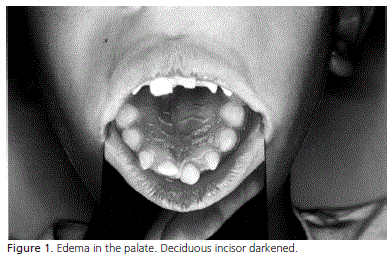
<body><![CDATA[<p><font size="2" face="Verdana, Arial, Helvetica, sans-serif">The technique of marsupialization, or Partsch's technique, consists of removing a window from the lesion and suturing the surrounding mucoperiosteum to </font><font size="2" face="Verdana, Arial, Helvetica, sans-serif">the margins of the cyst wall. The ensuing cavity is filled with gauze, which is removed after seven to ten days. If necessary, the gauze is changed during this period. This technique has previously been described for surgical treatment of ranulas and is also used for treating bone cysts. </font></p>     <p><font size="2" face="Verdana, Arial, Helvetica, sans-serif">This procedure aims to reduce the size of the cyst: opening the cyst eliminates its osmotic pressure and bone apposition gradually occurs at the site previously occupied by the epithelial covering of the cyst. These procedures can be used as a single treatment for a cyst or as preliminary treatment for subsequent enucleation. However, part of the cystic capsule will remain histopathologically unexamined for a longer time<sup>8</sup>. </font></p>     <p><font size="2" face="Verdana, Arial, Helvetica, sans-serif">Thomas<sup>9</sup> modified Partsch's method. In this modification, a small opening is made in the defect and a soft metal or polyethylene tube for drainage is inserted and fixed at the site by attaching it to an adjacent tooth. This technique was termed decompression.</font></p>     <p><font size="2" face="Verdana, Arial, Helvetica, sans-serif"> Neaverth &amp; Burg<sup>10</sup> presented an alternative treatment in which a tube was inserted inside the cystic cavity. This tube was periodically reduced in length as the lesion decreased in size. Several types and sizes of radiopaque tubes were used, such as: angiographic catheter, percutaneous catheter, urethral catheter and umbilical arterial catheter. All cases were in the maxilla. Full excision was not performed to avoid postoperative sequelae, such as devitalization of adjacent teeth, patient apprehension and discomfort, loss of bone support and paresthesia.</font></p>     <p><font size="2" face="Verdana, Arial, Helvetica, sans-serif"> Kruger<sup>11</sup> presented a modified marsupialization technique. An acrylic button that can be crossed by a drill or a metal or plastic tube is used to keep the wound open, thereby allowing drainage and irrigation of the cystic cavity and keeping it clean. These devices are removed when radiographic examination shows that new bone has formed. </font></p>     <p><font size="2" face="Verdana, Arial, Helvetica, sans-serif">The indications for these procedures are as follows<sup>7,12</sup>: a) tissue damage - enucleation of a large cyst close to vital structures may cause damage such as oroantral or oronasal fistulas, devitalization of healthy teeth and nerve injuries (for example, of the inferior alveolar nerve); b) surgical access - when the access is very difficult, there may be recurrences; c) preservation of an impacted tooth - when an unerupted, impacted, permanent tooth is affected by, e.g., a dentigerous cyst, this procedure may guide the eruption; d) surgery extent - for debilitated, unhealthy or elderly patients, this is a less invasive and stressful procedure; e) cyst size - with very voluminous cysts, the risk of fracturing the mandible is always present. It may be better to first marsupialize or decompress the cyst, with subsequent enucleation once satisfactory bone neoformation has occurred.</font></p>     <p>&nbsp;</p>      <p><font size="3" face="Verdana, Arial, Helvetica, sans-serif"><b>CASE REPORT</b> </font></p>     <p><font size="2" face="Verdana, Arial, Helvetica, sans-serif">The patient J.A.S., a six-year-old white boy, came to the Ephata Ear, Nose and Throat Clinic complaining mainly of facial asymmetry, with displacement of the nasal cavity. Clinical examination showed an extensive lesion completely covered by a shiny skin (<a href="#fig01">Figure 1</a>) in the left side of the maxilla displacing the nasal cavity. The maxillary vestibular face was also expanded.</font></p>     <p><font size="2" face="Verdana, Arial, Helvetica, sans-serif"> An interdisciplinary course of treatment was indicated for this patient to preserve the impacted permanent tooth inside the lesion. </font></p>     ]]></body>
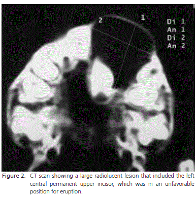
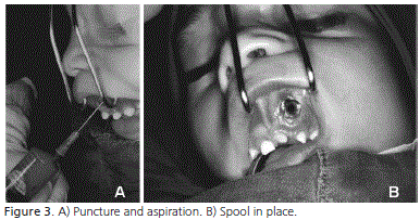
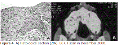
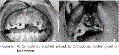
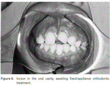
<body><![CDATA[<p><font size="2" face="Verdana, Arial, Helvetica, sans-serif">A panoramic radiograph showed an extensive radiolucent area that included the left central permanent upper incisor. The left central deciduous incisor had rotated in such a way that its apex was in contact with the confines of the lesion. A computed tomography (CT) scan was then taken to better evaluate the lesion and plan the surgery (<a href="#fig02">Figure 2</a>).</font></p>     <p><font size="2" face="Verdana, Arial, Helvetica, sans-serif"> A decompression surgery would be done first. The deciduous teeth were extracted and a polyethylene spool piece was inserted (<a href="#fig03">Figure 3</a>) with the patient under general anesthesia. The procedure was performed at the Ear, Nose and Throat Clinic of the Penido Burnier Institute, on February 25, 2000.</font></p>     <p><font size="2" face="Verdana, Arial, Helvetica, sans-serif"> An incisional biopsy was performed (Figure 3) and around 0.5 cm of oral mucosa was removed from the site where the lesion-decompression spool had been placed. Histopathological analysis of this specimen revealed a cyst covered with squamous epithelium histologically compatible with a maxillary dentigerous cyst (<a href="#fig04">Figure 4</a>). The spool was left in place for 15 days. The site was irrigated weekly with 0.2% chlorhexidine for six months, until August 2000.</font></p>     <p><font size="2" face="Verdana, Arial, Helvetica, sans-serif"> In August 2000, the fibrous gingiva that had been impeding the eruption of the right central upper incisor was removed. This was an outpatient procedure. A panoramic radiograph was requested and the regression of the lesion continued to be observed. </font></p>     <p><font size="2" face="Verdana, Arial, Helvetica, sans-serif">In December 2000, a new CT scan of the same area imaged by the baseline scan was done. It showed that the cystic lesion on the left side of the maxilla had completely regressed (Figure 4).</font></p>     <p><font size="2" face="Verdana, Arial, Helvetica, sans-serif"> In February 2001, panoramic and periapical radiographs were taken once more. Follow-up visits were done at every six months to ensure the space for the left central upper incisor to erupt. </font></p>     <p><font size="2" face="Verdana, Arial, Helvetica, sans-serif">In January 2002, another CT scan was done. There were no changes since the previous CT scan in December 2000. Clinically, the right permanent tooth was in the correct position, the left lateral incisor presented little displacement and only the left central permanent incisor was still missing.</font></p>     <p><font size="2" face="Verdana, Arial, Helvetica, sans-serif"> In December 2002, after orthodontic leveling and recovery of a small amount of space (<a href="#fig05">Figure 5</a>), a second operation under local anesthesia was performed for disimpacting the tooth, gluing a button and performing surgical exposure and orthodontic traction. The aim was to pull the impacted tooth into the occlusal plane (Figure 5).</font></p>     <p><font size="2" face="Verdana, Arial, Helvetica, sans-serif"> By October 2003, the patient's incisor had been rotated in the oral cavity. He presented mixed dentition and was awaiting the implementation of fixed-appliance corrective orthodontic treatment (<a href="#fig06">Figure 6</a>). </font></p>     <p><font size="2" face="Verdana, Arial, Helvetica, sans-serif">Case report: patient J.A.S., a four-year-old white boy.</font></p>     ]]></body>
<body><![CDATA[<p>&nbsp;</p>     <p><a name="fig01"></a></p>     <p>&nbsp; </p>     <p align="center"><img src="/img/revistas/rgo/v59n2/a19fig01.jpg">     <p>&nbsp;</p>     <p><a name="fig02"></a></p>     <p>&nbsp; </p>     <p align="center"><img src="/img/revistas/rgo/v59n2/a19fig02.jpg">     <p>&nbsp;</p>     <p><a name="fig03"></a></p>     ]]></body>
<body><![CDATA[<p>&nbsp; </p>     <p align="center"><img src="/img/revistas/rgo/v59n2/a19fig03.jpg">     <p>&nbsp;</p>     <p><a name="fig04"></a></p>     <p>&nbsp; </p>     <p align="center"><img src="/img/revistas/rgo/v59n2/a19fig04.jpg">     <p>&nbsp;</p>     <p><a name="fig05"></a></p>     <p>&nbsp; </p>     <p align="center"><img src="/img/revistas/rgo/v59n2/a19fig05.jpg">     ]]></body>
<body><![CDATA[<p>&nbsp;</p>     <p><a name="fig06"></a></p>     <p>&nbsp; </p>     <p align="center"><img src="/img/revistas/rgo/v59n2/a19fig06.jpg">     <p>&nbsp;</p> <font size="3" face="Verdana, Arial, Helvetica, sans-serif"><b>DISCUSSION</b> </font></p>      <p><font size="2" face="Verdana, Arial, Helvetica, sans-serif">Dentigerous cysts are the most common type of maxillary cysts in the first decade of life<sup>2</sup>. The latter author reported 17 cases in the first decade of life: six cases </font></p>     <p><font size="2" face="Verdana, Arial, Helvetica, sans-serif">involving the first lower premolar; four cases involving the second lower premolar; two cases involving the first lower molar; and one case of each of the central upper incisor, lateral upper incisor, upper canine, first upper premolar and second upper premolar. Nonetheless, only four cases of these lesions, associated with deciduous teeth, had been reported until 1992<sup>13</sup>. </font></p>     <p><font size="2" face="Verdana, Arial, Helvetica, sans-serif">In the present case, the central deciduous upper incisor appeared darkened. This may have been caused by trauma and a subsequent lack of any specific dental treatment. The necrosis seen on this tooth and its progression may have been the cause of the lesion, with the tooth retained inside the lesion.</font></p>     <p><font size="2" face="Verdana, Arial, Helvetica, sans-serif"> Aldama<sup>14</sup>believed that the treatment for such cases should be radical and stated that the prognosis was good when the cystic capsule was fully removed. Kruger<sup>11</sup> warned that development cysts could undergo histological transformations. Therefore, specimens need to be carefully examined after surgery.</font></p>     <p><font size="2" face="Verdana, Arial, Helvetica, sans-serif"> Archer<sup>12</sup>advocated the technique of marsupialization in order to prevent the loss of the teeth involved, along with other complications that result in socalled dental mutilation. This author presented statistical data to support his argument: few benign or malignant tumors develop in cystic coverings that are thus maintained. </font></p>     ]]></body>
<body><![CDATA[<p><font size="2" face="Verdana, Arial, Helvetica, sans-serif">The decompression technique is used prior to definitive excision in cases of large cysts. Olson et al.<sup>15</sup> reported excellent results regarding a case of a large odontogenic keratocyst when Partsch's technique was used, delaying enucleation. No recurrence was observed during a 4-year follow-up. </font></p>     <p><font size="2" face="Verdana, Arial, Helvetica, sans-serif">Sverzut et al.<sup>16</sup> described a clinical case of mandibular dentigerous cyst. Treatment was done in two stages: firstly decompression and then enucleation 142 days later. Thirteen months later, the area was clinically and radiographically normal. </font></p>     <p><font size="2" face="Verdana, Arial, Helvetica, sans-serif">Kronfeld &amp; Boyle<sup>17</sup> recommended extraction of an unerupted tooth if normal eruption was considered impossible. On the other hand, they recommended that a tooth in a favorable position should be allowed to erupt unaided once the cystic liquid had been eliminated. </font></p>     <p><font size="2" face="Verdana, Arial, Helvetica, sans-serif">Shear<sup>2</sup> believed that orthodontic care should always be available. However, Vieira et al.<sup>18</sup> presented the case of an eight-year-old patient who underwent marsupialization of the lesion and maintenance of the length of the dental arch. This allowed the associated permanent teeth to erupt without orthodontic traction. </font></p>     <p><font size="2" face="Verdana, Arial, Helvetica, sans-serif">Neaverth &amp; Burg<sup>10</sup> published four cases of decompression with plastic tubes in the anterior region of the maxilla, with excellent results. In these four cases, the tubes were removed after four weeks, five weeks, four months and one year. </font></p>     <p><font size="2" face="Verdana, Arial, Helvetica, sans-serif">The present clinical case corroborates the findings of previous authors who believed in the viability of the procedure. Despite the unfavorable position of the incisor, decompression was done in hopes that the impacted permanent incisor would be able to assume its place in the dental arch. Once this decision was made, regular clinical and radiographic follow-ups were necessary because dentigerous cysts may turn into keratocysts. However, according to Branon<sup>19</sup>, this is very unlikely. Pathological changes, such as degeneration to ameloblastomas, may occur and should be investigated in these cases by histopathology<sup>16</sup>.</font><font size="2" face="Verdana, Arial, Helvetica, sans-serif"></font></p>     <p>&nbsp;</p>  <font size="3" face="Verdana, Arial, Helvetica, sans-serif"><b>CONCLUSION</b> </font></p>      <p><font size="2" face="Verdana, Arial, Helvetica, sans-serif">Decompression resulted in significant reduction of cyst size, so subsequent enucleation was unnecessary. No remnants of the cyst were detected during the second operation, which was done to promote eruption. </font></p>     <p><font size="2" face="Verdana, Arial, Helvetica, sans-serif">This indicates that the lesion has completely regressed. This patient benefited greatly from the multidisciplinary cooperation between medical and dental experts. This approach should always be borne in mind.</font></p>     <p>&nbsp;</p> <font size="3" face="Verdana, Arial, Helvetica, sans-serif"><b>Colaborators</b> </font></p>      ]]></body>
<body><![CDATA[<p><font size="2" face="Verdana, Arial, Helvetica, sans-serif"> RG TEIXEIRA, PC MORAES, CRP JODAS, MJS BÖNECKER and DP TACCHELLI, were responsible for the development of all the stages of this article. </font></p>     <p>&nbsp;</p> <font size="3" face="Verdana, Arial, Helvetica, sans-serif"><b>REFER&Ecirc;NCIAS</b> </font></p>      <!-- ref --><p><font size="2" face="Verdana, Arial, Helvetica, sans-serif">1. Shafer WG, Hine MK, Levv BM. Cistos e tumores de origem odontog&ecirc;nica. In: Shafer WG, Hine MK, Levv BM. Tratado de patologia bucal. 4&ordf; ed. Rio de Janeiro: Interamericana, 1985. p. 239-270.    &nbsp;&nbsp;&nbsp;&nbsp;&nbsp;&nbsp;&nbsp;&nbsp;[&#160;<a href="javascript:void(0);" onclick="javascript: window.open('/scielo.php?script=sci_nlinks&ref=218939&pid=S1981-8637201100020001900001&lng=','','width=640,height=500,resizable=yes,scrollbars=1,menubar=yes,');">Links</a>&#160;]<!-- end-ref --></font></p>     <p><font size="2" face="Verdana, Arial, Helvetica, sans-serif"> 2. Shear M. Cisto dent&iacute;gero (folicular). In: Shear M. Cistos da regi&atilde;o bucomaxilofacial. S&atilde;o Paulo: Santos; 1989. p. 72- 96.</font><font size="2" face="Verdana, Arial, Helvetica, sans-serif"> </font></p>     <p><font size="2" face="Verdana, Arial, Helvetica, sans-serif">3. Regezi JA, Sciubba JJ. Cistos da boca. In: Regezi JA, Sciubba JJ. Patologia bucal. Rio de Janeiro: Guanabara Koogan; 1991. p. 223-7. </font></p>     <p><font size="2" face="Verdana, Arial, Helvetica, sans-serif">4. Smith G. Two dentigerous cysts in the mandible of one patient: case report. Aust Dent J. 1996;41(5):291-3. </font></p>     <p><font size="2" face="Verdana, Arial, Helvetica, sans-serif">5. Carvalho PI, Kumagai LT, Cacavalle AC. Dentigerous cyst associated with an unerupted mandibular third molar. Dentomaxillofac Radiol. 1997;26(2):137.</font></p>     <p><font size="2" face="Verdana, Arial, Helvetica, sans-serif"> 6. Waldron CA. Cistos e tumores odontog&ecirc;nicos. In: Neville BW, Damm D, Bouquot J, Allen C. Patologia oral &amp; maxilo facial. Rio de Janeiro: Guanabara Koogan; 1998. p. 481-527. </font></p>     <p><font size="2" face="Verdana, Arial, Helvetica, sans-serif">7. Ellis E. Tratamento cir&uacute;rgico das les&otilde;es orais patol&oacute;gicas. In: Peterson LJ. Cirurgia oral e maxilo facial contempor&acirc;nea. Rio de Janeiro: Guanabara Koogan; 1996. p. 467-90. </font></p>     ]]></body>
<body><![CDATA[<p><font size="2" face="Verdana, Arial, Helvetica, sans-serif">8. Takase T, Wada M, Nagahama F, Yamazaki M. Treatment of large radicular cysts by modified marsupialization. J Nihon Univ Sch Dent. 1996;38(4):161-8.</font></p>     <p><font size="2" face="Verdana, Arial, Helvetica, sans-serif"> 9. Thomas E. Saving involved vital teeth by tube drainage. J Oral Surg. 1947;5(1):1-9. </font></p>     <p><font size="2" face="Verdana, Arial, Helvetica, sans-serif">10. Neaverth EJ, Burg HA. Decompression of large periapical cystic lesions. J Endod. 1982;8(4):175-82. </font></p>     <p><font size="2" face="Verdana, Arial, Helvetica, sans-serif">11. Kruger GO. Cistos dos ossos, tecidos moles e estruturas adjacentes da cavidade bucal. In: Hupp JR, Ellis E, Myron R. Cirurgia bucal e maxilo facial. Rio de Janeiro: Guanabara Koogan; 1984. p.188-93. </font></p>     <p><font size="2" face="Verdana, Arial, Helvetica, sans-serif">12. Archer W. Oral surgery. 4th ed. Philadelphia: W.B. Saunders Co.; 1966.</font></p>     <p><font size="2" face="Verdana, Arial, Helvetica, sans-serif"> 13. Kusukawa J, Irie K, Morimatsu M, Koyanagi S, Kameyama T. Dentigerous cyst associated with deciduous tooth. Oral Surg Oral Med Oral Pathol. 1992;73(4):415-8. </font></p>     <p><font size="2" face="Verdana, Arial, Helvetica, sans-serif">14. Aldama JS. Quiste dent&iacute;gero: presentacion de um caso poco frecuente. Rev Cub Estomatol. 1988;25(3):81-5. </font></p>     <p><font size="2" face="Verdana, Arial, Helvetica, sans-serif">15. Olson RE, Thomsen S, Li-Min L. Odontogenic and keratocysts treated by the Partsch operation and delayed enucleation: report of a case. J Am Dent Assoc. 1977;94(2):321-5. </font></p>     <p><font size="2" face="Verdana, Arial, Helvetica, sans-serif">16. Sverzut CE, Barros VMR, Brentegani LG, Gomes PP. Descompress&atilde;o e enuclea&ccedil;&atilde;o de cisto dent&iacute;gero mandibular. Rev Assoc Paul Cir Dent. 2003;57(4):299-302.</font></p>     <p><font size="2" face="Verdana, Arial, Helvetica, sans-serif">17. Kronfeld R, Boyle PE. Dentes inclusos: cistos dent&iacute;geros. In: Kronfeld R, Boyle PE. Histopatologia dos dentes. 3&ordf; ed. Rio de Janeiro: Cient&iacute;fica; 1955. p. 464-7. </font></p>     ]]></body>
<body><![CDATA[<p><font size="2" face="Verdana, Arial, Helvetica, sans-serif">18. Vieira AR, Modesto A, Soares VR. Tratamento cir&uacute;rgico conservador de cisto dent&iacute;gero. Rev Assoc Paul Cir Dent. 1995;49(5):380-3. </font></p>     <p><font size="2" face="Verdana, Arial, Helvetica, sans-serif">19. Brannon RB. The odontogenic keratocyst: a clinicopathologic study of 312 cases. Part 1. Clinical features. Oral Surg Oral Med Oral Pathol. 1976;42(1):54-72.</font></p>     <p>&nbsp;</p>     <p>&nbsp;</p>     <p><font size="2" face="Verdana, Arial, Helvetica, sans-serif"><a name="back"/></a><a href="#top"><img src="/img/revistas/rfo/v18n1/seta.jpg" border="0" align="absmiddle"/></a><b>Endere&ccedil;o para correspond&ecirc;ncia:</b>    <br>   </font><font size="2" face="Verdana, Arial, Helvetica, sans-serif">RG TEIXEIRA     <br>   Faculdade S&atilde;o Leopoldo Mandic,     <br>Curso de Odontologia.    <br> Rua Jos&eacute; Rocha Junqueira, 13, Swift,     <br>   13045-755, Campinas, SP, Brasil.   </font><font size="2" face="Verdana, Arial, Helvetica, sans-serif">    ]]></body>
<body><![CDATA[<br>   e-mail: </font><font size="2" face="Verdana, Arial, Helvetica, sans-serif"><a href="mailto:rgte@terra.com.br" target="_blank">rgte@terra.com.br</a></font></p>     <p><font face="Verdana, Arial, Helvetica, sans-serif" size="2"><b>Recebido: :14/4/2010    <br> Aceito: 9/10/2010</b></font></p>       <p>&nbsp;</p>      ]]></body>
<back>
<ref-list>
<ref id="B1">
<label>1</label><nlm-citation citation-type="book">
<person-group person-group-type="author">
<name>
<surname><![CDATA[Shafer]]></surname>
<given-names><![CDATA[WG]]></given-names>
</name>
<name>
<surname><![CDATA[Hine]]></surname>
<given-names><![CDATA[MK]]></given-names>
</name>
<name>
<surname><![CDATA[Levv]]></surname>
<given-names><![CDATA[BM]]></given-names>
</name>
</person-group>
<article-title xml:lang="pt"><![CDATA[Cistos e tumores de origem odontogênica]]></article-title>
<person-group person-group-type="editor">
<name>
<surname><![CDATA[Shafer]]></surname>
<given-names><![CDATA[WG]]></given-names>
</name>
<name>
<surname><![CDATA[Hine]]></surname>
<given-names><![CDATA[MK]]></given-names>
</name>
<name>
<surname><![CDATA[Levv]]></surname>
<given-names><![CDATA[BM]]></given-names>
</name>
</person-group>
<source><![CDATA[Tratado de patologia bucal]]></source>
<year>1985</year>
<edition>4ª ed</edition>
<page-range>239-270</page-range><publisher-loc><![CDATA[Rio de Janeiro ]]></publisher-loc>
<publisher-name><![CDATA[Interamericana]]></publisher-name>
</nlm-citation>
</ref>
</ref-list>
</back>
</article>