Effect of different types of coronal flaring on the circularity and area of mandibular first molar mesial root canals
Efeito de diferentes tipos de preparo cervical na circularidade e na área de canais mesiais dos primeiros molares inferiores
Etevaldo Matos MAIA FILHOI; Cláudia de Castro RIZZII; José Martins CARNEIRO NETOI; Rejane Christine de Sousa QUEIROZI; Erick Miranda SOUZAII
I Universidade Ceuma, Curso de Odontologia. Rua Josué Montello, 1, Renascença II, 65075120, Sao Luis, MA, Brasil ]]> II Instituto Florence de Ensino Superio, Curso de Odontologia. São Luís, MA, Brasil.
ABSTRACT
Objective
This study assessed the effect of coronal flaring with the Gates-Glidden (GG) bur, La Axxess (LA) bur, and SX file on the area, circularity, and residual dentin thickness in the furcal direction of mandibular first molar root canals.
Methods
The mesiolingual and mesiobuccal canals of twenty-one mandibular first molars were instrumented. The teeth were embedded in acrylic resin, and after coronal flaring, the roots were cut perpendicularly to their long axis 2 mm below the cementoenamel junction. The sections were photographed before and after flaring with GG bur no. 3, LA bur no. 1 and SX file. The area, circularity, and residual dentin thickness in the furcal direction were calculated by the software Image J.
Conclusion
Coronal flaring with the study instruments was safe. The SX file achieved the smallest area. Circularity and residual dentin thickness in the furcal direction were not affected by the different instruments. No instrument was capable of achieving a circularity greater than 0.5.
Indexing terms: Dental pulp cavity. Endodontics. Instrumentation.
RESUMO
Objetivo
Avaliar o efeito no preparo cervical da broca Gates-Glidden (GG), La Axxess (LA) e lima SX na área, circularidade e na espessura residual dentinária voltada para a região de furca de canais radiculares dos primeiros molares inferiores.
Métodos ]]> Foram utilizados canais mésio-vestibulares e os mésio-linguais de vinte e um primeiros molares inferiores. Os dentes foram incluídos em resina acrílica e após a abertura coronária foram seccionados 2mm abaixo da junção amelo-cementária. As secções foram fotografadas antes e após o preparo cervical usando GG n. 3, LA n. 1 e SX. A área, a circularidade e o remanescente de dentina voltada para a furca foram calculados com auxílio do programa computacional Image J.
Resultados
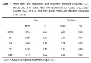
Antes do preparo cervical os canais apresentaram 0,56mm2 (±0,37) para área e 0,31 (±0,08) para circularidade. Após o uso da GG, SX e LA os valores médios para área foram 0,68 mm2 (±0,20), 0,64 mm2 (±0,18) e 0,85 mm2 (±0,23) e para circularidade de 0,39 (±0,08), 0,35 (±0,08) e 0,39 (±0,06), respectivamente. Houve diferença estatística significante entre a área para LA e SX (p<0,05). Não houve diferença para à circularidade (p>0,05) e nem para à espessura residual de dentina da furca (p>0,05).
Conclusão
O preparo cervical realizado com diferentes instrumentos apresentou adequada segurança sendo que a lima SX apresentou os menores valores de área. A circularidade e a quantidade de dentina residual na região de furca não foram influenciadas pelos diferentes instrumentos. Nenhum instrumento resultou em valores de circularidade maior que 0.5.
Termos de indexação: Cavidade pulpar. Endodontia. Instrumentação.
INTRODUCTION
Proper cleaning and shaping of the root canal requires free and direct access to the apical region. Coronalapical instrumentation techniques1 enable early flaring of the coronal third for subsequent access to the apical region. Consequently, inappropriate flaring of the coronal third will compromise access to the apical third, and in curved roots, such as the mesial roots of mandibular molars and buccal roots of maxillary molars, access is harder.
]]> Anticurvature filing aims to rectify the curvature of the coronal and middle thirds to facilitate access to the apical third2, increasing the efficacy of irrigants and sealing quality3, and reducing the incidence of instrument fracture4. Additionally, this maneuver must preserve the risk zone and make the canal as circular as possible without perforating the furcation.Because of their low price and high efficiency, Gates-Glidden (GG) burs have long been the instrument of choice for coronal flaring, and because of their shape, they tend to flare the canal uniformly, instead of conically5.
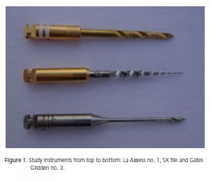
La Axxess burs, manufactured by Sybron Endo (USA), are made in sizes 20, 35, and 45, with a taper of 0.06. Studies with La Axxess burs have shown that when these burs are used for coronal flaring, they achieve better amplification of the apical third3.
The ProTaper (Dentsply Maillefer, Ballaigues, Switzerland) instrumentation system incorporates in its series of nickel-titanium instruments, the file SX, whose objective is to flare the coronal portion of the root canal with subsequent shaping of the apical region. However, the ability of these instruments to preserve furcal integrity is still questioned, and there are no studies in the literature that assess root canal circularity after the use of these instruments for coronal flaring.
The objective of this study was to assess the effect of coronal flaring with the Gates-Glidden bur number 3, La Axxess bur number 1, and SX file on the area, circularity, and residual dentin thickness in the furcal direction of the mesial roots of mandibular first molars.
METHODS
The present study was approved by the Research Ethics Committee of the University Center of Maranhão under protocol number 00781/11. Twenty-one human mandibular first molars were selected for instrumentation of the mesiolingual and mesiobuccal roots. The teeth were cleaned by leaving them in an aqueous solution of 5% NaOCl for 24 hours. The periodontal tissue and calculi were carefully removed, and the teeth were rinsed with tap water, dried, and stored in a 10% formalin solution.
The mesial roots were explored with K-files 08 and 10 without flaring. Patency was determined by a number 10 file (Dentsply Maillefer, Ballaigues, Switzerland).
Two preliminary radiographs of each mesial root were taken with a number 10 file inside the root canal, one in the buccolingual sense and the other in the mesiodistal sense. Radiographic exposure time and development technique were standardized. The radiographs were analyzed in a dark room by an x-ray illuminator and a 3.5x hand magnifier. Only teeth with curvatures of 10°-35° (Schneider6 method) were used.
]]> Once the cementoenamel junction was identified, the teeth were embedded in acrylic resin using a stainless steel muffle furnace similar to that described by Kuttler et al.7. The long axis of the tooth was positioned perpendicular to the horizontal plane. After the resin cured, the teeth were allowed to rest at room temperature for 48 hours. The tooth was opened with the cylindrical bur 1016 (KG Sorensen®, São Paulo, Brazil) under water irrigation, complemented with the Endo-Z bur (Dentsply Maillefer, Ballaigues, Switzerland).A low speed diamond saw (Extec® Labcut 1010, EXTEC Corp., Enfield, USA) was used for cutting the blocks perpendicularly to the long axis of the tooth, 2 mm below the cementoenamel junction.
The sections were photographed a digital camera (PowerShot G12, Canon, Japan) with a resolution of seven megapixels coupled with a surgical microscope (Opto, São Carlos, Brazil) with 20x magnification. The captured images, saved in the jpeg format, were digitally processed by the software Adobe Photoshop CS3 (Adobe System Incorporated, USA) to delimit the canal area before the procedures. The software Image J (http://rsbweb.nih. gov/ij/) was then used for calculating the area of the root canals, the circularity, and the smallest distance between each canal and the furcation.
After each tooth was sectioned and photographed, it was embedded again in the muffle furnace and the patency was tested with a number 10 K-File to probe for obstructions caused by the sectioning.
An experienced operator and equally experienced with the instruments flared the coronal third of all samples. The techniques were performed in a preestablished sequence, alternating the instruments so that each technique was used on 14 canals, that is, 7 mesiolingual and 7 mesiobuccal canals, totaling 42 canals.
The instruments were used in a 1:1 handpiece (Kavo, Rio de Janeiro, Brazil) together with an endodontic electric motor (Endo Pro Torque - Driller, USA). The Gates- Glidden and La Axxess burs were used at a speed of 6000rpm, while the SX files were used at 300rpm. When the La Axxess bur was used, pressure was made toward the safety area, as recommended by the manufacturer.
After flaring, the canals were irrigated with 1% sodium hypochlorite using a syringe with a 31 gauge needle (Ultradent Inc., South Jordan, UT, USA). After flaring, the sections were removed from the muffle furnace and photographed again, with subsequent calculation of the root canal areas, circularities, and residual dentin thicknesses in the furcal direction.
The increase in area and circularity was calculated by subtracting the area and circularity before flaring from those after flaring.
The data were tabulated and treated by the software SPSS version 17.00 (SPSS Inc. Chicago, IL, USA) with a significance level of 5%.
To verify whether the areas and circularities of the groups differed, the data were first tested for normality and homogeneity. The hypotheses of normality (Shapiro- Wilk test) and homogeneity (Levene test) were supported (p>0.05). One-way analysis of variance (one-way ANOVA) was then used for determining whether the areas, circularities, and residual dentin thicknesses in the furcal direction differed between the groups.
]]>
RESULTS
Table 1 shows the mean areas and circularities and their standard deviations before and after coronal flaring with the Gates-Glidden, SX, and La Axxess instruments.
One-way ANOVA was used for determining whether the mean areas and circularities of the groups differed significantly. A significant difference was found between two of the areas (F=4.061; p=0.025), but not between the circularities (F=1.156; p=0.325). The Tukey test confirmed that the areas obtained with the La Axxess and SX instruments differed significantly (p<0.05).
]]>
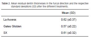
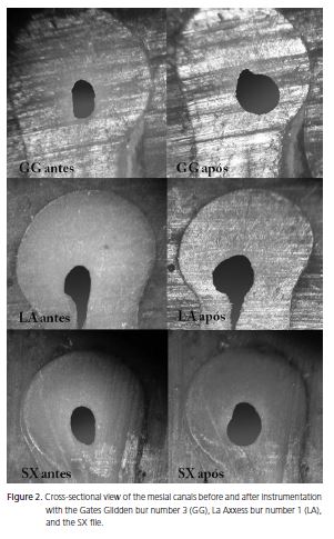
The mean residual dentin thicknesses in the furcal direction and the respective standard deviations are shown in Table 2. There was no significant difference between the treatments (p>0.05). Figure 2 shows the comparison between the groups.
]]>
DISCUSSION
Biomechanical flaring should be able to clean and shape the root canal properly, giving it a conic shape and circularity that facilitate filling. Coronal flaring is essential, since it provides direct access to the apical third, ensures enough room for the instruments, and reduces the risk of fractures4. This study is the first to assess the effect of coronal flaring with different instruments not only on canal area and but also on canal circularity.
Many methods have been employed to assess the effect of flaring on the shape of a root canal and residual dentin thickness, such as resin blocks8, radiographs9, scanning electron microscopy10, and computed tomography11-12. The methods used in the present study followed those proposed by Bramante et al.13 and modified by Kuttler et al.7, which have been used by many studies and enable the comparison of the thickness before and after coronal flaring5,14-17.
According to Bower18, the furcal region in the mandibular molars is two millimeters below the cementoenamel junction. Based on this information, the teeth were sectioned at that height. A similar approach was made by Wu et al.5.
The mean baseline area was 0.56mm, with a standard deviation of 0.37mm. This high standard deviation reflects the great anatomical diversity of root canals. Great area variability was also reported by Coutinho-Filho et al.19. The standard deviation fell to approximately half of the baseline value after coronal flaring, showing that flaring tends to homogenize the area, regardless of instrument.
The study results showed that the bur La Axxess 20.06, when used as recommended by the manufacturer, that is, by applying pressure towards the safety zone, resulted in a greater area without reducing the residual dentin thickness in the furcal direction, and resulted in a circularity similar to those of the other groups.
The SX file was used at a speed of 300rpm while the burs GG and La Axxess were used at 6000rpm. This may explain the smaller dentin removal by the SX file. Additionally, the taper of the SX file varies from 0.035 to 0.190mm/mm and it is superelastic20, making flaring safe without danger of transportation.
]]> The mean circularity before coronal flaring was 0.31±0.08, and after, 0.38±0.08. This shows that, although the area of the canals varied greatly, circularity varied little. Given that circularity varies from 0 (straight) to 1 (perfect circle), the numbers show that canal circularity improved moderately after flaring. The burs La Axxess and Gates Glidden achieved greater circularities, but the differences were not significant.The Gates-Glidden bur number 3 is cylindrical and has a diameter of 0.90mm. Wu et al.5 showed that these burs can cause stripping and the use of anti-curvature motion did not reduce their potential for perforation. However, the present study results showed that the areas, circularities, and residual dentin thicknesses in the furcal direction achieved by the Gates Glidden bur did not differ significantly from those achieved by the other instruments.
The circularities of the groups were similar, but no instrument was capable of achieving a circularity greater than 0.5. In other words, the study instruments were incapable of impressing their shape on the canal.
CONCLUSION
Coronal flaring with different instruments proved to be safe. The SX file achieved the smallest areas. The circularities and residual dentin thicknesses in the furcal direction achieved by different instruments were not significantly different. None of the study instruments achieved a root canal circularity greater than 0.5.
Collaborators
EM MAIA FILHO participated in all the confection stages of the study. CC RIZZI helped with the experimental procedures and manuscript writing. JM CARNEIRO NETO helped to review the references, followed the procedures, and wrote the manuscript. RCS QUEIROZ performed the statistical analyses. EM SOUZA helped to write the manuscript.
REFERENCES
1. Buchanan LS. The standardized-taper root canal preparation, Part 1: Concepts for variably tapered shaping instruments. Dent Today. 1998;17(5):54-60. doi: 10.1046/j.1365-2591.2000.00384.x. [ Links ]
]]> 2. Abou-Rass M, Frank AL, Glick DH. The anticurvature filing method to prepare the curved root canal. J Am Dent Assoc. 1980;101(5):792-4.3. Barroso JM, Guerisoli DM, Capelli A, Saquy PC, Pecora JD. Influence of cervical preflaring on determination of apical file size in maxillary premolars: SEM analysis. Braz Dent J. 2005;16(1):30- 4.
4. Ehrhardt IC, Zuolo ML, Cunha RS, De Martin AS, Kherlakian D, de Carvalho MC, et al. Assessment of the separation incidence of Mtwo files used with preflaring: prospective clinical study. J Endod. 2012;38(8):1078-81. doi: doi: 10.1016/j.joen.2012.05.001.
5. Wu MK, van der Sluis LW, Wesselink PR. The risk of furcal perforation in mandibular molars using Gates-Glidden drills with anticurvature pressure. Oral Surg Oral Med Oral Pathol Oral Radiol Endod. 2005;99(3):378-82. doi: 10.1016/j.tripleo.2004.07.008.
6. Schneider SW. A comparison of canal preparations in straight and curved root canals. Oral Surg Oral Med Oral Pathol. 1971;32(2):271-5. doi: 10.1016/0030-4220(71)90230-1.
7. Kuttler S, Garala M, Perez R, Dorn SO. The endodontic cube: a system designed for evaluation of root canal anatomy and canal preparation. J Endod. 2001;27(8):533-6. doi: 10.1097/00004770- 200108000-00008.
8. Weine FS, Kelly RF, Lio PJ. The effect of preparation procedures on original canal shape and on apical foramen shape. J Endod. 1975;1(8):255-62. doi: 10.1016/S0099-2399(75)80037-9.
9. Southard DW, Oswald RJ, Natkin E. Instrumentation of curved molar root canals with the Roane technique. J Endod. 1987;13(10):479-89. doi: 10.1016/S0099-2399(87)80015-8.
10. Mizrahi SJ, Tucker JW, Seltzer S. A scanning electron microscopic study of the efficacy of various endodontic instruments. J Endod. 1975;1(10):324-33. doi: 10.1016/S0099-2399(75)80012-4.
11. Sanfelice CM, da Costa FB, Reis So MV, Vier-Pelisser F, Souza Bier CA, Grecca FS. Effects of four instruments on coronal preenlargement by using cone beam computed tomography. J Endod. 2010;36(5):858-61. doi: doi: 10.1016/j.joen.2009.12.003.
]]> 12. Elayouti A, Dima E, Judenhofer MS, Lost C, Pichler BJ. Increased apical enlargement contributes to excessive dentin removal in curved root canals: a stepwise microcomputed tomography study. J Endod. 2011;37(11):1580-4. doi: doi: 10.1016/j. joen.2011.08.019.13. Bramante CM, Berbert A, Borges RP. A methodology for evaluation of root canal instrumentation. J Endod. 1987;13(5):243-5. doi: doi:10.1016/S0099-2399(87)80099-7.
14. Pilo R, Corcino G, Tamse A. Residual dentin thickness in mandibular premolars prepared with hand and rotatory instruments. J Endod. 1998;24(6):401-4.
15. Pilo R, Shapenco E, Lewinstein I. Residual dentin thickness in bifurcated maxillary first premolars after root canal and post space preparation with parallel-sided drills. J Prosthet Dent. 2008;99(4):267-73. doi: 10.1016/S0022-3913(08)60059-1.
16. Pilo R, Tamse A. Residual dentin thickness in mandibular premolars prepared with Gates Glidden and ParaPost drills. J Prosthet Dent. 2000; 83(6): 617-23.
17. Souza EM, do Nascimento LM, Maia Filho EM, Alves CM. The impact of post preparation on the residual dentin thickness of maxillary molars. J Prosthet Dent. 2011;106(3):184-90. doi: 10.1016/S0022-3913(11)60119-4.
18. Bower RC. Furcation morphology relative to periodontal treatment. Furcation entrance architecture. J Periodontol. 1979;50(1):23-7. doi: doi:10.1902/jop.1979.50.1.23.
19. Coutinho-Filho T, De-Deus G, Gurgel-Filho ED, Rocha-Lima AC, Dias KR, Barbosa CA. Evaluation of the risk of a stripping perforation with Gates-Glidden drills: serial versus crown-down sequences. Braz Oral Res. 2008; 22(1): 18-24.
20. Walia HM, Brantley WA, Gerstein H. An initial investigation of the bending and torsional properties of Nitinol root canal files. J Endod. 1988; 14(7): 346-51.
]]>
 Endereço para correspondência:
EM MAIA FILHO
e-mail: rizzimaia@yahoo.com.br
Recebido: 30/7/2012
Final version resubmitted on: 16/10/2012
Aceito: 30/11/2012
]]>