<?xml version="1.0" encoding="ISO-8859-1"?><article xmlns:mml="http://www.w3.org/1998/Math/MathML" xmlns:xlink="http://www.w3.org/1999/xlink" xmlns:xsi="http://www.w3.org/2001/XMLSchema-instance">
<front>
<journal-meta>
<journal-id>1981-8637</journal-id>
<journal-title><![CDATA[RGO.Revista Gaúcha de Odontologia (Online)]]></journal-title>
<abbrev-journal-title><![CDATA[RGO, Rev. gaúch. odontol. (Online)]]></abbrev-journal-title>
<issn>1981-8637</issn>
<publisher>
<publisher-name><![CDATA[Mundi Brasil Gráfica e Editora Ltda.]]></publisher-name>
</publisher>
</journal-meta>
<article-meta>
<article-id>S1981-86372017000100014</article-id>
<title-group>
<article-title xml:lang="en"><![CDATA[Adenomatoid odontogenic tumor simulating periapical cyst: case report]]></article-title>
<article-title xml:lang="pt"><![CDATA[Tumor odontogênico adenomatóide simulando cisto periapical: relato de caso clínico]]></article-title>
</title-group>
<contrib-group>
<contrib contrib-type="author">
<name>
<surname><![CDATA[MAIA]]></surname>
<given-names><![CDATA[Francisco Paulo Araújo]]></given-names>
</name>
<xref ref-type="aff" rid="A01"/>
</contrib>
<contrib contrib-type="author">
<name>
<surname><![CDATA[PINTO]]></surname>
<given-names><![CDATA[Priscilla Sarmento]]></given-names>
</name>
<xref ref-type="aff" rid="A02"/>
</contrib>
<contrib contrib-type="author">
<name>
<surname><![CDATA[LUNA]]></surname>
<given-names><![CDATA[Anibal Henrique Barbosa]]></given-names>
</name>
<xref ref-type="aff" rid="A02"/>
</contrib>
</contrib-group>
<aff id="A01">
<institution><![CDATA[,Universidade Federal da Paraíba Hospital Universitário Lauro Wanderley ]]></institution>
<addr-line><![CDATA[João Pessoa PB]]></addr-line>
<country>Brasil</country>
</aff>
<aff id="A02">
<institution><![CDATA[,Universidade Federal da Paraíba Faculdade de Odontologia ]]></institution>
<addr-line><![CDATA[João Pessoa PB]]></addr-line>
<country>Brasil</country>
</aff>
<pub-date pub-type="pub">
<day>00</day>
<month>03</month>
<year>2017</year>
</pub-date>
<pub-date pub-type="epub">
<day>00</day>
<month>03</month>
<year>2017</year>
</pub-date>
<volume>65</volume>
<numero>1</numero>
<fpage>92</fpage>
<lpage>99</lpage>
<copyright-statement/>
<copyright-year/>
<self-uri xlink:href="http://revodonto.bvsalud.org/scielo.php?script=sci_arttext&amp;pid=S1981-86372017000100014&amp;lng=en&amp;nrm=iso"></self-uri><self-uri xlink:href="http://revodonto.bvsalud.org/scielo.php?script=sci_abstract&amp;pid=S1981-86372017000100014&amp;lng=en&amp;nrm=iso"></self-uri><self-uri xlink:href="http://revodonto.bvsalud.org/scielo.php?script=sci_pdf&amp;pid=S1981-86372017000100014&amp;lng=en&amp;nrm=iso"></self-uri><abstract abstract-type="short" xml:lang="en"><p><![CDATA[The adenomatoid odontogenic tumor is a benign, non-invasive tumor and has a slow growth. Its preferred location is the anterior maxillary area, most often causing root displacement, more rarely resorption and mostly affects young women. Being asymptomatic, this injury is discovered upon routine radiographic examination, in which, in most cases, a unilocular, radiolucent image is observed and it may have some degree of calcification within the lesion. This work aims to report the case of a 14-year-old patient who attended the Lauro Wanderley University Hospital (UFPB), complaining of increased volume over a period of 8 months, in the region of non-vital tooth 21. The CT scan showed well-delimited unilocular, radiolucent lesion, with buccal cortex resorption. The proposed treatment plan involved enucleation, followed by peripheral ostectomy and bone graft with alloplastic materials. Because of its benign character, encapsulated and slow growing, the treatment of choice for adenomatoid odontogenic tumor is conservative, the surgical enucleation of the injury being advocated. The excised piece was sent for histopathological analysis, where the AOT diagnosis was confirmed. After 7 months, new bone formation was observed without signs of recurrence.]]></p></abstract>
<abstract abstract-type="short" xml:lang="pt"><p><![CDATA[O Tumor Odontogênico Adenomatóide é um tumor benigno, não invasivo e de crescimento lento. Acomete principalmente mulheres jovens, na região anterior da maxila, causando na maioria das vezes deslocamento radicular e raramente reabsorção. Por ser assintomática, essa lesão é geralmente descoberta em exames radiográficos de rotina, no qual se observa na maioria dos casos uma imagem radiolúcida unilocular, podendo apresentar algum grau de calcificação no interior da lesão. Este trabalho tem como objetivo relatar o caso clínico de um paciente de 14 anos, gênero feminino, que compareceu ao Hospital Universitário Lauro Wanderley - Universidade Federal da Paraíba, com queixa de aumento de volume de 8 meses de evolução, em região de elemento 21 não vital. Ao exame tomográfico observou-se lesão radiolúcida, unilocular, bem delimitada, com reabsorção da cortical vestibular. O plano de tratamento proposto foi a enucleação seguida de ostectomia periférica e enxerto ósseo com material aloplásico. A análise histopatológica confirmou o diagnóstico de Tumor Odontogênico Adenomatóide. Após sete meses observa-se nova formação óssea e sem sinais de recidiva.]]></p></abstract>
<kwd-group>
<kwd lng="en"><![CDATA[Maxilla.]]></kwd>
<kwd lng="en"><![CDATA[Oral pathology.]]></kwd>
<kwd lng="en"><![CDATA[Radicular cyst.]]></kwd>
<kwd lng="pt"><![CDATA[Maxila.]]></kwd>
<kwd lng="pt"><![CDATA[Patologia bucal.]]></kwd>
<kwd lng="pt"><![CDATA[Cisto radicular.]]></kwd>
</kwd-group>
</article-meta>
</front><body><![CDATA[ <p align="right"><font size="2" face="Verdana, Arial, Helvetica, sans-serif"><b>CL&Iacute;NICO</b> / CLINICAL</font></p>     <p>&nbsp;</p>     <p><font size="4" face="Verdana, Arial, Helvetica, sans-serif"><a name="top"/></a><B>Adenomatoid odontogenic tumor simulating periapical cyst: case report</B></font></p>     <p>&nbsp;</p>     <p><font size="3" face="Verdana, Arial, Helvetica, sans-serif"><b>Tumor odontog&ecirc;nico adenomat&oacute;ide simulando cisto periapical: relato de caso cl&iacute;nico</b></font></p>     <p>&nbsp;</p>     <p><font size="2" face="Verdana, Arial, Helvetica, sans-serif"><b>Francisco Paulo Ara&uacute;jo MAIA<sup>I</sup></b>;   <b>Priscilla Sarmento PINTO<sup>II</sup></b>; <b>Anibal Henrique Barbosa LUNA<sup>II</sup></b></font></p>     <p>&nbsp;</p>     <p><font size="2" face="Verdana, Arial, Helvetica, sans-serif"><sup>I</sup> Universidade Federal da Para&iacute;ba, Hospital Universit&aacute;rio Lauro Wanderley. Cidade Universit&aacute;ria, s/n, Campus I, Cidade Universit&aacute;ria, 58059-900, Jo&atilde;o Pessoa, PB, Brasil</font>    <br> <font size="2" face="Verdana, Arial, Helvetica, sans-serif"><sup>II</sup> </font><font size="2" face="Verdana, Arial, Helvetica, sans-serif">Universidade Federal da Para&iacute;ba, Faculdade de Odontologia. Jo&atilde;o Pessoa, PB, Brasil</font>    ]]></body>
<body><![CDATA[<br> </p>     <br>     <p><font size="2" face="Verdana, Arial, Helvetica, sans-serif"><a href="#back">Correspondence:</a></font></p>     <p>&nbsp;</p>     <p>&nbsp;</p> <hr size="1" noshade>     <p><font size="3" face="Verdana, Arial, Helvetica, sans-serif"><b>ABSTRACT</b> </font></p>     <p><font size="2" face="Verdana, Arial, Helvetica, sans-serif">The adenomatoid odontogenic tumor is a benign, non-invasive tumor and has a slow growth. Its preferred location is the anterior maxillary area, most often causing root displacement, more rarely resorption and mostly affects young women. Being asymptomatic, this injury is discovered upon routine radiographic examination, in which, in most cases, a unilocular, radiolucent image is observed and it may have some degree of calcification within the lesion. This work aims to report the case of a 14-year-old patient who attended the Lauro Wanderley University Hospital (UFPB), complaining of increased volume over a period of 8 months, in the region of non-vital tooth 21. The CT scan showed well-delimited unilocular, radiolucent lesion, with buccal cortex resorption. The proposed treatment plan involved enucleation, followed by peripheral ostectomy and bone graft with alloplastic materials. Because of its benign character, encapsulated and slow growing, the treatment of choice for adenomatoid odontogenic tumor is conservative, the surgical enucleation of the injury being advocated. The excised piece was sent for histopathological analysis, where the AOT diagnosis was confirmed. After 7 months, new bone formation was observed without signs of recurrence.</font></p>     <p><font size="2" face="Verdana, Arial, Helvetica, sans-serif"><B>Indexing terms: </B>Maxilla. Oral pathology. Radicular cyst.</font></p> <hr size="1" noshade>     <p><font size="3" face="Verdana, Arial, Helvetica, sans-serif"><B>RESUMO</B></font></p>     <p><font size="2" face="Verdana, Arial, Helvetica, sans-serif">O Tumor Odontog&ecirc;nico Adenomat&oacute;ide &eacute; um tumor benigno, n&atilde;o invasivo e de crescimento lento. Acomete principalmente mulheres jovens, na regi&atilde;o anterior da maxila, causando na maioria das vezes deslocamento radicular e raramente reabsor&ccedil;&atilde;o. Por ser assintom&aacute;tica, essa les&atilde;o &eacute; geralmente descoberta em exames radiogr&aacute;ficos de rotina, no qual se observa na maioria dos casos uma imagem radiol&uacute;cida unilocular, podendo apresentar algum grau de calcifica&ccedil;&atilde;o no interior da les&atilde;o. Este trabalho tem como objetivo relatar o caso cl&iacute;nico de um paciente de 14 anos, g&ecirc;nero feminino, que compareceu ao Hospital Universit&aacute;rio Lauro Wanderley - Universidade Federal da Para&iacute;ba, com queixa de aumento de volume de 8 meses de evolu&ccedil;&atilde;o, em regi&atilde;o de elemento 21 n&atilde;o vital. Ao exame tomogr&aacute;fico observou-se les&atilde;o radiol&uacute;cida, unilocular, bem delimitada, com reabsor&ccedil;&atilde;o da cortical vestibular. O plano de tratamento proposto foi a enuclea&ccedil;&atilde;o seguida de ostectomia perif&eacute;rica e enxerto &oacute;sseo com material alopl&aacute;sico. A an&aacute;lise histopatol&oacute;gica confirmou o diagn&oacute;stico de Tumor Odontog&ecirc;nico Adenomat&oacute;ide. Ap&oacute;s sete meses observa-se nova forma&ccedil;&atilde;o &oacute;ssea e sem sinais de recidiva.</font><font size="2" face="Verdana, Arial, Helvetica, sans-serif"> </font></p>     ]]></body>
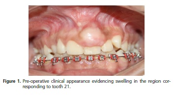
<body><![CDATA[<p><font size="2" face="Verdana, Arial, Helvetica, sans-serif"><B>Termos de indexa&ccedil;&atilde;o: </B>Maxila. Patologia bucal. Cisto radicular.</font></p> <hr noshade size="1">     <p>&nbsp;</p>     <p><font size="3" face="Verdana, Arial, Helvetica, sans-serif"><B> INTRODUCTION</B></font></p>     <p><font size="2" face="Verdana, Arial, Helvetica, sans-serif">The adenomatoid odontogenic tumor (AOT) is a not very common benign epithelial tumor, first described in 1907 by Dreibaldt as a pseudo-adenoameloblastoma<sup>1</sup>, classified as an odontogenic tumor by the World Health Organization (WHO) in 1971<sup>2</sup>. In 2005, the WHO defined AOT as being composed of a variety of histological patterns of odontogenic epithelium embedded in conjunctive tissue stroma characterized by its slow, progressive growth<sup>3</sup>. </font></p>     <p><font size="2" face="Verdana, Arial, Helvetica, sans-serif">The AOT is usually an asymptomatic lesion. When it takes on larger proportions, it presents clinically with increased volume and a firm consistency on palpation. Displacement of adjacent teeth due to the growth of the tumor is more common than root resorption<sup>4</sup>. </font></p>     <p><font size="2" face="Verdana, Arial, Helvetica, sans-serif">The adenomatoid odontogenic tumor is a slowgrowing, benign and non-invasive lesion frequently discovered during routine radiographic examination<sup>5</sup>. The AOT has three clinicopathological variants: intraosseous follicular (pericoronal), intraosseous follicular (extracoronal) and extraosseous (peripheral)6. Normally, the radiographic characteristics present radiolucent, singlechambered lesions enfolding the crown of impacted teeth, although erupted teeth may also be found associated with this lesion. </font></p>     <p><font size="2" face="Verdana, Arial, Helvetica, sans-serif">The aim of this study is to report a case of AOT involving an erupted tooth that, due to its size, was treated during the diagnostic procedure (excisional biopsy), performing enucleation with peripheral ostectomy and a bone graft for alveolar reconstruction.</font></p>     <p>&nbsp;</p>     <p><font size="3" face="Verdana, Arial, Helvetica, sans-serif"><B>CASE REPORT</B></font></p>     <p><font size="2" face="Verdana, Arial, Helvetica, sans-serif">White, 14-year-old female patient came into the Oral and Maxillofacial Surgery Service of the Lauro Wanderley University Hospital (UFPB), complaining of increased volume in the region of the maxilla. Her historical record of the current disease reported the discovery of the lesion associated with tooth 21, non-vital, without pain, having evolved over 8 months. Endodontic treatment had been performed previously. In the intraoral physical examination, an increased volume, firm to the touch, and gingival hyperplasia was found in the region relating to tooth 21 (<a href="#fig01">Figure 1</a>). </font></p>     ]]></body>
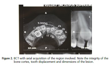
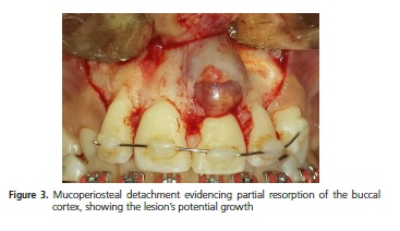
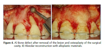
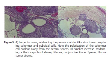
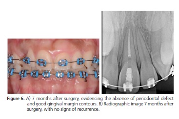
<body><![CDATA[<p><font size="2" face="Verdana, Arial, Helvetica, sans-serif">The patient had undergone a tomographic examination of the region under another professional, which revealed a radiolucent image with a radiopaque halo measuring approximately 9.3 mm x 1 cm at its largest diameter, causing partial resorption of the buccal cortex with displacement of adjacent teeth (<a href="#fig02">Figure 2</a>), and a preemptive orthodontic retainer was fitted to immobilize the teeth involved during the period of repair. An excisional biopsy was scheduled to clarify the diagnosis of the lesion and its definitive treatment. Aspiration biopsy revealed the absence of amber-colored liquid, thereby eliminating the possibility of it being a cystic lesion. The surgical treatment of choice was enucleation of the lesion by means of a trapezoid flap incision carried out in the region of elements 11, 12, 21 and 22, followed by mucoperiosteal detachment (<a href="#fig03">Figure 3</a>) and ostectomy using a no. 6 surgical spherical bur (Beavers Dental&reg;, Morrisburg, Canada) with copious irrigation in a 0.9% sterile saline solution, conducted under local anesthetic. Having removed the lesion, a peripheral ostectomy was performed using a spherical diamond bur (KG Sorensen&reg;, S&atilde;o Paulo, Brazil) (<a href="#fig04">Figure 4a</a>). Given the defect in the portion of buccal bone resulting from the enucleation of the lesion, a graft was performed using lyophilized cortical bone (CORTBONE&reg;, Jo&atilde;o Pessoa, Brazil) (<a href="#fig04">Figure 4b</a>), and the operated region was sutured using Vicryl 4-0 absorbable thread (ETHICON&reg;, S&atilde;o Paulo, Brazil). </font></p>     <p><font size="2" face="Verdana, Arial, Helvetica, sans-serif">The histological cuts were examined under microscope, dyed using hematoxylin and eosin, and fragments were found of benign neoplasia of odontogenic origin, characterized by the proliferation of ductlike structures with a nodular, cribiform pattern (<a href="#fig05">Figure 5a</a>), as well as the deposition of basophilic structures consistent with osteodentine, added to the fibrous conjunctive tissue capsule with loose collagen fibers (<a href="#fig05">Figure 5b</a>), with a slight inflammatory infiltrate and slight vascularization, confirming the AOT diagnosis.  </font></p>     <p><font size="2" face="Verdana, Arial, Helvetica, sans-serif">At the present time, the patient has completed 7 months since the surgery took place, noting the repair to the bone, normal probing depth with satisfactory gingival contours (<a href="#fig06">Figure 6a</a>), and absence of any sign of recurrence in the routine radiographic examination (<a href="#fig06">Figure 6b</a>).</font></p>     <p>&nbsp;</p>     <p><a name="fig01"></a></p>     <p>&nbsp; </p>     <p align="center"><img src="/img/revistas/rgo/v65n1/a14fig01.jpg">     <p>&nbsp;</p>     <p><a name="fig02"></a></p>     <p>&nbsp; </p>     ]]></body>
<body><![CDATA[<p align="center"><img src="/img/revistas/rgo/v65n1/a14fig02.jpg">     <p>&nbsp;</p>     <p><a name="fig03"></a></p>     <p>&nbsp; </p>     <p align="center"><img src="/img/revistas/rgo/v65n1/a14fig03.jpg">     <p>&nbsp;</p>     <p><a name="fig04"></a></p>     <p>&nbsp; </p>     <p align="center"><img src="/img/revistas/rgo/v65n1/a14fig04.jpg">     <p>&nbsp;</p>     ]]></body>
<body><![CDATA[<p><a name="fig05"></a></p>     <p>&nbsp; </p>     <p align="center"><img src="/img/revistas/rgo/v65n1/a14fig05.jpg">     <p>&nbsp;</p>     <p><a name="fig06"></a></p>     <p>&nbsp; </p>     <p align="center"><img src="/img/revistas/rgo/v65n1/a14fig06.jpg">     <p>&nbsp;</p>     <p><font size="3" face="Verdana, Arial, Helvetica, sans-serif"><B>DISCUSSION</B></font></p>     <p><font size="2" face="Verdana, Arial, Helvetica, sans-serif">AOT is a rare lesion, representing around 1.7% of all odontogenic tumors<sup5></sup>.It has a preference for the maxillary anterior region, in roughly two-thirds of cases afflicting the anterior portion of the maxilla. It is usually associated with impacted teeth, most commonly the canine<sup>7-8</sup>. It mainly affects females, in a ratio of 1.9:1, and this preference is even higher in the Asian population, the highest incidence being in Sri Lanka (3.2:1) and Japan (3:1)<sup>9</sup>. As with all alterations, AOT often causes expansion of the involved bone and the displacement of adjacent teeth<sup>10</sup>. </font></p>     ]]></body>
<body><![CDATA[<p><font size="2" face="Verdana, Arial, Helvetica, sans-serif">In a study conducted by Chindasombatjaroen et al. (2012), 75% of cases presented with root displacement and 12.5% with resorption. These findings show that AOT most frequently causes root displacement and more rarely, resorption, corroborating the present study. It is important to emphasize that in this case the crown of tooth 21 remained in place. Root displacement was only detected in the tomographic examination, a finding that may not have been made using conventional radiography. In a study conducted by Becker, which analyzed 272 cases of AOT between 1950 and 2010, follicular lesions represented 69% of all cases, extrafollicular 27% and the peripheral variant 5%, and in the current case the variant found was of the extrafollicular type. As far as the radiographic characteristics are concerned, in 91% of cases a unilocular, radiolucent lesion was observed while only 3% were multilocular, and 77% of the lesions exhibited some degree of calcification. Chindasombatjaroen et al. (2012), in a retrospective study carried out between 2000 and 2011, reported that 67% of the cases presented radiopaque areas inside the lesion, which is not consistent with that presented in the case described.</font></p>     <p><font size="2" face="Verdana, Arial, Helvetica, sans-serif"> The radiographic characteristics of AOT lead to a differential diagnosis with a variety of odontogenic lesions. The follicular variant is linked to the crown and often a part of the root of an impacted tooth, usually the upper canine and just occasionally the permanent molars. Accordingly it is often initially considered to be a dentigerous cyst based on a clinical and radiographic evaluation<sup>2</sup>. The keratocystic odontogenic tumor and the unicystic ameloblastoma are also similar to the follicular variant when located in the pericoronal region, however they are more common in the posterior region of the mandible, unlike AOT<sup>6</sup>, where it is more common in the anterior region of the maxilla. Given the endodontic history and being a small, incipient lesion not associated with an impacted tooth, the diagnostic hypothesis was a periapical cyst. Periapical cysts usually occur in non-vital teeth, presenting a certain degree of mobility as the lesion grows and with the displacement of adjacent teeth, associated with loss of lamina dura<sup>5</sup>. In a retrospective study conducted by Akinyamoju et al.<sup>11</sup>, the upper left central incisor was the tooth most affected in periapical lesions<sup>1</sup>, corroborating the diagnostic hypothesis of the present case. </font></p>     <p><font size="2" face="Verdana, Arial, Helvetica, sans-serif">The histological aspects of AOT, according to Neville et al.<sup>6</sup>, revealed a well-circumscribed lesion surrounded by thick, fibrous capsule, presenting with spindle-shaped epithelial cells forming structures similar to rosettes around a central area, whether or not filled with eosinophilic material. In a study carried out by Leon et al.<sup>5</sup> which analyzed the histopathological characteristics of 39 lesions diagnosed as AOT, they observed that all the lesions had different rosette parts composed of cuboidal-shaped, epithelial cells, containing eosinophilic material, many of the lesions exhibiting calcified material, corroborating the histopathological findings found in the case described. </font></p>     <p><font size="2" face="Verdana, Arial, Helvetica, sans-serif">The adenomatoid odontogenic tumor has a low rate of recurrence, surgical enucleation of the lesion being the treatment of choice<sup>12</sup>. This is explained by the fact that AOT is usually delimited by a capsule of well developed conjunctive tissue, and may be a solid mass, or have varying degrees of cystic degeneration. The presence of this capsule permits easy and complete removal, this being one of the factors that justifies conservative surgical treatment as the advocated treatment<sup>13</sup>. Nevertheless, the present case presented as a lesion that was small in size, indicating an excisional biopsy and leading to potential esthetic repercussions due to the periodontal defects. Moreover, not knowing the definitive diagnosis of the lesion, which presented with a perforation of the cortical bone and the need for osteoplasty to normalize the bone margins, a peripheral ostectomy was carried out to minimize the risk of possible recurrence. As this related to a diagnosis of AOT, a more conservative approach could be used without harm to the patient, as there is a low tendency towards recurrence. The enucleation of the lesion and the peripheral ostectomy resulted in an alveolar defect in the buccal region of tooth 21, indicating a graft for reconstruction and better esthetic and functional results. </font></p>     <p>&nbsp;</p>     <p><b><font size="3 " face="Verdana, Arial, Helvetica, sans-serif">CONCLUSION</font></b></p>     <p><font size="2" face="Verdana, Arial, Helvetica, sans-serif"> AOT is a slow-growing, benign lesion which is usually discovered during routine radiographic examination and, due to its fibrous capsule, may be easily enucleated from the bone, taking into account the best treatment option, recurrence being rare after enucleation. Control x-rays are performed to observe the new bone formation and the absence of recurrence.</font></p>     <p>&nbsp;</p>     <p><font size="3 " face="Verdana, Arial, Helvetica, sans-serif"><B>Collaborators</B></font></p>       <p><font size="2" face="Verdana, Arial, Helvetica, sans-serif">   FPA MAIA, case surgeon and manuscript author. PS PINTO, case assistant and manuscript author. AHB LUNA, lecturer supervising the case and manuscript reviewer.</font></p>     ]]></body>
<body><![CDATA[<p>&nbsp;</p>     <p><font size="3" face="Verdana, Arial, Helvetica, sans-serif"><B>REFERENCES </B></font></p>     <!-- ref --><p><font size="2" face="Verdana, Arial, Helvetica, sans-serif">1. John JB, John RR. Adenomatoid odontogenic associated with dentigerous cyst in posterior maxilla: a case report and review of literature. J Oral Maxillofac Pathol. 2012; 14(2): 59-62.    &nbsp;&nbsp;&nbsp;&nbsp;&nbsp;&nbsp;&nbsp;&nbsp;[&#160;<a href="javascript:void(0);" onclick="javascript: window.open('/scielo.php?script=sci_nlinks&ref=260240&pid=S1981-8637201700010001400001&lng=','','width=640,height=500,resizable=yes,scrollbars=1,menubar=yes,');">Links</a>&#160;]<!-- end-ref --> </font></p>     <p><font size="2" face="Verdana, Arial, Helvetica, sans-serif">2. Phillips MD, Closmann JJ, Baus MR, Torske KR, Williams SB. Hybrid odontogenic tumor with features of ameloblastic fibroodontoma, calcifying odontogenic cyst, and adenomatoid odontogenic tumor: a case report and review of the literature. J Oral Maxillofac Surg. 2010 Feb;68(2):470-4. doi: 10.1016/j. joms.2009.04.118</font></p>     <p><font size="2" face="Verdana, Arial, Helvetica, sans-serif"> 3. Philipsen HP, Brin H. The adenomatoid odontogenic tumor, ameloblastic adenomatoid tumor or adeno-ameloblastoma. Acta Pathol Microbiol Scand. 1969;75;375-398. </font></p>     <p><font size="2" face="Verdana, Arial, Helvetica, sans-serif">4. Taylor AM. New findings and controversies in Odontogenic tumors. Med Oral Patol Oral Cir Bucal. 2008;13(9):555-8. </font></p>     <p><font size="2" face="Verdana, Arial, Helvetica, sans-serif">5. Leon JE, Mata GM, Fregnani ER, Carlos-Bregni R, de Almeida OP, Mosqueda-Taylor A, et al. Clinicopathological and immunohistochemical study of 39 cases of Adenomatoid Odontogenic Tumour: a multicentric study. Oral Oncol. 2005 Sep;41(8):835-42. </font></p>     <p><font size="2" face="Verdana, Arial, Helvetica, sans-serif">6. Neville BW, Damm DD, Allen CM, Bouquot JE. Oral and maxillofacial pathology. 2nd ed. Philadelphia: WB Saunders; 2012. </font></p>     <p><font size="2" face="Verdana, Arial, Helvetica, sans-serif">7. Garg D, Palaskar S, Shetty VP, Bhushan A. Adenomatoid odontogenic tumor-hamartoma or true neoplasm: a case report. J Oral Sci. 2009 Mar;51(1):155-9. </font></p>     ]]></body>
<body><![CDATA[<p><font size="2" face="Verdana, Arial, Helvetica, sans-serif">8. Baskaran P, Misra S, Kumar MS, Mirthra R. Adenomatoid Odontogenic Tumor &ndash; A Report Of Two Cases with Histopathology Correlation. J Clin Imaging Sci. 2011;1: 64. </font></p>     <p><font size="2" face="Verdana, Arial, Helvetica, sans-serif">9. Neville BW, Damm DD, Allen CM, Bouquot JE. Oral and maxillofacial pathology. 3rd ed. London: Saunders, Elsevier; 2009. </font></p>     <p><font size="2" face="Verdana, Arial, Helvetica, sans-serif">10. Batra P, Prasad S, Parkash H. Adenomatoid odontogenic tumour: review and case report. J Can Dent Assoc. 2005 Apr;71(4):250- 3.</font></p>     <p><font size="2" face="Verdana, Arial, Helvetica, sans-serif"> 11. Akinyamoju AO, Gbadebo SO, Adeyemi BF. Periapical lesions of the jaws: A review of 104 cases in Ibadan. Ann Ib Postgrad Med. 2014 Dec;12(2):115-119. </font></p>     <p><font size="2" face="Verdana, Arial, Helvetica, sans-serif">12. Nigam S, Gupta SK, Chaturvedi KU. Adenomatoid odontogenic tumor &ndash; a rare cause of jaw swelling. Braz Dent J. 2005;16:251- 253. doi: 10.1590/S0103-64402005000300015 </font></p>     <p><font size="2" face="Verdana, Arial, Helvetica, sans-serif">13. Philipsen HP, Nikai H, Barnes L, Eveson JW, Reichart P, Sidransky D. Adenomatoid odontogenic tumor. In: Barnes L, John W. Eveson JW, Reichart P, Sidransky D. Pathology and genetics of head and neck tumors. Lyon: IARC Press; 2005. p. 304-305.</font></p>     <p>&nbsp;</p>     <p>&nbsp;</p>     <p><font size="2" face="Verdana, Arial, Helvetica, sans-serif"><a name="back"/></a><a href="#top"><img src="/img/revistas/rgo/v65n1/seta.jpg" border="0" align="absmiddle"/></a><b>Correspondence to:</b>    <br>   FPA MAIA</font>    ]]></body>
<body><![CDATA[<BR>   <font size="2" face="Verdana, Arial, Helvetica, sans-serif">Cidade Universit&aacute;ria, s/n, Campus I    <br>    Cidade Universit&aacute;ria    <br> 58059-900, Jo&atilde;o Pessoa    <br> PB, Brasil    <br>    </font><font size="2" face="Verdana, Arial, Helvetica, sans-serif">   e-mail: <a href="mailto:fcopaulomaia@gmail.com" target="_blank">fcopaulomaia@gmail.com</a></font></p>     <p>&nbsp;</p>       <p><font face="Verdana, Arial, Helvetica, sans-serif" size="2"><b>Received on:</b> 17/2/2016<br/> <b>Final version resubmitted on:</b> 2/9/2016<br/> <b>Approved on:</b> 12/9/2016</font></p>     <p>&nbsp;</p>      ]]></body>
<back>
<ref-list>
<ref id="B1">
<label>1</label><nlm-citation citation-type="journal">
<person-group person-group-type="author">
<name>
<surname><![CDATA[John]]></surname>
<given-names><![CDATA[JB]]></given-names>
</name>
<name>
<surname><![CDATA[John]]></surname>
<given-names><![CDATA[RR.]]></given-names>
</name>
</person-group>
<article-title xml:lang="en"><![CDATA[Adenomatoid odontogenic associated with dentigerous cyst in posterior maxilla: a case report and review of literature.]]></article-title>
<source><![CDATA[J Oral Maxillofac Pathol.]]></source>
<year>2012</year>
<volume>14</volume>
<numero>2</numero>
<issue>2</issue>
<page-range>59-62.</page-range></nlm-citation>
</ref>
</ref-list>
</back>
</article>