


Services on Demand
Article
 pdf in English
 Article in xml format
 Article references
 Send this article by e-mail
Related links
Share

 Permalink
Stomatos
Print version ISSN 1519-4442
Stomatos vol.19 n.36 Canoas Jan./Jun. 2013
CASE REPORT / RELATO DE CASO
Bilateral severe root resorption associated with impacted maxillary canines: details on diagnosis and treatment plan – A case report
Reabsorção radicular severa associada a caninos maxilares impactados: detalhes sobre o diagnóstico e plano de tratamento – Relato de caso
Annarosa ScapiniI; Ivana Ardenghi VargasII; Carlos Alberto FeldensIII; Paulo Floriani KramerIII; Aline Estades BertelliIV; Maria Perpétua Mota FreitasII
I Master in Pediatric Dentistry, Lutheran University of Brazil, Canoas, Brazil
II Professors of Orthodontics, Lutheran University of Brazil, Canoas, Brazil
III Professors of Pediatric Dentistry, Lutheran University of Brazil, Canoas, Brazil
IV Postgraduate student (Master's degree), Lutheran University of Brazil, Canoas, Brazil
ABSTRACT
Maxillary canines are the second most frequently impacted teeth in the dental arch. Root resorptions are often found in teeth adjacent to an impacted maxillary canine (IMC) and may potentially lead to tooth loss. The treatment is often complicated when diagnosis is established at a later stage. Computed tomography (CT) has been proven to be superior in determining the presence and degree of root resorption in teeth adjacent to IMC to support treatment plans and clinical decisions. This report describes the case of a patient with bilaterally impacted maxillary canines associated with severe root resorption of the right first premolar and the left lateral incisor and focuses on the importance of CT imaging for diagnosis and treatment plan.
Keywords: Cuspid, Impacted, Root Resorption.
RESUMO
Caninos maxilares estão em segundo lugar entre os dentes mais frequentemente impactados nos arcos dentários. Reabsorções radiculares são achados comuns em dentes adjacentes ao canino maxilar impactado (CMI) e podem potencialmente causar a perda dentária. O tratamento geralmente é complicado quando o diagnóstico é dado em estágios avançados. A tomografia computadorizada (TC) tem provado ser superior na determinação da presença e grau de reabsorção radicular no dente adjacente ao CMI, auxiliando nos planos de tratamento e nas decisões clínicas. O presente artigo descreve o caso clínico de um paciente com impacção bilateral de caninos maxilares associada a reabsorção radicular severa do primeiro pré-molar superior direito e do incisivo lateral superior esquerdo, focando na importância do uso da TC para o diagnóstico e plano de tratamento.
Palavras-chave: Dente Canino, Dente Impactado, Reabsorção da Raiz.
INTRODUCTION
Permanent maxillary canines are the most frequently impacted teeth in the dental arch after third molars, with a frequency of impaction ranging from 1% to 3% 1-3. Resorptions are often found in teeth adjacent to impacted maxillary canines (IMC) and may be asymptomatic. As a result, they may already be at an advanced stage when clinically diagnosed, potentially leading to tooth loss 4. The lateral incisor root is most commonly resorbed in cases of IMC, although central incisors may also be affected 1. Premolar root resorptions is a rare event 5,6.
Conventional periapical and panoramic radiography, usually the first imaging choice when eruption disorders are suspected, is an inaccurate method for the diagnosis of root resorption 2,4,7. Computed tomography (CT) has proven to be superior in determining the presence and degree of root resorption in teeth adjacent to IMC 8,9. However, CT scanning should be limited to special situations because of the risks of exposure to high doses of radiation in young patients 10.
This report describes a patient with bilaterally impacted maxillary canines associated with severe root resorption of the left lateral incisor and the right first premolar, focusing on the importance of CT imaging for the establishment of diagnosis and for the definition of a treatment plan.
CASE REPORT
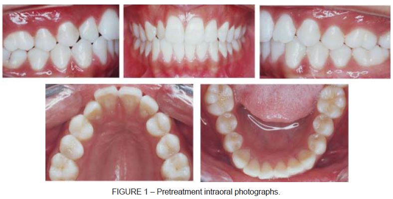
A 15-year-old girl came for an orthodontic treatment at the Orthodontics Department of the Lutheran University of Brazil. The patient had no other complaints. Intraoral examination revealed permanent dentition, absent maxillary canines, crossbite of the maxillary right lateral incisor and Class II molar relationship on both sides. The maxillary arch showed insufficient space to accommodate the permanent canines, whereas the mandibular arch presented tooth crowding, at a negative discrepancy of 3 mm. The dental midline was coincident with the facial midline. The patient had a Class I skeletal pattern, a convex profile and good lip competence (Figure 1).
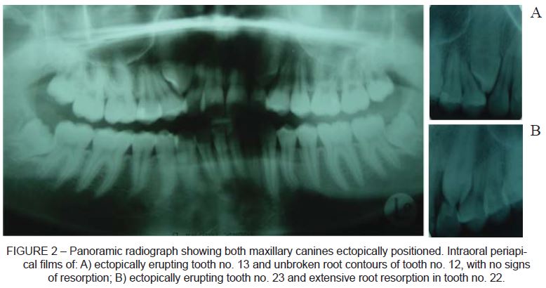
Radiographs revealed impaction of both maxillary canines and severe root resorption of the maxillary left lateral incisor (Figure 2). CT was then required to assess the degree of root resorption in adjacent teeth. Axial and sagital CT scans confirmed the presence of severe root resorption of the left lateral incisor and also revealed resorption involving the apical and middle thirds of the right first premolar. Both maxillary impacted canines were positioned buccally. After careful evaluation and discussion with the patient and her parents, we made a decision to extract the two resorbed teeth (left lateral incisor and right first premolar) and to use orthodontic therapy to induce the eruption of maxillary canines.
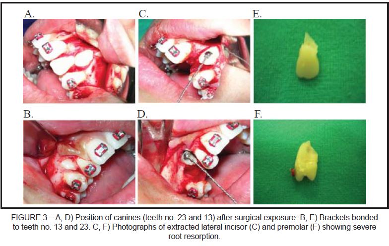
For the orthodontic treatment, upper and lower preadjusted multibrackets appliances were used. The patient was referred to the Oral and Maxillofacial Surgery Department to extract the maxillary left lateral incisor and the maxillary right first premolar and to attach the orthodontic traction devices to the crown of the maxillary canines. The orthodontic device was a retainer with twisted Aciflex wires (Ethicon, Norderstedt, Germany) (Figure 3).
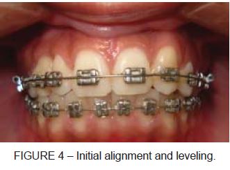
The initial alignment and leveling was achieved using a sequence of 0.014" to 0.020" steel wires. After that, the anchorage unit was set, and 0.014", 0.016" and 0.018" nickel titanium arch wires were superimposed to induce movement of maxillary canines in the oclusal direction. When the oclusal plane was reached, continuous arch wires were used for final teeth alignment and leveling (Figure 4).
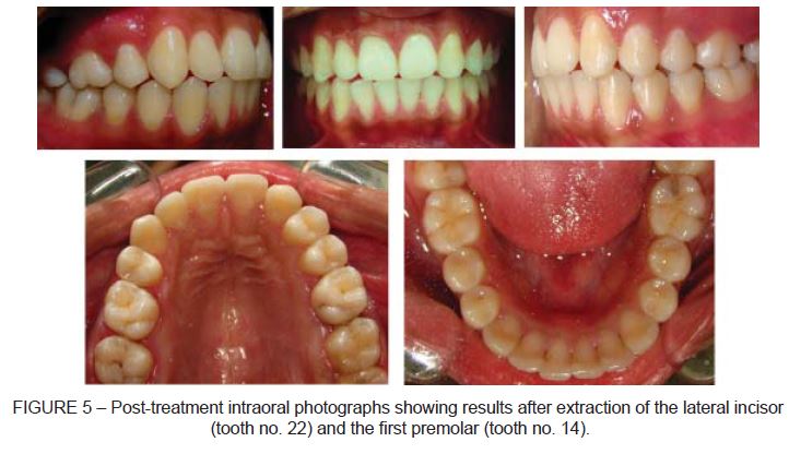
Twenty months after the beginning of orthodontic treatment, the patient was satisfied with the results obtained and requested the orthodontic appliance to be removed before ideal results were able to be achieved. She was referred to the Department of Esthetic Dentistry for volumetric reconstruction of the maxillary left canine. Figure 5 present the clinical characteristics after treatment, demonstrating the satisfactory aesthetic and functional results.
DISCUSSION
Early detection of IMC is important in avoiding damage in adjacent teeth and the analysis of the degree of root resorption is essential for treatment decision-making. In cases of severe root resorption, the risk of losing the tooth should be considered both during and after treatment 1-3.
In this case, periapical and panoramic radiographs revealed IMC and resorption in left lateral incisors. Although root resorption was advanced, no clinical signs or symptoms were detected at the time of diagnosis, which is in agreement with other studies 1,2,4,7. CT images were fundamental to determine the occurrence and degree of root resorption in the first premolar and to confirm the extent of root resorption in the left lateral incisor. In this case, the maxillary arch did not have sufficient space for canine eruption, and extractions were recommended. After analyzing the CT scans, we made a decision to extract the right first premolar and the left lateral incisor, which presented severe resorption as a result of IMC. After the orthodontic treatment and removal of fixed appliances, the anatomy of the crown of the maxillary left canine was reconstructed to aesthetically resemble a lateral incisor. Midline position, overjet and overbite were adequate. Periodontal health was not compromised, and gingival attachment in the canine region was intact. The aesthetic result obtained with the treatment was satisfactory for the patient.
The present report reinforces the importance of early detection of IMC to prevent resorption of adjacent teeth. However, when resorption of adjacent teeth is already present, CT imaging seems to be the best method to analyze its severity, establish a prognosis and define the most appropriate treatment plan 8,9. In such cases, orthodontic treatment may be a more adequate intervention, because it allows to replace teeth that have undergone resorption due to impacted canines and to recover an aesthetic appearance, function and long-term stability.
REFERENCES
1. Alqerban A, Jacobs R, Lambrechts P, Loozen G, Willems G. Root resorption of the maxillary lateral incisor caused by impacted canine: a literature review. Clin Oral Investig. 2009;13:247-55. [ Links ]
2. Ericson S, Kurol J. Radiographic assessment of maxillary canine eruption in children with clinical signs of eruption disturbance. Eur J Orthod. 1986;8:133-40. [ Links ]
3. Shah RM, Boyd MA, Vakil TF. Studies of permanent tooth anomalies in 7,886 Canadian individuals. I: impacted teeth. Dent J. 1978;44:262-4. [ Links ]
4. Ericson S, Kurol J. Incisor resorption caused by maxillary cuspids. A radiographic study. Angle Orthod. 1987;57:332-46. [ Links ]
5. Cooke ME, Nute SJ. Maxillary premolar resorption by canines: three case reports. Int J Paediatr Dent. 2005;15:210-2. [ Links ]
6. Postlethwaite KM. Resorption of premolar roots by ectopic canines. Br Dent J. 1989;167:397-8. [ Links ]
7. Katsnelson A, Flick WG, Susarla S, Tartakovsky JV, Miloro M. Use of panoramic x-ray to determine position of impacted maxillary canines. J Oral Maxillofac Surg. 2010;68:996-1000. [ Links ]
8. Bjerklin K, Ericson S. How a computerized tomography examination changed the treatment plans of 80 children with retained and ectopically positioned maxillary canines. Angle Orthod. 2006;76:43-51. [ Links ]
9. Ericson S, Kurol PJ. Resorption of incisors after ectopic eruption of maxillary canines: a CT study. Angle Orthod. 2000;70:415-23. [ Links ]
10. Brenner D, Elliston C, Hall E, Berdon W. Estimated risks of radiation-induced fatal cancer from pediatric CT. AJR Am J Roentgenol. 2001;176:289-96. [ Links ]
 Corresponding Author:
Carlos Alberto Feldens, PhD
Rua João Telles 185/1301
CEP 90035-121, Porto Alegre, RS
Brazil
E-mail:cafeldens@terra.com.br
