


Services on Demand
Article
 pdf in English
 Article in xml format
 Article references
 Send this article by e-mail
Related links
Share

 Permalink
IJD. International Journal of Dentistry
On-line version ISSN 1806-146X
IJD, Int. j. dent. vol.9 n.1 Recife Jan./Mar. 2010
ORIGINAL ARTICLE ARTIGO ORIGINAL
In vitro evaluation of professional fluoride therapies for improving softening resistance against dental erosion
Avaliação in vitro de terapias de aplicação profissional de flúor para aumento da resistência à desmineralização erosiva
Heitor Marques HonórioI; Daniela RiosII; Ana Carolina MagalhãesIII; Fernanda Herrera StancariIV; Marília Afonso Rabelo BuzalafV
IPhD, Assistant Professor, Department of Pediatric Dentistry, Alfenas Federal University
IIPhD; Assistant Professor, Department Department of Pediatric Dentistry, Bauru School of Dentistry, University of São Paulo
IIIPhD; Assistant Professor, Department of Biological Sciences, Bauru School of Dentistry, University of São Paulo
IVUnder Graduate Student, Bauru School of Dentistry, University of São Paulo
VPhD, Full Professor; Department of Biological Sciences, Bauru School of Dentistry, University of São Paulo
ABSTRACT
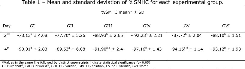
This in vitro study evaluated the effect of professional fluoride therapies against enamel erosion. Ninety bovine enamel blocks were randomly allocated to each group: GI-Duraphat® (NaF, 2.26%F), GII-Duofluorid® (NaF, 2.71% F), GIII-TiF4 varnish (2.45% F), GIV- TiF4 solution (2.45% F), GV-no fluoride varnish and GVI-water (control). The varnishes, the solution and water were applied onto the enamel. The blocks were subjected to 6 sequential erosive cycles (cola drink for 10 min and artificial saliva for 50 min, each) per day, during 4 days. After the erosive cycles, the blocks were maintained in artificial saliva for 18 h. Enamel alterations were determined in the 2nd and 4th days, by microhardness test (%SMHC). Data were tested using ANOVA and Tukey's tests (p<0.05). The mean %SMHC (±SD) in the 2nd and 4th days was, respectively: GI (-78.13±4.08ª and -90.01±2.83ª), GII (-77.70±5.26ª and -89.63±6.08ª), GIII (-88.93±2.65b and -91.90±2.4A,B), GIV (-92.23±2.21b and -97.16±1.43C), GV (-87.72±2.04b and -94.16±1.14B,C) and GVI (-88.10±1.51b and -93.12±1.93B). The data from this in vitro study show that NaF varnishes were able to partially prevent softening from dental erosion until the 4th day of erosive cycle.
Key words: Erosion; Prevention; Dental enamel; Fluoride; Microhardness
RESUMO
O presente estudo in vitro avaliou o efeito de terapias de aplicação profissional de flúor para aumento da resistência à desmineralização erosiva. Noventa blocos de esmalte bovino foram aleatoriamente alocados nos seguintes grupos: GI-Duraphat® (NaF, 2.26%F), GII-Duofluorid® (NaF, 2.71% F), GIII-verniz de TiF4 (2.45% F), GIV-solução de TiF4 (2.45% F), GV- verniz sem flúor e GVI-água (controle). Os vernizes, a solução e a água foram aplicados sobre o esmalte dentário. Os blocos foram submetidos a 6 ciclos seqüenciais de erosão (bebida a base de cola por 10min e saliva artificial por 50min, cada) por dia, durante 4 dias. Após as ciclagens erosiva os blocos foram mantidos em saliva artificial por 18h. As alterações do esmalte foram avaliadas no 2° e 4° dias, por testes de microdureza superficial (%PDS). Os dados foram analisados por ANOVA e teste de Tukey (p<0.05). A média da %PDS (±DP) no 2° e 4° dias foi respectivamente: GI (-78,13±4,08ª e -90,01±2,83ª), GII (-77,70±5,26ª e -89,63±6,08ª), GIII (-88,93±2,65b e -91,90±2,4A,B), GIV (-92,23±2.21b e -97,16±1,43C), GV (-87,72±2,04b e -94,16±1,14B,C) e GVI (-88,10±1,51b e -93,12±1,93 B). Os resultados deste estudo mostram que o verniz de NaF foi capaz de prevenir parcialmente o amolecimento do esmalte pela erosão até o 4º dia de ciclagem erosiva.
Palavras-chave: Erosão; Prevenção; Esmalte dentário; Flúor; Microdureza
INTRODUCTION
Dental erosion is a well recognized dental problem that is apparently increasing among the younger population in the last decades1. Erosion involves a chemical removal of superficial hard tissue from the tooth surface by acids from soft drinks and fruit juices or from eating disorders and gastric reflux2.
The best solution for erosion is its primary prevention by the treatment and elimination of the cause before the lesions on teeth occur3. However, until the cause is not eliminated, several preventive measures have been proposed, including topical fluoride applications3-6. Although the preventive action of fluoride on dental caries is well known7, its role in erosion is still controversial4, since the deposited calcium fluoride-like material from topical fluoride application is supposed to dissolve readily in most acidic drinks4. However, high-concentrated fluoride applications, like oral rinses, acidic gels or varnishes, have been demonstrated, in some cases, to decrease the development of erosion in enamel5,6,8,9. More acidic and concentrated fluoride preparations, which form thicker calcium fluoride-like layer, might offer additional minerals to be dissolved during the erosive challenge, before the subjacent enamel is attacked.
Titanium tetrafluoride (TiF4) solution have also been investigated as a concentrated fluoride product for erosion prevention10-13. The literature describes that after TiF4 application there is a glaze like layer formation that could have an inhibitory effect on erosion10,12. On the other hand, other experiments did not demonstrate promising results in increasing enamel resistance and decreasing the rate of dental erosion development13,14. Similarly to the solution, the titanium tetrafluoride varnish also showed controversial results, with limited15 and significant effect against erosion16.
In view of the above considerations, the purpose of this in vitro study was to test the capacity of different concentrated professional fluoride therapies in improving softening resistance against enamel erosion.
METHODS
Experimental design
Ninety enamel blocks were obtained from bovine teeth, polished and subjected to initial surface microhardness analysis, for selection. Enamel with microhardness ranging from 332 to 377 KHN was randomly distributed into 6 groups: GI- Duraphat® (NaF, 2.26%F, pH 4.5; Colgate, São Bernardo, São Paulo, Brazil, n = 15), GII-Duofluorid® (2.71%F as NaF, 2.92%F as CaF2, pH 8.0; FGM, Joinvile, Santa Catarina, Brasil, n=15), GIII- TiF4 varnish (2.45% F, pH 1.0; FGM, Joinvile, Santa Catarina, Brasil, n = 15), GIV- TiF4 solution (2.45% F, pH 1.0), GV- no-fluoride varnish (FGM, Joinvile, Santa Catarina, Brasil, n=15) and GVI- water (control). The varnishes used in GIII and GV had a composition identical to GII, except for the presence of TiF4 instead of NaF and the absence of fluoride, respectively. The varnishes and the solutions were applied onto the enamel surfaces. Thus, the blocks were subjected to 6 erosive cycles per day, in an oven at 25º C, during 4 days. In each cycle, demineralization and remineralization were performed by immersion in cola drink (10 min) and artificial saliva (50 min), respectively. Each day, the 6 cycles were conducted sequentially (totalizing 6 h) and the blocks were then immersed in artificial saliva for 18 h. Enamel softening was determined in the 2nd and 4th days, using microhardness tests (%SMHC).
Enamel blocks preparation
Enamel blocks (4X4X2.5mm) were prepared from incisor bovine teeth, freshly extracted, sterilized by storage in 2% formaldehyde solution (pH 7.0) for 30 days at room temperature. The enamel surface of the blocks was ground flat with water-cooled carborundum discs (320, 600 and 1200 grades of Al2O3 papers; Buehler, Lake Bluff, IL, USA), and polished with felt paper wet by diamond spray (1 u.m; Buehler), resulting in removal of about 100 u.m depth of the enamel which was controlled with a micrometer. The surface microhardness determination was performed by five indentations (Knoop diamond, 25 g, 5 s, HMV-2000; Shimadzu Corporation, Tokyo, Japan). The enamel blocks with microhardness ranging from 332 to 377 KHN were randomly distributed into 6 groups.
Treatment and erosive cycling
A thin layer of the varnishes was applied with microbrush on enamel surface. After 6 hours, the varnishes were removed carefully using a surgical blade. Removal was completed with cotton swabs with soaked in acetone. For preparing the 4% (2.45% F) TiF4 solution, solid TiF4 (Aldrich Chemical Company, Milwaukee, WI, USA) was dissolved in deionized water. The pH of solution was 1.2. The application was made in drops with cotton roll, during 1 min. The drop was left undisturbed until the surface appeared dry. Additional drops were applied in the same manner until 1 min had elapsed. The water was applied in the same manner.
The blocks were subjected to a erosive cycling model, during 4 days. The 6 erosive cyclings were performed by day. In separate containers, the blocks were immersed in cola drink (Spal, Porto Real, RJ, Brazil) at room temperature, for 10 min (30 mL per block) and in artificial saliva [1.5 mmolL-1 Ca(NO3)2.4H2O, 0.9 mmolL-1 NaH2PO4.2 H2O, 150 mmolL-1 KCl, 0.1 molL-1 Tris buffer, 0.03 ppm F, pH 7.0] for 50 min (15 mL per block).17 Each day, the 6 cycles were conducted sequentially (totalizing 6 h) and the blocks were then maintained in artificial saliva for 18 h.
Microhardness determination
At 2nd and 4th days, the final microhardness test (SMH1) was made as described earlier (Knoop diamond, 25 g, 10 s, HMV-2000; Shimadzu Corporation, Tokyo, Japan). Five indentations, at distances of 200 m.m from each other, were made in the center of enamel blocks (SMH). The percentage of surface microhardness change was calculated for both days: 100(SMH1 -SMH)/SMH.
Statistical analysis
The assumptions of equality of variances and normal distribution of errors were checked. Since the assumptions were satisfied, ANOVA and Tukey's test were carried out for statistical comparisons and the significance limit was set at 5%.
RESULTS0
The data showed that at the 2nd experimental day, GI (Duraphat) and GII (Duofluorid) significantly reduced softening when compared to the other groups (p<0.001). These, did not differ significantly from each other (Table 1).
At the 4th experimental day, the softening increased for all the groups. GI (Duraphat) and GII (Duofluorid) significantly reduced softening when compared to GIV (TiF4 solution), GV (no F varnish) and GVI (control water)(p<0.001). The 4% titanium tetrafluoride varnish (GIII) showed an effect similar to the other commercial fluoride varnishes (GI and GII). However, this varnish (GIII) also showed no significant difference when compared to the no-fluoride varnish (GV) and the control water (GVI) (Table 1).
DISCUSSION
This in vitro experimental study included an intensive erosive attack to simulate what occurs in the clinical situation with patients that show eating disorders, gastric reflux or excessive acid diet. In these cases the high-concentrated fluoride applications have been demonstrated to decrease the development of erosion in enamel5,6. However these applications must be done by the professional and are not suitable for daily home usage. Considering these aspects, only one application of the test products was employed along with a high erosive challenge.
The single fluoride application and the high erosive challenge could explain the limited softening prevention shown by GI and GII when compared to other studies in the literature, which tested the effect of commercial fluoride varnishes and found a better reduction of erosion in vitro8,9,13,18. Another important aspect to consider is the removal of the varnish, which was done six hours after the pH cycling had been initiated, in order to mimic the clinical situation when there is a natural removal of the varnish by tooth brushing, mastication and tongue friction15,16,18. Vieira et al.14 found that the mechanical barrier formed by the varnish played a fundamental role in the protection mechanism against erosion of the product, but in this case a less pronounced erosive attack was accomplished (three 10 min demineralization cycles followed by 2 h of remineralization) and during the experiment the varnish remained on the blocks. Opposite results were found in the present study, because the mechanical protection conferred by the varnish was not present. This rendered GV not different from the control water (GVI) on the 2nd and 4th days.
According to the literature, the beneficial action of TiF4 solution on dental erosion has been attributed to its low pH (around 1.2), favoring the linking between titanium and oxygen of the group phosphate, thus leading to the formation of a titanium dioxide glazelike layer on the surface10,12. In the present study, the formation of this layer was not able to prevent softening of enamel surface. This result is in agreement with Vieira et al.14 that showed in vitro that the 4% TiF4 solution did not have a significant protective effect against erosion.
The varnish allows a higher contact time of fluoride with enamel, thus increasing the formation of calcium fluoride-like deposits.19 It was expected that the incorporation of TiF4 into the varnish would allow the formation of a more resistant compound which in turn would enhance the prevention of erosion, but this was not the case. In fact, both forms of use of TiF4 did not show a significant protective effect against erosion, which is in agreement to two other studies13,14,15 However, other studies evaluating the application of a TiF4 solution or varnish showed better results10,16,20,21, but the methodology was different, with multiple applications or with mild erosive conditions.
The extrapolation of our data to the in vivo condition must be done carefully. For convenience, bovine enamel was used. Although bovine enamel has been widely used in dental research as a substitute for human enamel, morphological differences such as higher porosity exist when compared to human enamel, which result in higher rates of erosion formation22. An enhancement of erosion formation rate may also have been expected because polished surfaces were used and they are more susceptible to acid challenges than natural surfaces.22 In addition, the salivary protection cannot be completely reproduced in the in vitro condition23. Furthermore, it must not be forgotten that in the mouth mechanical factors such as abrasion and attrition may act synergistically with erosion.
CONCLUSION
In conclusion, using this in vitro protocol with a high erosive challenge, the NaF varnishes were able to partially prevent softening from dental erosion until the 4th day of erosive cycle.
Acknowledgements
This study was supported by FAPESP (Proc. 05/54203-3, 2006/00263-8, 2006/045872).
REFERENCES
1. Jaeggi T, Lussi A. Prevalence, incidence and distribution of erosion. Monogr Oral Sci 2006;20:44-65. [ Links ]
2. Imfeld T. Dental erosion. Definition, classification and links. Europ J Oral Sci 1996;104:151-5. [ Links ]
3. Lussi A, Hellwig E. Risk assessment and preventive measures. Monogr Oral Sci 2006;20:190-9. [ Links ]
4. Wiegand A, Attin T. Influence of fluoride on the prevention of erosive lesions - a review. Oral Health Prev Dent 2003;1:245-53. [ Links ]
5. Buchalla W, Lagerwey M, Kohnke S, Becker K, Lennon AM, Attin T. Fluoride is able to reduce erosive and erosive/abrasive enamel loss under severe erosive conditions. Caries Res 2004;38(Suppl):291. [ Links ]
6. Ganss C, Klimek J, Brune V, Schumann A. Effects of two fluoridation measures in erosion progression on enamel and dentine in situ. Caries Res 2004;38:561-6. [ Links ]
7. Ten Cate JM. Review on fluoride, with special emphasis on calcium fluoride mechanisms in caries prevention. Europ J Oral Sci 1997;105:461-5. [ Links ]
8. Sorvari R, Meurman JH, Alakuijala P, Frank RM. Effect of fluoride varnish and solution on enamel erosion in vitro. Caries Res 1994;28:227-32. [ Links ]
9. Vieira A, Jager DHJ, Ruben JL, Huysmans MCDNJM. Inhibition of erosive wear by fluoride varnish. Caries Res 2007;41:61-7. [ Links ]
10. Buyukyilmaz T, Ogaard B, Rolla G. The resistance of titanium tetrafluoride-treated human enamel to strong hydrochloric acid. Eur J Oral Sci 1997;105:473-7. [ Links ]
11. Tveit AB, Hals E, Isrenn R, Totdal B. Highly acid SnF2 and TiF4 solution. Caries Res 1983;17:412-8. [ Links ]
12. Vieira A, Ruben JL, Stokroos I, Huysmans MCDNJM: Surface and surface analysis of eroded bovine enamel pretreated with fluoride. Caries Res 2004;38 (Suppl):391. [ Links ]
13. Vieira A, Ruben JL, Huysmans MC. Effect of titanium tetrafluoride, amine fluoride and fluoride varnish on enamel erosion in vitro. Caries Res 2005;39:371-9. [ Links ]
14. Vieira A, Lugtenborg M, Ruben JL, Huysmans MC. Brushing abrasion of eroded bovine enamel pretreated with topical fluorides. Caries Res 2006;40:224-30. [ Links ]
15. Magalhães AC, Stancari FH, Rios D, Buzalaf MAR. Effect of an experimental 4% titanium tetrafluoride varnish on dental erosion by a soft drink. J Dent 2007;35:858-61. [ Links ]
16. Magalhães AC, Kato MT, Rios D, Wiegand A, Attin T, Buzalaf MAR. The effect of an experimental 4% TiF4 varnish compared to NaF varnishes and 4% TiF4 solution on dental erosion in vitro. Caries Res 2008;42:269-74. [ Links ]
17. Vieira AEM, Delbem ACB, Sassaki KT, Rodrigues E, Cury JA, Cunha RF. Fluoride dose response in pH-cycling models using bovine enamel. Caries Res 2005;39:514-20. [ Links ]
18. Murakami C, Bönecker M, Corrêa MSNP, Mendes FM, Rodrigues CRMD. Effect of fluoride varnish and gel on dental erosion in primary and permanent teeth. Arch Oral Biol 2009, in press. [ Links ]
19. Seppã, L. Effects of a sodium fluoride solution and a varnish with different fluoride concentrations on enamel remineralization in vitro. Scand J Dent Res 1988;96:304-9. [ Links ]
20. Hove L, Holme B, Ogaard B, Willumsen T, Tveit AB. The protective effect of TiF4, SnF2, and NaF on erosion of enamel by hydrochloric acid in vitro measured by white light interferometry. Caries Res 2006;40:444-3. [ Links ]
21. Schlueter N, Ganss C, Mueller U, Klimek J. Effect of TiF4 and NaF on erosion progression in enamel and dentine in vitro. Caries Res 2007;41:141-5. [ Links ]
22. Meurman JH, Frank RM. Progression and surface ultrastructure of in vitro caused erosive lesions in human and bovine enamel. Caries Res 1991;25:81-7. [ Links ]
23. Hall AF, Buchanan CA, Millett DT, Creanor SL, Strang R, Foye RH. The effect of saliva on enamel and dentine erosion. J Dent 1999;27:333-9. [ Links ]
 Correspondence:
Heitor Marques Honório
Rua Gabriel Monteiro da Silva, 714
Alfenas/MG 37130-000, Brazil
Telephone: 55 35 32991424
e-mail: heitorhonorio@yahoo.com.br
Recebido em 27/02/2010
Aprovado em 18/03/2010
