Services on Demand
Article
Related links
Share
RGO.Revista Gaúcha de Odontologia (Online)
On-line version ISSN 1981-8637
RGO, Rev. gaúch. odontol. (Online) vol.59 n.3 Porto Alegre Jul./Sep. 2011
ARTIGO ORIGINAL / ORIGINAL ARTICLE
Comparison of radiopacity in luting resin cements using conventional and digital radiographic systems
Comparação da radiopacidade de cimentos resinosos pelos sistemas de radiografia convencional e digital
Giovana Mongruel GOMESI; Gislaine Cristine MARTINSI ;Bruna Fortes BITTENCOURTI; Osnara Maria Mongruel GOMESI; João Carlos GOMESI; Abraham Lincoln CALIXTOI
I Universidade Estadual de Ponta Grossa, Faculdade de Odontologia
ABSTRACT
Objective
To evaluate the radiopacity of three different resin composite luting cements using the histogram method (conventional radiography) and pixel counting method (digital radiography).
Methods
Fifteen specimens were divided into 3 different resin composite luting cement groups: G-I) Cement-Post (Ângelus®, Londrina, Brazil), G-II) RelyX ARC (3MESPE, St. Paul, Minnesota, USA) and G-III) Variolink II (Ivoclar/Vivadent, Schaan, Liechtenstein). After 24 hours, conventional x-rays of the specimens were taken with a lead gauge, making a visual evaluation by scores, to classify the specimen's radiopacity according to the scale shade (control); they were then scanned for the purposes of analyzing the histogram using Adobe Photoshop CS2, version 8.0. Using the same specimens, x-rays were taken using a Digital X-RAY Intra-Oral System (Gnatus DSR, Ribeirão Preto, Brazil). In this case, the capture of the digital images was performed using Cygnus Imaging® software and the digital radiopacity was measured by counting pixels, using the Image Tool® software (UTHSCSA).
Results
The non-parametric Kruskal-Wallis test and Dunn's post-test (p<0.05) showed that the means and respective standard deviation percentages of white pixels in the digital x-rays were: G-I) 48.94 ±3.16, G-II) 60.22 ±3.86 and G-III) 69.36 ±5.32. As for the conventional x-rays, the means and standard deviations of the histogram analyses that evaluated gray tones were: G-I) 71.98 ±13.02; G-II) 85.40 ±4.47; G-III) 130.51 ±5.82.
Conclusion
In conclusion, regardless of the method used (conventional or digital x-rays), G-III obtained the largest radiopacity value.
Termos de indexação: Cementation. Dental cements. X-Rays.
RESUMO
Objetivo
Avaliar a radiopacidade de cimentos resinosos pelo método do histograma (radiografia convencional) e da contagem de pixels (radiografia digital).
Métodos
Foram utilizados 15 corpos-de-prova divididos em três grupos de cimentos resinosos: Grupo I) Cement-Post (Ângelus®, Londrina, Brasil), Grupo II) RelyX ARC (3MESPE, St. Paul, USA) e Grupo III) Variolink II (Ivoclar/Vivadent, Schaan, Liechtenstein). Após 24 horas, foram realizadas radiografias convencionais dos corpos-de-prova juntamente com uma escala de chumbo, realizando assim uma avaliação visual por escores para classificar a radiopacidade dos corpos-de-prova de acordo com a tonalidade da escala (controle); e em seguida, foram escaneadas para análise do histograma no software Adobe Photoshop CS2 versão 8.0. Com os mesmos corpos-de-prova, realizaram-se tomadas radiográficas empregando-se o Sistema Intra-oral de Raio-X Digital (Gnatus, Ribeirão Preto, Brasil), sendo a captura das imagens digitais realizadas com o software Cygnus Imaging® e a radiopacidade digital mensurada pela contagem de pixels com o software Image Tool® (UTHSCSA).
Resultados
O teste não-paramétrico de Kruskal-Wallis e Dunn (p<0,05) mostrou que as médias das porcentagens de pixels brancos das radiografias digitais e os seus respectivos desvios-padrão foram: Grupo I) 48,94 ±3,16; Grupo II) 60,22 ±3,86 e Grupo III) 69,36 ±5,32. Já nas radiografias convencionais, as médias e os desvios-padrão das análises do histograma avaliando tons de cinza foram: Grupo I) 71,98 ±13,02; Grupo II) 85,40 ±4,47 e Grupo III) 13,51 ±5,82.
Conclusão
Pode-se concluir que utilizando tanto o sistema de radiografia convencional quanto o sistema digital, o Grupo III obteve uma maior radiopacidade.
Termos de indexação: Cimentação. Cimentos dentários. Radiografia.
INTRODUCTION
Dental cements include a broad category of materials used for bonding and sealing dental restorations to the teeth, widely used for cementation of fixed crowns and intra-canal posts. New luting agents, particularly those with adhesive capacity, have been introduced into the market, and the choice of luting agent depends on the clinical situation in which this agent may be used, as well as its physical, biological and manipulative properties1.
For more than a century, zinc phosphate cement has been widely used as a cementing agent, despite its oft-documented disadvantages, including high solubility, lack of adhesion and low initial pH1. Recently, there has been considerable interest in luting materials with adhesive capacity and nowadays there are resin cements that are highly desirable for use in clinical practice, since they are insoluble in the oral environment and may produce high bond strength values on dentin and enamel2. So, the application of luting cements has grown considerably in recent years, mainly due to their superior mechanical properties and increased retentive capacity1.
Basically, the composition of resin cements is similar to that of composite resins: an organic matrix and silica and glass-based inorganic fillers, joined together via the silane agent and their physical properties are determined by the filler type, distribution and content, presenting different opacities, colors and polymerization methods. They are recommended for luting veneers, ceramic crowns and indirect restorations and are also available in three different forms of curing: chemically cured, photocured and dual cured3. Despite all the improvements already made, the use of dual cured luting resins should be approached with caution, due to the limited action of their chemical polymerization agent over time. Therefore, a rigorous photoactivation is required3.
An essential property of luting agents is radiopacity. This material requirement permits a contrast with the tooth structure in radiography4, and is essential to detect proximal excesses, check the marginal adaptation of the marginal adaptation of prosthetic elements and for longitudinal accompaniment of recurrent caries. It is important that these cements have a higher radiopacity than dentin, as problems of interpretation, such as secondary caries or gap formation near the restorations, may be avoided; moreover, it is difficult to detect the cement line radiographically when the material is less radiopaque than dentin5. However, the level of radiopacity varies considerably with the composition of the material, especially with regard to the type and amount of mineral content4,6; thus, it is necessary to evaluate and to compare the radiopacity of some materials that are available in the market, which may be evaluated by different radiographic methods.
As a general rule, photodensitometers are used for reading optical densities on radiographic films, in accordance with the recommendations of the ADA (American Dental Association), and the radiopacity of a luting agent is often compared to a device with aluminum step wedges, i.e. aluminum equivalence, measured in mm7.
Conventional radiographic film has been widely used as an option for recording images, however it possesses several drawbacks, which determine that it could be replaced. Studies using digital radiography only began in the late 1980s. This device was able to facilitate and speed up comparative analysis. Thus, information technology is used to facilitate the acquisition of images with the use of lower doses of radiation, and also eliminates chemical radiographic processing, which is responsible for a great percentage of errors that affect image quality, promoting a better view of density and contrast, depending on the program used. Digital radiography determines gray levels from 0 to 255, with nuances, where "0" is black and "255" is white8.
The digital image is codified by a finite set of values and it is represented in two dimensions by means of digital values, called pixels. Therefore a pixel is the smallest element, or value of an image, representing the digital equivalent of the silver crystal of conventional x-rays. The number of gray levels which it is possible for a pixel of a scanned image to display is called the dynamic range. This dynamic range of scanned images does not exceed that of conventional film. Human beings can only detect between 14 and 16 tones of gray, on rare occasions reaching 30 or 40. Thus, one of the applications of the digital system, which displays a scale of 255 gray tones, is the measurement of the gray level of image areas; this means determine the numerical value that corresponds to the average gray scale of pixels in a given area8. The value (pixel) may also be called the intensity of the image and it represents a property, such as color, tone, brightness, etc.
So, in conventional radiography, radiopacity may be assessed by the number of gray levels that make up the image, while in digital radiography, the radiopacity may be measured by counting pixels. In this way, this study proposed to evaluate the radiopacity of three resin cements comparing different radiographic methods: conventional radiography (score and histogram) and digital image (pixel counting).
METHODS
This study was approved by the Ethical Research Committee of the State University of Ponta Grossa, UEPG, Brazil (protocol # 18097/2008). Using conventional dental radiology and digital dental radiology, the radiopacity of three resin luting cements was evaluated: G-I) Cement- Post (Ângelus®, Londrina, Brazil) (chemically cured), G-II) RelyX ARC (3MESPE, St. Paul, USA) (dual cured) and G-III) Variolink II (Ivoclar/Vivadent, Schaan, Liechtenstein) (light-cured). The materials were handled following the manufacturer's instructions, and 15 specimens (SP) were created, n=5 for each group. Specimens were formed with the aid of a teflon matrix (8 mm in diameter and 2 mm thick), in accordance with ADA guidelines (ISO 4049).
After 24 hours, in conditions of darkness and with a relative humidity at 37ºC, the specimen radiographs were carried out in a dental X-ray device (Gnatus, Ribeirão Preto, Brazil), with 70 kV (kilovolts) and 8 mA (milliamps), using D radiographic films (Kodak velocity D, Kodak Ultra, Eastman Kodak, NY, USA), and placed together with a densitometric lead gauge. Then, the films were developed in an automatic X-ray processor (Revell, Del Grandi Produtos Radiológicos Ltda., São Paulo, Brazil) at 27ºC ± 3ºC with specific developers and fixers for the processing (Prograd, Curitiba, Brazil). A visual evaluation was performed using scoring (1 to 4) to rate the specimen's radiopacity, according to the tone of the densitometric scale (control), where a score of 1 represented the tone with the least radiopacity and a score of 4 corresponded to the highest level of radiopacity. This visual observation was performed after scanning the films (Color Page HR7X Slim Scanner), using Photoshop CS2, version 8.0, to identify the color (tone) on its scale. In addition, an analysis was performed of the histogram generated in Adobe Photoshop CS2 version 8.0 to check for different tones in the specimens.
Using the same specimens, x-rays were taken using the Digital X-Ray Intra-Oral System (Timex-70 DRS, Gnatus, Ribeirão Preto, Brazil), with the same electrical specifications used in conventional radiographic systems (70 kV and 8 mA). Instead of the radiographic film, an imaging plate (sensor) was used and the capture of the digital images was carried out using Cygnus Imaging software. Each specimen's radiopacity was evaluated by a digital imaging system, determining gray levels measured by Image Tool® software (UTHSCSA). The results were obtained by measuring the pixels (px) contained in these images.
The X-ray devices were calibrated with an exposure time of 0.2s (conventional radiography) and 0.06s (digital radiography) and the radiographic position was standardized (the central beam of the x-ray focusing at a 90º angle to the surface of the image receptor, and at a focus-object distance of 10 cm and with the sensor and the specimen in parallel).
Data were analyzed by descriptive statistical analysis and by the non-parametric Kruskal-Wallis test and Dunn's post-test (p<0.05).
RESULTS
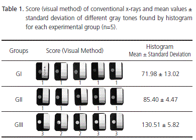
Using the conventional radiography method, it may be demonstrated that, with visual analysis and the use of scores, G-III presented the highest radiopacity and with the analysis obtained from the histogram, G-III also obtained the highest mean (130.51 ± 5.82). However, statistical analysis only demonstrated a significant difference between G-III and G-I (Table 1).


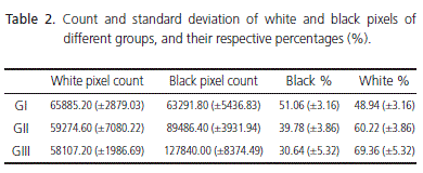
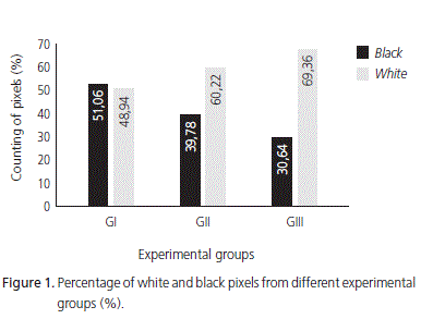
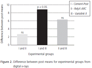
The non-parametric Kruskall-Wallis test (p<0.05) with multiple comparisons and Dunns post-test (p=0.0060) showed that the percentage mean and standard deviation of white pixels from digital x-rays were: G-I) 48.94 ±3.16, G-II) 60.22 ±3.86 and G-III) 69.36 ±5.32; it was also observed that G-III obtained the highest percentages of white pixels, which are responsible for the radiopacity of the material. In Figure 2, differences can be seen between the post means of experimental groups, noting that G-III demonstrated a significant difference only in relation to G-I (p<0.05)

DISCUSSION
Luting cements must have certain properties in order to fulfill their proper clinical use, such as flow, adequate working time, no deterioration in dental fluids, adhesion to dentin walls, tissue tolerance, antimicrobial activity and radiopacity. Therefore, it is essential that these materials have sufficient radiopacity to detect excess cement or the lack of it in the marginal adaptation of prosthetic crowns, using radiographic techniques8. It is important that these cements have higher radiopacity values than dentin, since problems such as erroneous interpretation concerning the presence of secondary caries or gap formation near to marginal restorations can be avoided9.
It is well known that the material radiopacity depends on its composition and the proportion of radiopacifier component. Hence, in the current study, different luting resins were used to compare the digital radiographic method and the conventional method. Potential filler particles that provide radiopacity to zinc phosphate cements, glass ionomer cements and luting resins include zinc oxide, magnesium oxide, fluoridealuminum silicate glass, barium glass, strontium, zirconia and bismuth. In this study, the resin cement that showed the lowest radiopacity was the chemically activated one (G-I), rejecting the null hypothesis, also reported by other authors10 who evaluated the radiopacity of several resin luting cements and found that the chemically activated cement also obtained lower radiopacity values than the others; the use of these relatively radiolucent cements should be contraindicated in certain situations where dental margins are located in areas that are difficult to access and prone to periodic failure; in other words, it becomes difficult to detect these resin cements in these conditions of recurrent failure, thus impairing effective diagnosis9.
Since the conventional radiographic method is still used by most dentists in their clinical practices, the images obtained by this technique were evaluated in this study as a control for comparison / validation of the measurement of radiopacity obtained with digital images.
The digital image has lower spatial resolution than radiographic film and higher contrast resolution. The spatial resolution increases as the number of pixels in the image increases and contrast is defined by the number displayed on the grayscale - the fewer the shades of gray, the greater the contrast. Digitization promotes lower resolution than radiographic film, but sufficient to detect an object of 0.08 mm8. However, the measurement of radiopacity was easily detected using the digital radiographic method, since, after taking the x-ray, the image is already obtained and the measurement may be taken immediately.
Dubrez et al.11, comparing digital analysis and photodensitometry, found that both have the same accuracy of results, which was also observed in the current study, where both the conventional radiography and digital method show the same results. G-III showed the highest radiopacity, since higher values of white pixels, which are responsible for the radiopacity of matter, were obtained. Thus, digitization, a sensitive analytical technique, is able to compensate for the lower spatial resolution, providing accurate and reliable values8. It was possible to corroborate this fact in this study; although the results were similar, it was shown that digital analysis was more sensitive and detailed, bearing out the studies of other researchers12, who concluded that the direct digital image may be as efficient as conventional film in dental practice. Digital radiography is an alternative which offers advantages, such as reduced exposure time without affecting image quality13. In addition, use of the computer permits additional options such as digital image storage and exchange of radiological information. Image manipulation and automatic analysis may benefit the diagnosis. Thus, the digital image certainly has great potential, especially with regard to the increased quality of diagnosis and the automatic analysis of the image8.
CONCLUSION
Within the limitations of this study, it was concluded that there were no differences between the results obtained using conventional or digital radiographic system; increased radiopacity was found in G-III, followed by G-II and G-I, respectively, for both radiographic systems.
REFERÊNCIAS
1. Hill EE, Lott J. A clinically focused discussion of luting materials. Aus Dent J. 2011;56(Suppl 1):67-76. [ Links ]
2. Reis A, Loguercio AD. Materiais dentários diretos: dos fundamentos à aplicação clínica. São Paulo: Santos; 2009.
3. Ruttermann S, Alberts I, Raab WH, Janda RR. Physical properties of self-, dual-, and light-cured direct core materials. Clin Oral Investig. 2011;15(4):597-603.
4. Hotta M, Yamamoto K. Comparative radiopacity of bonding agents. J Adhes Dent. 2009;11(3):207-12. 5. Rosenstiel SF, Land MF, Crispin BJ. Dental luting agents: a review of the current literature. J Prosthet Dent. 1998;80(3):280-301.
6. Tsuge T. Radiopacity of conventional, resin-modified glassionomer, and resin-based luting materials. J Oral Sci. 2009;51(2):223-30.
7. Baksi GB, Sen BH, Eyuboglu TF. Differences in aluminum equivalent values of endodontic sealer: conventional versus digital radiography. J Endod. 2008;34(9):1101-4.
8. Carvalho JR Jr, Correr-Sobrinho L, Correr AB, Sinhoreti MA, Consani S, Sousa-Neto MD. Radiopacity of root filling materials using digital radiography. Int Endod J. 2007;40(7):514-20.
9. Pedrosa R, Brasileiro I, Dos Anjos Pontual M, Dos Anjos Pontual A, Silveira M. Influence of materials radiopacity in the radiographic diagnosis of secondary caries: evaluation in film and two digital systems. Dentomaxillofac Radiol. 2011 Sep;40(6):344-50.
10. Attar N, Tam LE, McComb D. Mechanical and physical properties of contemporary dental luting agents. J Prosthet Dent. 2003;89(2):127-34.
11. Dubrez B, Jacot-Descombes A, Pun T, Cimasoni G. Comparison of photodensitometric with high-resolution digital analysis of bone density from serial dental radiographs. Dentomaxillofac Radiol. 1992;21(1):40-4.
12. Anbiaee N, Mohassel AR, Imanimoghaddam M, Moazzami SM. A comparison of the accuracy of digital and conventional radiography in the diagnosis of recurrent caries. J Contemp Dent Pract. 2010;11(6):25-32.
13. Giusti EC, Fernandes KPS, Marques JLL. Medidas eletrônica e radiográfica digital na odontometria: análise in vivo. RGO - Rev Gaúcha Odontol. 2007;55(3):239-46.
 Endereço para correspondência:
GM GOMES
Universidade Estadual de Ponta Grossa,
Faculdade de Odontologia.
Av. General Carlos Cavalcanti,
Uvaranas, 84030-000, Ponta Grossa, Brasil.
e-mail: giomongruel@gmail.com
Recebido: 12/3/2010
Aceito: 15/1/2011


