


Services on Demand
Article
 pdf in English
 Article in xml format
 Article references
 Send this article by e-mail
Related links
Share

 Permalink
RGO.Revista Gaúcha de Odontologia (Online)
On-line version ISSN 1981-8637
RGO, Rev. gaúch. odontol. (Online) vol.60 n.2 Porto Alegre Apr./Jun. 2012
ORIGINAL / ORIGINAL
In vitro penetration of two cements into artificially created lateral canals
Análise in vitro da obturação de canais laterais simulados utilizando dois tipos de cimentos
Erika Mendonça VIANA I; Eduardo Diogo GURGEL FILHO I; Camila Correia Lima Siqueira VALENTE I; Manuela Delny de Araújo LEITE I; Sâmia Mendonça MONTEZUMA I; Danna Mota MOREIRA II
I Universidade de Fortaleza, Curso de Odontologia. Av. Washington Soares, 1321, Edson Queiroz, 60811-905, Fortaleza, CE, Brasil
II Universidade Estadual de Campinas, Faculdade de Odontologia, Departamento de Endodontia. Piracicaba, SP, Brasil
ABSTRACT
Objective
The aim of this study was to assess the penetration of two obturation materials, Guta-percha (Konne Ind. e Com. Ltda., Belo Horizonte, Brazil) / Pulp Canal Sealer (Kerr Sybron, Romulus, USA) and Resilon (Resilon Research LLC, Madison, CT, USA) / Epiphany (Pentron Clinical Technologies, Wallingford, CT, USA), into artificially created lateral canals.
Methods
Twenty upper, single-rooted human teeth with full or partial crown, straight root and radiograph suggesting one root canal were selected. Six lateral canals were created on each root, two on each third. The teeth were then divided randomly into 2 groups of 10 teeth each. The continuous wave of condensation technique was used to obturate all canals. After obturation, the roots were sectioned cross-sectionally into 3 segments with a diamond disc to expose the lateral canals. The segments were fixed in a fast curing epoxy resin and micrographs of the lateral canals magnified 20 times were taken. The software Carnoy 2.0 was then used to measure obturation material penetration into each lateral canal. The data were treated by the non-parametric Kruskal-Wallis test.
Results
The extent of the lateral canals penetrated by the two obturation materials did not differ significantly in any of the root thirds (p>0.05).
Conclusion
Both obturation materials, Guta-percha (Konne Ind. e Com. Ltda., Belo Horizonte, Brazil) / Pulp Canal Sealer (Kerr Sybron, Romulus, USA) and Resilon (Resilon Research LLC, Madison, CT, USA) / Epiphany (Pentron Clinical Technologies, Wallingford, CT, USA), presented good penetration, which did not differ significantly.
Indexing terms: Dental pulp cavity. Root canal filling materials. Root canal obturation.
RESUMO
Objetivo
Avaliar a capacidade de selamento de canais laterais simulados frente a dois sistemas obturadores: Guta-percha (Konne Ind. e Com. Ltda., Belo Horizonte, Brasil) e cimento Pulp Canal Sealer (Kerr Sybron, Romulus, USA) e Resilon (Resilon Research LLC, Madison, CT, USA) com Epiphany (Pentron Clinical Technologies, Wallingford, CT, USA).
Métodos
Foram selecionados 20 dentes humanos unirradiculares superiores, com coroas totais ou parcialmente íntegras, raízes retas e imagem radiográfica sugestiva de um canal. Foram confeccionados seis canais laterais, com a utilização de uma broca Long Neck ½ (Dentsply-Maillefer, Ballaigues, Suíça), em cada espécime, sendo igualmente distribuído nos 3 terços radiculares. Posteriormente, os dentes foram divididos aleatoriamente em 2 grupos de 10 dentes. A técnica de obturação utilizada foi a Onda Contínua de Condensação. Após as obturações, as raízes foram seccionadas transversalmente com um disco de diamante em 3 segmentos, que possibilitou a visualização dos canais laterais. Os segmentos foram incluídos em uma resina epóxica de presa rápida. As imagens dos terços radiculares inseridos em resina epóxica foram capturadas em lupa estereomicroscópica, com aumento de 20 vezes. Foram realizadas medições lineares das obturações em cada um dos canais dos diferentes segmentos dos dentes de cada grupo através do programa Carnoy 2.0. Os dados foram submetidos à análise estatística utilizando o teste não paramétrico de Kruskall-Wallis.
Resultados
A análise dos resultados demonstrou não haver diferença estatística no escoamento entre os materiais obturadores e seus terços apicais, médios e cervicais (p > 0,05).
Conclusão
Concluiu-se que os dois materiais obturadores, apresentaram um bom escoamento nos canais laterais, não havendo diferença estatística entre eles.
Termos de indexação: Cavidade pulpar. Materiais restauradores do canal radicular. Obturação do canal radicular.
INTRODUCTION
Periapical tissue repair is the main objective of endodontic therapy. There is a consensus that the cleaning, shaping and obturation of the root canal system is the main triad for a successful endodontic treatment1.
Many studies have reported endodontic treatment failure because of the presence of lateral canals2-3, relating treatment success or failure, respectively, to the obturation or not of these canals4.
Hermetic obturation is necessary to prevent the leaking of irritating substances beyond the apical foramen or lateral canals, disrupting periodontal ligament integrity5. A poorly fitting obturation may compromise treatment prognosis regardless of how well the other stages of endodontic therapy were done6.
A successful endodontic treatment depends on many factors and one of them is correct obturation. The obturation must fill the entire system of root canals and seal the apical foramen and lateral canals perfectly with an inert, dimensionally stable and biologically compatible material6.
The cement fills the surface irregularities and acts as lubricant for the gutta-percha cone, improving its fitting. It also obturates the additional open canals and multiple apical foramens7. Studies on gutta-percha and endodontic cements reported that isolated use of these materials compromises performance. However, when combined, they complement each other. If, on the one hand, studies confirm the essentiality of endodontic cement, on the other, its solubility limits its use to the root canal, always as a thin layer8.
Schilder9 helped to improve the three-dimensional understanding of obturations by demonstrating a new way of reducing leakage by warming and condensing gutta-percha inside the root canal. The warm gutta-percha technique has achieved a better fit against the walls of the main and lateral canals when compared with cold lateral condensation9.
In 2004, a new canal obturation material was introduced in the market called Resilon, along with a new endodontic adhesive system, the Epiphany cement10.
Resilon, a biocompatible thermoplastic, contains synthetic polyester polymers. The use of the cone is similar to that of gutta-percha and it is also retractable, since it is soluble in chloroform. It is based on composite resins that can bind to the walls of the canal when used with a self-etching primer and dentinal adhesive11-12. Resilon is the obturation core and contains bioactive crystals, in addition to bismuth oxide and barium sulfate.
Resilon and gutta-percha points are identical and available in ISO calibers and 0.04 and 0.06 conicities, and also in the form of accessory points from size XL to L11-12.
Epiphany Self-Etch Primer can be used with all obturation techniques, that is, cold lateral condensation, vertical condensation and the continuous wave of condensation described by Buchanan13 using System B and the gun Obtura II11-12.
The primer was removed from the composition of Epiphany SE Self-Etch but its properties and instructions for use the same. The new Epiphany SE Self-Etch is the newest advance for endodontic treatments from Pentron Clinical Technologies. When used with any points or pellets, Resilon from Epiphany SE Sealer provides reliable sealing14.
The objective of this study was to assess the ability of two different obturation materials, gutta-percha/ Kerr Pulp Canal Sealer and Resilon/Epiphany, to penetrate lateral root canals.
METHODS
Tooth selection and preparation
Twenty upper, single-rooted human teeth with full or partial crowns, straight roots and radiographic images suggesting the presence of one canal were selected. The teeth were donated by a private practice dental surgeon who took responsibility for the patients' privacy. All teeth were extracted for therapeutic reasons, namely, periodontal problems. The circumstances associated with the extractions are recorded in the patients' records.
The study complied with Resolution 196/96 of the National Council of Health, which incorporates, under the perspective of individuals and the public, four basic bioethical principles: autonomy, non-maleficence, beneficence and justice. These principles aim to ensure the rights and obligations of the scientific community towards research subjects and the State. The study was approved by the University of Fortaleza Research Ethics Committee, protocol number 183/08. All teeth were preserved in 10% formaldehyde and rinsed for 10 minutes with running water immediately before use. The crowns were cut cross-sectionally one millimeter above the cementoenamel junction by a watercooled silicon carbide disc mounted on a straight handpiece with a low-speed micromotor. A single person performed all the stages of the procedure.
Shaping and cleaning
Shaping and cleaning followed the crown-down technique. First, the file Kerr #15 (Dentsply-Maillefer, Ballaigues, Switzerland) was used to explore the root and establish patency. Then, the coronal and middle thirds were shaped by the drills Gates Glidden (Dentsply-Maillefer, Ballaigues, Switzerland) using the instruments from large to small sequence (GG#5, GG#4, GG#3, GG#2). Working length was determined visually, 1mm short of the root length, which was determined by patency once the file crossed the foramen.
The roots were shaped with the files Flexofile 1st series (Dentsply-Maillefer, Ballaigues, Switzerland) and Kerr 2nd series (Dentsply-Maillefer, Ballaigues, Switzerland). The roots of all specimens were standardized with the file Kerr #45, followed by the 3 subsequent files.
The roots were irrigated with 5ml of a 2.5% sodium hypochlorite solution (Biodinâmica Química, São Paulo, Brazil), always making sure to maintain patency after each instrumentation.
Creation of lateral canals
After instrumentation of the main canal, lateral canals were created by the drill Long Neck ½ (Dentsply- Maillefer, Ballaigues, Switzerland). The drill was mounted on a straight handpiece with a low-speed micromotor. Six lateral canals were drilled in each specimen, equally distributed along the 3 root thirds, on the mesial and distal surfaces. All specimens were radiographed and and the radiographs were processed automatically.
Obturation of different groups
The teeth were randomly distributed into 2 groups of 10 teeth, and each group was obturated with a different system: Group I - gutta-percha (Konne Ind. e Com. Ltda., Belo Horizonte, Brazil)/Kerr Pulp Canal Sealer (Kerr Sybron, Romulus, USA); Group II – Resilon (Resilon Research LLC, Madison, CT, USA) / Epiphany (Pentron Clinical Technologies, Wallingford, CT, USA). The canals were irrigated with 17% trisodium ethylenediaminetetraacetic acid (EDTA) (Biodinâmica Química, São Paulo, Brazil) for 5 minutes. This solution was then rinsed out with saline (Gaspar Viana, Fortaleza, Brazil).
The canals were then blotted with paper points. All specimens were obturated by the continuous wave of condensation technique, described by Buchanan (1996), using the device System B (SybronEndo, Orange, CA, USA). The canals were backfilled with the system Obtura II (Obtura Corporation, Fenton, MO, USA). The main cones used were the medium-sized accessory cones Taper 06 (Konne Ind. e Com. Ltda., Belo Horizonte, Brazil), calibrated to size 45 using an endodontic calibration ruler (Dentsply Maillefer, Ballaigues, Switzerland) and cement Kerr Pulp Canal Sealer (Kerr Sybron, Romulus, USA), used according to the manufacturer's instructions.
For group II, the Resilon/EpiphanyY cone and cement were used. The medium-sized cone Taper 06 was calibrated to size 45 using an endodontic calibration ruler and placed in the canal with the cement.
Tooth imaging
After obturation, the teeth were radiographed using a standardized exposure time. The films were processed automatically and filed.
The roots were cross-sectioned into 3 segments by a diamond disc, exposing the lateral canals. The segments were fixed in a fast curing epoxy resin. The blocks were then sanded with wet and dry sandpapers of different grit sizes (400, 800 and 1200, descending grit sizes) and polished by a felt disc with 1.4 μm diamond paste mounted on a lathe spindle.
Micrographs of the root thirds fixed in epoxy resin were taken by the stereomicroscope Leica MZ-5 (Leica Microsystems GmbH, Wetzlar, Hesse, Germany) with 20x magnification. The software Carnoy 2.0 was used for measuring the penetration lengths of the obturation materials in the canal segments of each group. An image of a millimeter ruler is used for software calibration and pasted on each micrograph. The mouse is then used to draw a line over the lateral canal and respective obturation and the software calculates their lengths.
Statistical treatment of the data was done by the software Biostatic 5.0 using the non-parametric Kruskal- Wallis test. All tests were repeated to verify method error. Ten percent of the samples were used and then checked to see if the results matched.
RESULTS
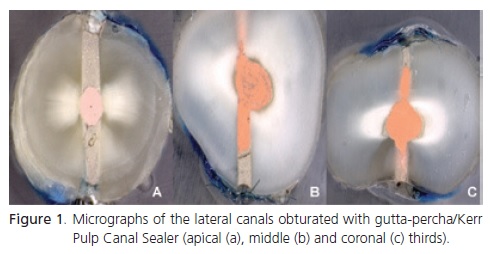
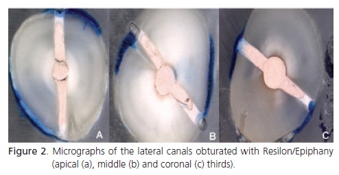
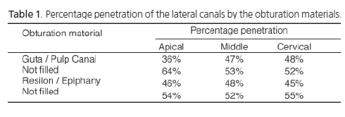
Penetration into the lateral canals by the study obturation materials was measured and compared, as shown by Figures 1 and 2.
Gutta-percha/Kerr Pulp Canal Sealer filled 36% of the lateral canals in the apical third, 47% in the middle third, and 48% in the coronal third. Hence, 64%, 53% and 52% of the lateral canals in the apical, middle and coronal thirds, respectively, were not filled.
Resilon/Epiphany filled 46% of the lateral canals in the apical third, 48% in the middle third, and 45% in the coronal third. Hence, 54%, 52% and 55% of the lateral canals in the apical, middle and coronal thirds, respectively, were not filled. The data is presented in Table 1.
Penetration into the lateral canals in the apical, middle and coronal thirds of the roots by the two obturation materials did not differ significantly (p>0.05).
DISCUSSION
Many studies have reported successful endodontic treatments in teeth with lateral canals4,15-16. The objective of this study was to assess the penetration of two obturation materials, gutta-percha/Kerr Pulp Canal Sealer and Resilon/ Epiphany, into lateral canals.
Some authors have used resin blocks to assess penetration of different obturation materials, but these have limitations when compared with extracted teeth since they do not have dentin and smear layer17-20. The present study used extracted human teeth. Six lateral canals with an approximate diameter of 150 μm were created on each tooth. This diameter is similar to the diameter of lateral canals reported by some studies19,21-23. The creation of lateral canals has been recommended to prevent misshaping the obturation while sectioning the tooth or exposing the filling material in teeth that have been chemically decalcified23-24.
Brothman25 has shown that vertical condensation of warm gutta-percha practically doubles the number of obturated lateral canals when compared with cold lateral condensation of gutta-percha, justifying the choice of the continuous wave of condensation technique. Many studies have reported that vertical condensation fills main canal and lateral canal irregularities better than lateral condensation17-21,23,26.
The penetration of the two study obturation materials, gutta-percha/Kerr Pulp Canal Sealer and Resilon/ Epiphany, into the lateral canals of the three root thirds did not differ significantly. Gutta-percha/Kerr Pulp Canal Sealer and Resilon/Epiphany obturated 36% and 46%, respectively, of the lateral canals in the apical third, 47% and 48%, respectively, of the lateral canals in the middle third, and 48% and 45%, respectively, of the lateral canals in the coronal third.
Almeida et al.23 compared the cements Kerr Pulp Canal Sealer and Epiphany and found that both cements achieved high penetration into the three thirds of the canals, suggesting that both cements could penetrate lateral canals. They did not find significant penetration differences between these two cements, confirming the results of the present study.
Alicia Karr et al.22 compared the penetration of gutta-percha and Resilon into lateral grooves and depressions. Both obturation materials presented similar penetration into lateral dentinal grooves and depressions 3, 5 and 7 millimeters short of the working lengths but better penetration of gutta-percha 1 millimeter short of the working length when the cursor of System B was used 3 millimeters short of the working length.
Karabucak et al.24 compared the penetration of gutta-percha and Resilon into lateral canals using thermoplastic obturation techniques and found that both systems penetrated the lateral canals well, corroborating the results of this study.
The present study found that both cements penetrated the lateral canals well, meaning that both can be used successfully in clinical practice.
Future studies should investigate the penetration of different obturation materials into lateral canals with different variable analysis or the use of temporary fillings applied with different obturation techniques, helping dental surgeons to make better choices in clinical practice and improve treatment outcomes.
CONCLUSION
Considering the methodology and limitations of this study, there was no significant difference between the penetrations of gutta-percha/Kerr Pulp Canal Sealer and Resilon/Epiphany into artificially created lateral canals.
Collaborators
EM VIANA instrumented and obturated all the teeth, carried out the laboratory work and wrote the article. ED GURGEL FILHO supervised the study and helped to write the article. CCLS VALENTE, MDA LEITE and SM MONTEZUMA helped with the laboratory work and to write the article. DM MOREIRA took the micrographs of the specimens at the State University of Campinas School of Dentistry and helped to write the article.
REFERENCES
1. De Deus GA, Krebs RL, Gurgel-Filho ED, Coutinho-Filho T, Lopes MF. Avaliação do grau de limpeza obtido por duas técnicas de instrumentação. Rev Bras Odontol. 2000;57(6):354-8. [ Links ]
2. Nguyen NT. Obturação do sistema de canais radiculares. In: Cohen S, Burns RC. Caminhos da polpa. 6ª ed. Rio de Janeiro: Guanabara; 1997. p. 216-70.
3. Nicholls E. Lateral radicular disease due lateral branching of the root canal. Oral Surg Oral Med Oral Pathol. 1963;16(7):839-45.
4. Weine FS. The enigma of the lateral canal. Dent Clin Nort Am. 1984;28(4):833-53.
5. Lifshitz J, Schilder H, Pameijer CH. Scanning electron microscope study of the warm gutta-percha technique. J Endod. 1983;19(1):17-24.
6. De Deus QD. Endodontia. 5ª ed. Rio de Janeiro: Medsi; 1992.
7. Ruddle CJ. Obturação do sistema de canais radiculares. In: Cohen S, Burns RC. Caminhos da polpa. 6ª ed. Rio de Janeiro: Guanabara; 1997. p. 240-6.
8. Bhatti SA, Joshi R. Thermoplasticized gutta-percha obturation techniques. Dent Update. 1997;24(1):10-3.
9. Schilder H. Filling root canal in three dimension. Dent Clin Nort Am. 1967;11:723-44.
10. Paqué F, Sirtes G. Apical sealing ability of Resilon/Epiphany versus gutta-percha/Ah Plus: immediate and 16-months leakage. Int Endod J. 2007;40:722-9.
11. Morante PRT. ¿Es el Resilon el nuevo material de obturación endodóntica? Ciência. 2006;166:116-32.
12. Guelfand CS, Benítez MSR. Uso clínico del ResilonY: un nuevo material adhesivo para la obturación de los conductos radiculares. Ciência. 2006;167:92-108.
13. Buchanan LS. The continuous wave of condensation: centered condensation of warm gutta-percha in 12 seconds. Dent Today. 1996;15(1):60-7.
14. Pentron Clinical Technologies. Epiphany® soft resin endodontic obturation system: introducing the new standard in obturation [text on the Internet]. Wallingford: Pentron Clinical Technologies [cited 2010 Mar 15]. Available from: <http://www.pentron.com/Pentron/admindocs/kits_data_62.pdf.>.
15. Seltzer S. Endodontology: biological considerations in endodontics. New York: McGraw-Hill; 1981.
16. Xu Z, Zhang Z. Filling of the lateral canal: report of two cases. Oral Surg Oral Med Oral Pathol. 1984;58(2):221-4.
17. Dulac KA, Nielsen CJ, Tomazic TJ, Ferrillo PJ, Hatton JF. Comparison of the obturation of lateral canals by six techniques. J Endod. 1999;25(5):376-80.
18. Gurgel-Filho ED, Feitosa JPA, Gomes BPFA, Ferraz, CCR, Souza- Filho FJ, Teixeira FB. Assessment of different gutta-percha brands during the filling of simulated lateral canals. Int Endod J. 2006;39(2):113-8. doi: 10.1111/j.1365-2591.2006.01054.x.
19. Goldberg F, Artaza LP, De Silvio AC. Effectiveness of different obturation techniques in the filling of simulated lateral canals. J Endod. 2001;27(5):362-4.
20. Wolcott J, Himel VT, Powell W, Penney J. Effect of two obturation techniques on the filling of lateral canals and the main canal. J Endod. 1997;23(10):632-5. doi: 10.1016/S0099- 2399(97)80176-8.
21. Sousa BC, Gomes FA, Carvalho PRB, Ferreira CM, Gurgel-Filho ED, Albuquerque DS. Filling lateral canals: evaluation of different filling techniques. Eur J Dent. 2010;4(3):251-6.
22. Alicia Karr N, Braumgartener JC, Marshall JG. A comparison of gutta-percha and Resilon in the obturation of lateral grooves and depressions. J Endod. 2007;33(6):749-52. doi: 10.1016/j. joen.2007.02.017.
23. Almeida JFA, Gomes BPFA, Ferraz CCR, Sousa-Filho FJ, Zaia AA. Filling of artificial lateral canals and microleakage and flow of five endodontic sealers. Int Endod J. 2007;40(9):692-9. doi: 10.1111/j.1365-2591.2007.01268.
24. Karabucak B, Kim A, Chen V, Iqbal MK. The comparison of guttapercha and Resilon penetration into lateral canals with different thermoplastic delivery systems. J Endod. 2008;34(7):847-9. doi: 10.1016/j.joen.2008.03.024.
25. Brothman P. A comparative study of vertical and lateral condensation of gutta-percha. J Endod. 1981;7(1):27-30. doi: 10.1016/S0099-2399(81)80264-6.
26. Canalda-Sahli C, Berástegui-Jimeno E, Brau-Aguade E. Apical sealing using two thermoplasticized gutta-percha techniques compared with lateral condensation. J Endod. 1997;23(10):636- 8. doi: 10.1016/S0099-2399(97)80177-X.
 Correspondence to:
EM VIANA
e-mail: erika_mendovi@hotmail.com
Received on: 9/11/2010
Final version resubmitted on: 7/1/2011
Approved on: 11/6/2011
