


Services on Demand
Article
 pdf in English
 Article in xml format
 Article references
 Send this article by e-mail
Related links
Share

 Permalink
RSBO (Online)
On-line version ISSN 1984-5685
RSBO (Online) vol.7 n.4 Joinville Dec. 2010
ORIGINAL RESEARCH ARTICLE
Adhesive coronal seal of Syntac and Tetric flow following different dentine pretreatment protocols
Johannes EbertI; Roland FrankenbergerII; Christina KarlI; Anselm PetscheltII; Matthias Johannes RoggendorfII
IDental Clinic 1, Operative Dentistry and Periodontology, University of Erlangen-Nuremberg – Erlangen – Germany
IIDepartment of Operative Dentistry and Endodontics, Dental School, Philipps-University of Marburg – Marburg – Germany
ABSTRACT
INTRODUCTION: The purpose of this study was to test different sealer removal protocols following root canal filling before adhesive seal of access cavities.
MATERIAL AND METHODS: Forty single root teeth were selected for the study, prepared to size 60 taper .02, and filled with AH plus and a single gutta-percha cone size 55 taper .02. Excess sealer was removed with: ethanol-moisturised foam pellet (group 1), pellet and additional etch-and-rinse procedure (group 2), pellet and additional preparation with a water-cooled diamond bur (group 3) or by etch-and-rinse following temporary filling for one week (group 4). Syntac and Tetric flow were used as a secondary protective seal. A dye penetration test (centrifugation 3min / 30G; 5% methylene blue) was carried out. Results were analyzed statistically using PASW 18.0 (Kolmogorov-Smirnov-test, Kruskal-Wallis-test, Mann-Whitney-test; p < 0.05).
RESULTS: Groups 2, 3 and 4 revealed less leakage than group 1 (p < 0.05; Mann-Whitney-tests) and displayed no coloration exceeding the adhesive seal. Teeth with immediate (group 2) or delayed (group 4) adhesive seal showed similar results.
CONCLUSION: Acid etching or bur preparation may be recommended before adhesively sealing the access cavity in single-rooted teeth. There is no need to wait until the sealer has set.
Keywords: dentine bonding; etch-and-rinse; secondary protective seal.
Introduction
To date, no root canal filling method or material alone is able to prevent coronal bacterial leakage over an extended period of time. In laboratory tests, root canal fillings usually leak after one to three months of exposure to microorganisms [7, 11, 12, 19, 20]. Temporary restorations prevent the penetration of bacteria for not more than two weeks [2, 4]. Thus, an additional "secondary protective seal"over root canal fillings using dentine adhesives should be applied, as proposed by Belli et al. [5].
Dentine bonding agents are known to be relatively technique-sensitive [9]. Within the clinical situation of a freshly obturated root canal, remnants of gutta-percha and sealer are possible contaminants of dentine. Compared to adhesive luting of tooth-colored inlays [8], the effect of contamination on pulp chamber dentine is not fully understood. Thus, the aim of the present study was to investigate different sealer removal protocols prior to adhesive seal of access cavities. Furthermore, it should be found out if a temporary filling for one week is needed to allow the sealer to set. The null hypothesis tested was that the different pretreatments before the adhesive seal have no influence on microleakage of the coronal seal.
Material and methods
Forty teeth with one straight root canal were selected for the study. Teeth were stored in a 0.5% Chloramine-T solution (Merck, Darmstadt, Germany) and used within one month after extraction.
The coronal two thirds of the crowns were removed. Standardised access cavities of approximately 3 mm x 5 mm were cut into the coronal aspects of the teeth. The teeth were checked to be caries-free at this level to avoid a possible influence of cariously altered dentine. Root canals of all teeth were prepared to size 60 taper .02 by nickel-titanium instruments (FlexMaster, VDW, Munich, Germany) 1 mm short of the apex. All instrumentation was accompanied by irrigation with 1 mL of 3% NaOCl followed by 1 mL of 40% citric acid after each instrument. A final irrigation with 1 mL of 40% citric acid followed by 1 mL of 3% NaOCl and, finally, 1 mL of 70% ethanol was performed and the root canals were dried with paper points.
Each root canal was obturated with AH Plus (Dentsply), placed with a lentulo spiral size 40, and a single gutta-percha cone size 55 taper .02 (Coltène Whaledent, Langenau, Germany) adjusted to tug fit. No additional condensation was carried out. Excess gutta-percha was removed with a hot instrument by cutting off at the orifice of the canals.
Following this, specimens were randomly divided into 4 groups of 10 teeth each, according to different pretreatment protocols prior to application of the dentine bonding agent.
Group 1 (without etch/bur): The walls and the floor of the pulp chamber were cleaned of excess sealer with ethanol-moisturised foam pellets until it appeared to be clean as judged by the naked eye. The dentine was gently air-dried. Then a thin layer of Syntac Primer (Ivoclar-Vivadent, Schaan, Liechtenstein) was applied for 15 s and thoroughly air blown to remove solvent and water. The pulp chamber region was treated with Syntac Adhesive (Ivoclar-Vivadent) for 10 s and also air blown. Finally, a thin layer of Syntac Heliobond (Ivoclar-Vivadent) was applied with a brush, gently air blown to prevent pooling and photo-polymerized (Polylux II, KaVo, Biberach, Germany) for 40 s. Following this, Tetric flow (Ivoclar-Vivadent) was applied on the pulp chamber floor and root canal orifice in one portion and light cured for 40 s. This resulted in a layer of approximately 2.5 mm of composite.
Group 2 (etch-and-rinse): Following cleaning with ethanol-moisturised foam pellets, dentine was etched for 10 s with a 37% phosphoric acid gel, rinsed with water-spray for 30 s and gently air-dried. The coronal seal was performed like in group 1.
Group 3 (bur): Following the use of the pellets, a thin layer of pulp chamber wall dentine was ground away with a rough water-cooled diamond bur mounted on a high-speed handpiece (KaVo). After cleaning with ethanol and air-drying, the sealing was performed in the same way as in group 1.
Group 4 (temporary filling): A temporary filling (Cavit, 3M-ESPE, Seefeld, Germany) was placed for one week (37ºC, 100% humidity) to allow complete setting of the sealer. After removal of Cavit with a scaler, dentine pretreatment and adhesive sealing were performed like in group 2.
After that, all teeth were stored in 0.5% Chloramine-T solution again until the sealer had set. The coronal surfaces of the specimen were gently ground flat with wet 600-grit SiC paper to expose the cross-sectional area of the adhesive seal and its transition to dentine. The roots of the teeth were covered with two layers of nail varnish, leaving out the coronal surface. Then the teeth were placed into test tubes together with 5% methylene blue dye solution (Merck), pipetted to a height of 30mm. Coronal dye-penetration was performed using centrifugation for 3 min at 30 G (Varifuge-K, Heraeus Christ, Osterode, Germany; 400 rpm).
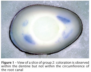
Each specimen was embedded in a resin material (Modralit-3K; Dreve Dentamid, Unna, Germany) and attached to a cutting machine (Buehler Isomet saw, Buehler, Düsseldorf, Germany), equipped with a sectioning blade with a thickness of 0.25 mm. The first, very thin (< 0.3 mm) slice of the coronal aspect was not included in the evaluation due to coloration of the dentinal surface, making it impossible to identify the coloration of the restoration-to-dentine interface. Transversal cuts were made perpendicular to the long axis under water cooling to obtain slices at 0.75 mm intervals (figure 1) and to harvest 6 slices for each tooth.
Dye penetration was scored along the restoration-dentine interface using a microscope (Wild stereomicroscope, Leica Geosystems AG, Heerbrugg, Switzerland) at x40 magnification. Linear dye penetration (simple yes / no decision per slice) and dye penetration area were measured. For the latter, a video camera attached to a computer was used. Pictures of the specimen slices were analyzed with the software Tifmess 1.7 [10]. In every slice, the length of dye penetration around the adhesive seal or the filled root canal system could be recorded. This value was multiplied with the distance between the sectional planes (0.75 mm). All such values per tooth were added up.
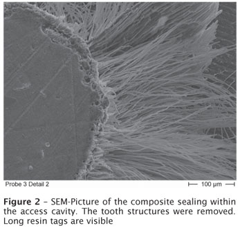
Samples of each group were mounted on small glass plates and subjected to a procedure to remove all tooth substance. To do that, the specimens were decalcified in 1 239;N HCl for 3 days followed by the removal of organic substances in NaOCl 5% for one day. The specimens were dried and gold sputtered (Balzers SCD 050, Balzers, Balzers, Liechtenstein). Then a microstructure analysis was performed under a Scanning electron microscope (SEM) (Leitz ISI Mini-SEM SR 50, Akashi, Tokyo, Japan).
Data were statistically analyzed with PASW 18.0 (SPSS, Chicago, IL, USA), using Kolmogorov-Smirnov test, Kruskal-Wallis test and Mann-Whitney test. The level of significance was set at = 0.05.
Results
Results for linear dye penetration and dye penetration area for each group are presented in table I. Kolmogorov-Smirnov test revealed normal distribution only for group 1. Thus, non-parametric test was applied. Statistically significant differences between groups were identified (Kruskal-Wallis test, p < 0.001). Much deeper dye penetration could be evaluated in group 1 compared to groups 2-4 (Mann-Whitney-tests, p < 0.05), where dye could mostly be discovered not even in the first slice. Results for linear dye penetration and penetration area were similar.

In some of the samples, coloration of dentinal tubules originating from the coronal aspect of the roots stopped some tenth of millimetres before reaching the dentine-restoration-interface (figure 1). An explanation for this finding can be found within the SEM-evaluation: long tags of composite resin were observed (figure 2), obviously preventing further spreading of the dye towards the adhesive sealing of the pulp chamber.
Discussion
For the results of the dye penetration test, statistical analysis revealed a significant impact of the pretreatment protocol. Thus, the null hypothesis was rejected.
The fluid movement model [16, 21] has achieved widespread use for apical leakage testing in Endodontics. However, within the present study, a combined system of root canal filling and adhesive seal was to be tested. By utilization of the fluid movement model it would have been difficult to detect voids, if only one of the two components would leak. Radioisotopes and dyes could also detect coronal microleakage within the coronal sealing alone. Dye penetration tests are fast and easy methods [17]. Methylene blue is a small molecule that penetrates further than other dyes [1] and radioisotopes [13]. It was also shown that dye penetration tests allow better distinction between groups than bacterial leakage tests [3]. Thus, dye penetration with methylene blue was chosen for the present study.
The inclusion of group four may seem odd, because an additional appointment only to perform the adhesive sealing would not be practical both for the dentist and the patient. The group was included to find out, whether it is necessary to wait until the sealer has set before applying the adhesive coronal seal. According to our results, this is not the case.
The results of the present study show that the use of an alcohol-moisturised foam pellet alone obviously was not sufficient to remove remnants of the sealer and that the dentine bonding thus was not able to provide a tight protective seal over the root canal filling. Both groups with the use of the etch-and-rinse method and the group with the use of the bur performed equally well (groups 2, 3 and 4) and better than group 1. On the other hand, in groups 2 and 3, the development of voids within the coronal aspect of the sealer and gutta-percha by the use of water spray and the mechanical action of the bur may have occurred because the sealer had not set yet. Obviously they were filled afterwards by the adhesively bonded flowable composite and thus represented no problem.
Within our study, the single cone technique was used, as in some recently published studies [6, 14, 15, 17, 18, 22]. In this way, a "worst-case scenario"should be simulated regarding gross contamination by sealer remnants. In further studies, it has to be evaluated if other root canal filling techniques may reveal better sealing of the protective layer even without etching or bur preparation due to less sealer contamination of the access cavities.
The results of the present study showed good potential of sealing for the combination of Syntac and Tetric flow. However, whether this is true for other dentine bonding systems and other composites has to be evaluated by further studies. It also has to be clarified, whether the results can be transferred to larger teeth, as molars. The greater dimensions within the access openings of these teeth may cause problems due to higher shrinkage forces. A further aspect of interest is, whether the adhesive seal is able to survive chewing forces, where normally no stabilizing restorations are placed into the teeth when the patient is referred from the endodontist back to his general dentist.
Conclusion
According to the results of the present study, at the end of an endodontic treatment an etch-and-rinse procedure or bur preparation may be recommended prior to application of a secondary protective seal by adhesively bonded flowable composite. There is no need for a second appointment to wait for sealer setting.
References
1. Ahlberg KM, Assavanop P, Tay WM. A comparison of the apical dye penetration patterns shown by methylene blue and India ink in root-filled teeth. Int Endod J. 1995 Jan;28(1):30-4. [ Links ]
2. Balto H, Al-Nazhan S, Al-Mansour K, Al-Otaibi M, Siddiqu Y. Microbial leakage of Cavit, IRM, and Temp Bond in post-prepared root canals using two methods of gutta-percha removal: an in vitro study. J Contemp Dent Pract. 2005 Aug;6(3):53-61. [ Links ]
3. Barthel CR, Moshonov J, Shuping G, Ørstavik D. Bacterial leakage versus dye leakage in obturated root canals. Int Endod J. 1999 Sep;32(5):370-5. [ Links ]
4. Barthel CR, Strobach A, Briedigkeit H, Gobel UB, Roulet JF. Leakage in roots coronally sealed with different temporary fillings. J Endod. 1999 Nov;25(11):731-4. [ Links ]
5. Belli S, Zhang Y, Pereira PN, Özer F, Pashley DH. Regional bond strengths of adhesive resins to pulp chamber dentine. J Endod. 2001 Aug;27(8):527-32. [ Links ]
6. Brackett MG, Martin R, Sword J, Oxford C, Rueggeberg FA, Tay FR et al. Comparison of seal after obturation techniques using a polydimethylsiloxane-based root canal sealer. J Endod. 2006 Dec;32(12):1188-90. [ Links ]
7. Chailertvanitkul P, Saunders WP, MacKenzie D. Coronal leakage of obturated root canals after long-term storage using a polymicrobial marker. J Endod. 1997 Oct;23(10):610-3. [ Links ]
8. Frankenberger R, Krämer N, Lohbauer U, Nikolaenko SA, Reich SM. Marginal integrity: is the clinical performance of bonded restorations predictable in vitro? J Adhes Dent. 2007;9(Suppl 1):107-16. [ Links ]
9. Frankenberger R, Krämer N, Petschelt A. Technique sensitivity of dentine bonding: effect of application mistakes on bond strength and marginal adaptation. Oper Dent. 2000 Jul-Aug;25(4):324-30. [ Links ]
10. Frankenberger R, Tay FR. Self-etch vs etch-and-rinse adhesives: effect of thermo-mechanical fatigue loading on marginal quality of bonded resin composite restorations. Dent Mater. 2005 May;21(5):397-412. [ Links ]
11. Khayat A, Lee SJ, Torabinejad M. Human saliva penetration of coronally unsealed obturated root canals. J Endod. 1993 Sep;19(9):458-61. [ Links ]
12. Magura ME, Kafrawy AH, Brown Jr CE, Newton CW. Human saliva coronal microleakage in obturated root canals: an in vitro study. J Endod. 1991 Jul;17(7):324-31. [ Links ]
13. Matloff IR, Jensen JR, Singer L, Tabibi A. A comparison of methods used in root canal sealability studies. Oral Surg Oral Med Oral Pathol. 1982 Feb;53(2):203-8. [ Links ]
14. Monticelli F, Sadek FT, Schuster GS, Volkmann KR, Looney SW, Ferrari M et al. Efficacy of two contemporary single-cone filling techniques in preventing bacterial leakage. J Endod. 2007 Mar;33(3):310-3. [ Links ]
15. Monticelli F, Sword J, Martin RL, Schuster GS, Weller RN, Ferrari M et al. Sealing properties of two contemporary single-cone obturation systems. Int Endod J. 2007 May;40(5):374-85. [ Links ]
16. Pashley DH, Thompson SM, Stewart FP. Dentine permeability: effects of temperature on hydraulic conductance. J Dent Res. 1983 Sep;62(9):956-9. [ Links ]
17. Roggendorf MJ, Ebert J, Petschelt A, Frankenberger R. Influence of moisture on the apical seal of root canal fillings with five different types of sealer. J Endod. 2007 Jan;33(1):31-3. [ Links ]
18. Sagsen B, Er O, Kahraman Y, Orucoglu H. Evaluation of microleakage of roots filled with different techniques with a computerized fluid filtration technique. J Endod. 2006 Dec;32(12):1168-70. [ Links ]
19. Torabinejad M, Ung B, Kettering JD. In vitro bacterial penetration of coronally unsealed endodontically treated teeth. J Endod. 1990 Dec;16(12):566-9. [ Links ]
20. Wolanek GA, Loushine RJ, Weller RN, Kimbrough WF, Volkmann KR. In vitro bacterial penetration of endodontically treated teeth coronally sealed with a dentine bonding agent. J Endod. 2001 May;27(5):354-7. [ Links ]
21. Wu MK, De Gee AJ, Wesselink PR, Moorer WR. Fluid transport and bacterial penetration along root canal fillings. Int Endod J. 1993 Jul;26(4):203-8. [ Links ]
22. Wu MK, van der Sluis LW, Wesselink PR. A 1-year follow-up study on leakage of single-cone fillings with RoekoRSA sealer. Oral Surg Oral Med Oral Pathol Oral Radiol Endod. 2006 May;101(5):662-7. [ Links ]
 Corresponding author:
Johannes Ebert
Dental Clinic 1 – Operative Dentistry and Periodontology –
Glueckstr. 11, 91054 Erlangen – Germany
E-mail: ebert@dent.uni-erlangen.de
Received for publication: August 11, 2010.
Accepted for publication: August 26, 2010.
